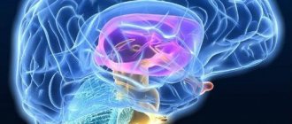
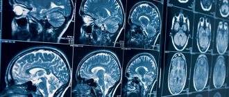
One of the most important human organs is the brain. There is no life without it, because it is the coordinator of the work of all departments of the body. Therefore, it is necessary to take timely care of its health and normal functioning. Today, MRI of the brain with contrast is considered the most highly informative method. Where is it better to do this scan and for what?
Magnetic resonance imaging is based on the principle of nuclear magnetic resonance, that is, the ability of the nuclei of certain substances to emit and absorb the energy of radio frequency pulses. But MRI of the brain with contrast is even more accurate. It helps to visualize pathologies that cannot be determined using X-rays, CT scans, or even conventional tomography. The advantages of this method are painlessness, non-invasiveness, safety, and the absence of a recovery period between checks.
MRI of the brain with contrast, which shows
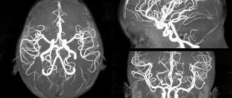
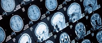
Magnetic resonance imaging allows you to visualize an accurate three-dimensional image of the brain; it can be used to examine in detail the deep layers of tissue and blood vessels of the head. And at the same time avoid overlapping the image of the skull bones. It shows disturbances in brain activity, helps detect tumors, aneurysms, and diseases of the nervous system. The quality of the images reveals a complete picture of all structures. The advantage of this technique is the ability to track dynamics, so it is often used before and after surgery.
MRI of the brain with and without contrast: differences
The main difference is the administration of a substance containing gadolinium ions or salts into the body. Whereas the usual procedure does not require the use of special drugs.
The contrast is a coloring liquid. Once injected, it spreads through the bloodstream and illuminates the tissues, making them clearer. The more intense the blood flow, the faster the contrast agent disperses. The magnetic field of the tomograph visualizes the organ being examined. Damaged tissues change color under its influence, allowing the specialist to determine the boundaries of the pathology as accurately as possible.
MRI of the brain with contrast helps to reveal a detailed picture of the state of the blood vessels, the processes occurring in the brain and its reaction to external stimuli. When examining neoplasms, metastases and their locations can be detected. In addition, this method makes it possible to diagnose tumors no larger than 1 millimeter in size. All changes that occur are monitored in real time.
The survey has two subtypes:
- The first involves gradual drip administration of the drug and is prescribed for diagnosing disorders of the vascular system.
- The second requires a bolus injection of dye. This method allows for a large volume of the drug to enter the vascular bed in one injection. It is designed to detect cancer and vascular accidents.
Since no preparation is required for a brain MRI with contrast, you can decide that examinations can be done as often as you like. In fact, there are a number of limiting factors:
- Contrast tolerance by the patient;
- Identifying side effects;
- Subjective well-being of the patient.
If tolerance is good, then adults can be examined once every seven days. In the event of side effects or a negative reaction, the procedure is carried out only if serious indicators are present.
It is worth remembering that the contrast substance itself cannot have a harmful effect, since it is excreted from the body in the urine during the day, which means it cannot accumulate and affect a person. Unpleasant sensations cause side effects, which include allergic reactions, shortness of breath, dizziness, itching in the eye area.
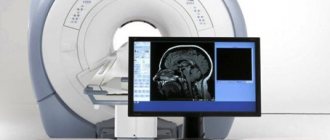
Immediately after scanning, the received data is decrypted. This takes about thirty to forty minutes, after which the radiologist hands over the received data or sends it to the treating doctor. Either X-ray paper or CDs are used as media. It contains indicators such as the characteristics of fluid flow in the spinal canal, the activity of the cortex under the influence of stimuli, the speed of blood flow, and the degree of tissue diffusion.
Gadolinium contrast
All contrast agents used for MRI contain gadolinium compounds. They are hypoallergenic and quickly eliminated from the body, so most often they do not cause side effects. In clinical practice, such paramagnetic contrast agents are used as: “Magnevist”, “Omniscan”, “Dotarem”, “Gadovist”, etc. Any of the above paramagnetic agents are always used in MRI of the mammary glands (if the purpose of the study is not to assess the condition of the implants) . Contrast contrast is often required for MRI of the pituitary gland, abdominal organs, pelvis and soft tissues.
MR angiography usually does not require additional examination, because in this case, the natural contrast is fast-moving blood.
If a tumor process or demyelinating disease is suspected, the attending physician, as a rule, pre-refers the patient for an MRI with contrast. But in some cases, the decision to administer contrast is made by the radiologist directly during the MRI - which is why his presence is necessary during the examination. At the slightest suspicion of a tumor process, the radiologist immediately prescribes an MRI with contrast to the patient.
The contrast agent is safe and is not a contraindication for MR imaging of women during breastfeeding, as well as in studies of children of all ages. However, MRI with contrast is contraindicated during pregnancy.
In addition, MRI with contrast is also contraindicated for patients with polyvalent allergies, acute and chronic renal and liver failure. You can learn more about the limitations of MRI with contrast in a special section of our website.
MRI of the brain with contrast for children
For children and adults, the indications for scanning are the same. Due to the impossibility of complete immobility for children under six years of age, they most often undergo the procedure under anesthesia with a laryngeal mask. You should not worry about the possible negative consequences of using anesthesia; according to statistics, complications occur no more often than in 1-2 percent of cases. In some cases, the anesthesiologist may recommend sedation, in which sleep is not as deep, which means breathing function is not impaired.
When performing brain MRIs with contrast on children, centers suggest replacing anesthesia with the presence of a significant adult who will reassure the baby, hold his hand, tell him a story, and ensure he remains still.
Contrast agents for MRI
MRI with contrast
During the procedure, drugs based on gadolinium salt are administered intravenously - Magnevist, Dotarem, Primovist, Omniscan. They are well tolerated and rarely cause intolerance. After being introduced into the bloodstream, the contrast spreads throughout the body, penetrating all tissues and structures and “illuminating” them.
As a result, the image obtained during MRI becomes brighter, more contrasty and more detailed. This allows the doctor to see the smallest pathological foci that are difficult to detect during a routine examination.
Indications for MRI of the brain with contrast
It is used to make or clarify a diagnosis. Among the indications:
- Frequent fainting;
- Headaches of unknown etiology;
- Tumors of the brain and cerebellopontine ganglion;
- Epilepsy;
- Abscess;
- Sinusitis;
- Hearing damage;
- Visual impairment;
- Aphasia;
- Anomalies and pathologies of blood vessels;
- Infectious diseases of the nervous system;
- Pituitary adenoma;
- Paroxysmal states;
- For head injuries and contusions accompanied by internal bleeding, an MRI of the brain with contrast is required;
- Neurodegenerative conditions;
- Multiple sclerosis;
- Senile dementia.
Indications for the procedure
Tomography is performed in the following cases:
- Memory impairment, attention problems, absent-mindedness.
- Pressing headache, nausea and vomiting, hallucinations.
- Possibility of pituitary adenoma
- Frequent fainting without reason.
- Impaired consciousness.
- Mood swings, irritability.
- Trembling hands, shortness of breath, dizziness and other autonomic changes.
It is worth noting that an MRI of the pituitary gland with contrast is always performed, since without the use of ascertaining formations are not detected.
As a result of the examination, it is possible to identify such abnormalities as vascular dissection, wall protrusion, inflammation of arteries and veins, atherosclerosis, the presence of tumors and metastases.
Contraindications
It is worth considering that intravenous administration of any substance has contraindications, so before agreeing to it, you need to check with your doctor about the possible risks. Be sure to inform if the patient has any types of allergies or kidney failure. This examination can be prescribed even to children; long-term observations have shown its safety for children. Of course, the price for an MRI of the brain with contrast in Moscow is higher than for a regular one, but health is not something you can save on.
Contraindications are absolute, these include:
- Wearing permanent implants made of metal or foreign metal bodies;
- Use of electronic pacemakers;
- The patient's weight is above 120 kilograms;
- Hematopoietic anemia.
Relative contraindications that will require additional preparation for MRI of the brain with contrast include:
- State of drug or alcohol intoxication;
- Heart failure;
- Tattoos with dyes containing metals;
- Panic attacks, claustrophobia, anxiety disorders;
- Availability of insulin pumps, prosthetic heart valves, non-ferromagnetic implants;
- Allergy;
- Pregnancy up to twelve weeks.
In the presence of these contraindications, scanning is done only on the direction of a doctor.
What is MRI
The method allows you to evaluate the organ layer by layer, where each layer is located a few microns from the previous one.
Due to this, it is possible to determine the pathology at the very initial stage. The work uses devices of different power and types. Thanks to the study, it is possible to assess the condition of organs and tissues, the presence of pathological foci, developmental anomalies, and so on. Tomography is a safe and most informative technique. Diagnostics is especially important for vascular disorders, injuries, tumors, inflammatory processes, metastases and cysts. To obtain more accurate results, your doctor may order an MRI with contrast. During the procedure, a substance sensitive to a magnetic field is introduced into the patient's body. The method of administration depends on the diagnostic purposes. Some substances are taken orally, others are administered intravenously. Once ingested, the substance illuminates the tissue. The picture is clearer. It is worth considering that the speed of spread of the contrast agent through the tissues depends on the speed of blood flow.
Contrast studies are carried out according to the following principles:
- After administration, the substance spreads through tissues and blood vessels.
- The affected tissues change color, which makes it possible to assess the size and location of the pathological focus.
- During the study, it is possible to identify distant metastases.
When performing the examination, it is important to take into account some nuances:
- There may be an allergy to the contrast agent; it should be excluded before the procedure begins to avoid unpleasant consequences.
- The procedure is not performed during pregnancy or suspicion of pregnancy. Instead, a regular MRI is prescribed in the 2-3 trimester.
- The presence of metal elements in the body is a general contraindication to performing a diagnostic procedure.
- The presence of a pacemaker, hearing implant, or pump in patients with diabetes mellitus is also a contraindication for both MRI with and without contrast.
- Mental disorders that do not allow you to maintain immobility during the entire procedure. In case of claustrophobia, it is possible to stabilize the patient’s condition due to the presence of a loved one in the office.
Paramagnetic drugs are used to carry out the procedure. They are not toxic to the body and do not contain iodine. Therefore, the likelihood of an allergic reaction is minimal. In addition, a minimal dosage of the substance is used. In most cases these are gadolinium salts. They dissolve well and do not cause side effects. For diagnostics, drugs such as Gadovist, Dotarem, Omniscan are used.
Price
For an MRI of the brain with contrast, the price in Moscow is about 6,000 rubles. If the procedure takes place under sedation or anesthesia, you will have to pay for the services of an anesthesiologist. The cost is also affected by the model of the device - more modern high-field silent devices will cost more. But the quality of images taken with such a tomograph is many times higher. Some medical centers offer the opportunity to do magnetic resonance imaging at night. This service will cost at an increased rate.
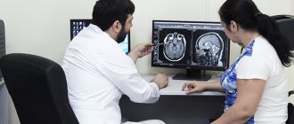
In which clinic can I get an MRI of the head with contrast?
Our information retrieval resource contains a database with all diagnostic institutions of the northern capital. The mrt-v-spb.ru team has gathered in one place dozens of clinics where you can undergo MRI for brain diseases. All medical centers presented on the portal have licensing permits, qualified radiologists, as well as powerful expert-class equipment.
To make it easier to select organizations, use:
- search system for metro stops and city districts;
- online map with verified addresses;
- panel sorting by rating and prices.
You can get a free informative consultation, help in choosing a clinic near your home or place of work, and also make an appointment by calling the hotline. Our operators take calls every day from 8 am to midnight. When you book a place for an examination through the service, you can get a discount on MRI of the brain of up to 1,000 rubles.
Bibliography:
- Nechipay, E.A. Possibilities of MR dynamic contrast enhancement in the differential diagnosis of primary and secondary brain tumors. / E.A. Nechipay, M.B. Dolgushin, I.N. Pronin, A.Kh. Bekyashev, E.A. Kobyakova, L.M. Fadeeva, E.I. Schultz // “Medical Imaging”. - No. 4. -2015.
- Serkov S.V. Diffusion-weighted MRI in the diagnosis of brain tumors: dis. ... medical candidate Sciences: 14.00.28 and 14.00.19 / Serkov Sergey Vladimirovich - M., 2005.
- Trufanov G.E. Radiation diagnostics: textbook: T. 1 / ed. prof. G.E. Trufanova. — 2011.
- Abe, T. Clinical significance of discrepancy between arterial spin labeling images and contrast-enhanced images in the diagnosis of brain tumors / Abe T., Mizobuchi Y., Sako W., Irahara S., Otomi Y., Obama Y., Nakajima K., Khashbat D., Majigsuren M., Kageji T., Nagahiro S., Harada M. // Magn Reson Med Sci. — 2015. V. 14. — Issue 4.
- Belenkov Yu.N., Ternovoy S.K., O.I. Belichenko // Clinical application of contrast-enhanced magnetic resonance imaging. VIDAR. Moscow, 1996.
- Tyutin L.A. Diagnostic capabilities of contrast magnetic resonance imaging / L.A. Tyutin, E.K. Yakovleva // Bulletin of Radiology. 1999. - No. 4. — P. 13-16.
- Baert AL Dynamic contrast-enhanced magnetic resonance imaging. In oncology / AL Baert, K. Sator. // Berlin etc.: Springer. - 2005. - N.6.
- Choi, HS Glioma Grading Capability: Comparisons among Parameters from Dynamic Contrast-Enhanced MRI and ADC Value on DWI / Choi HS, Kim AH, Ahn SS, Shin N., Kim J., Lee SK // Korean J Radiol. — 2013. — V. 14.
MRI of the brain with contrast: where is the best place to do it?
There are more than six dozen medical centers where this procedure is performed in the capital alone. Therefore, one must approach their choice responsibly. And focus not only on cost, but also on the quality and thickness of the sections. In order for them to be as thin as possible, it is worth giving preference to tomographs with a field of at least 1.5 Tesla. Such devices take photographs of tissues sequentially, obtaining at least 20 photographs at a time. The thickness of the sections is 4-5 millimeters. The leaders in the number of truly competent doctors who can perform MRI of the brain with contrast are Moscow centers. You can contact most of these clinics not only if you have an appointment, but also on your own.
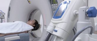
MRI with contrast: preparation for the study
There are no strict rules for preparing for the diagnosis of most organs. There are certain recommendations:
- It is recommended to come to the procedure with contrast on an empty stomach, limiting food intake two to three hours before the appointed time, this helps prevent the development of nausea;
- if intravenous sedation is planned, the patient must comply with the requirements of the anesthesiologist;
- before examining the pelvic and abdominal organs, it is necessary to adhere to a diet excluding gas-forming products;
- on the eve of diagnosis of the genitourinary and digestive systems, bowel preparation is required (a laxative or enema is used); The last meal should take place 6 hours before the visit to the clinic, liquids - 4 hours.
Before an MRI with contrast, you should refrain from eating and drinking
The contrast agent penetrates not only into the blood, but also into breast milk. During lactation, breastfeeding can be resumed no earlier than 24 hours after the injection, so you should prepare in advance and stock up on food for the baby. Milk that is unfit for consumption is expressed and discarded.