Carotid angiography is an invasive X-ray imaging procedure of the carotid arteries that is performed through a catheter inserted into a blood vessel in the arm or leg. A contrast agent is injected through the catheter, which is opaque to X-rays and therefore shows the internal lumen of the carotid arteries. This procedure is the gold standard for visualization of the carotid and cerebral vessels.
Preparing for the study
- Preparation for the study involves shaving the access site (usually the groin area).
- You should tell your doctor about any allergic reactions you have.
- If you are diabetic, tell your doctor so your medications can be adjusted on the day of the test.
- Tell your doctor if you are allergic to anything, especially iodine, shellfish, X-ray contrast, latex, or rubber products (such as rubber gloves).
- It is advisable to drink enough fluids before the examination to reduce the possible effects of contrast on the kidneys.
Preparing for the examination
Since the examination method is X-ray with the introduction of a contrast agent, you need to carefully prepare for cerebral angiography. Preparation includes the following steps:
- A maximum of 5 days before the examination, take a general blood and urine test (to determine the condition of the kidneys and exclude the presence of infectious diseases), a coagulogram (to determine the coagulation function of the blood).
- Do electrocardiography and phonocardiography (to exclude heart diseases).
- Do not drink alcohol for at least two weeks before the examination.
- Do not take medications that affect blood clotting for at least a week before angiography.
- 1-2 days before the examination, perform an allergy test with contrast, which is carried out by administering 0.1 ml of the drug to the patient and further observing the reaction on the skin. If redness, rash, and itching do not appear on the skin, then the test is negative and angiography is possible.
- Do not eat anything 8 hours before the examination and do not drink anything during the last 4 hours.
- It is possible to take tranquilizers or herbal sedatives if there is significant anxiety. However, it should be remembered that these medications can only be taken as prescribed by a doctor!
- If necessary, shave the contrast injection site.
- Remove all jewelry and other metal objects before angiography.
- Immediately before undergoing the examination, medical personnel must explain to the patient the methodology, goals and possible risks of this examination method.
After research
Tell your doctor or nurses if you feel:
- Itching, dry throat
- Nausea
- Chest discomfort
- Vision problems
- Weakness in one or more limbs
At the end of the study, you will be transferred to a ward. It is necessary to remain in bed for several hours. The doctor will periodically examine the access site and assess the general condition. If there are no problems, then discharge can be made the next day.
What will a CT scan of the cerebral arteries show?
CT scan of the cerebral arteries allows you to evaluate the anatomical structure and relative position of the vessels and detect deviations from the norm. The examination makes it possible not only to determine problems with the blood supply to the brain, but also, using software, to convert the obtained information into a three-dimensional spatial model.
CT scan of the cerebral arteries shows:
- vascular malformations (aneurysms, arteriovenous malformations);
- thrombosis and embolism;
- benign and malignant tumors;
- stenosis of cerebral vessels;
- bending, looping;
- cerebral vasculitis (inflammation of the vascular wall);
- hemorrhages into the cranial cavity and other vascular damage as a result of head injuries;
- angiopathy of various origins;
- acute cerebrovascular accidents of an ischemic and hemorrhagic nature, i.e. foci of ischemia or infarction (strokes);
- atherosclerosis of arteries.
Based on CT data of the cerebral arteries, the doctor makes a conclusion about the state of the lumen of the vessels, their walls and other indicators of the functioning of the circulatory system.
Possible complications of carotid angiography
Complications are rare - the risk is 0.5%. The most dangerous are complications from the brain. They can develop as a result of the effect of contrast or when pieces of plaque are torn off during catheter insertion. Complications include:
- Transient ischemic attack - short-term disturbance of cerebral circulation
- Ischemic stroke is a persistent disorder of cerebral circulation
- Bleeding from the arterial access site
- Carotid artery dissection with thrombosis
- Allergic reaction to contrast
Timely diagnosis of complications allows you to minimize their consequences.
How to prepare for a CT scan of the head and neck vessels
When examining the arteries, CT of the neck with contrast is used to obtain the most complete data. Without a contrast agent, the information content of the examination is extremely low.
Before undergoing diagnostics, there are several preparatory stages:
- do a blood biochemistry test to determine the level of urea and creatinine;
- inform your doctor in advance about the presence of allergic reactions to medications;
- do not eat 3 hours before undergoing a CT scan with contrast;
- provide the doctor with previous results of ultrasound, CT and MRI for review;
- remove all metal jewelry.
10. Stenosis and blockage of cerebral vessels
Stenosis is a narrowing of cerebral vessels that occurs due to the influence of unfavorable physiological factors, for example, deposits of calcium and cholesterol on the walls of blood vessels.
The narrowing of the intracranial arteries leads to the fact that certain areas of the brain cease to receive enough oxygen. The risk of complications increases when blood vessels narrow by more than 50%. The process causes strokes.
Signs of a problem depend on which part of the brain has undergone a pathological change (anterior, middle or posterior). The main symptoms of stenosis and occlusion include:
- unilateral or bilateral damage to the facial muscles;
- violation of speech functions;
- visual defects (flickering “fly spots”, split images);
- severe headaches that do not disappear after taking medications;
- increase in pressure.
The likelihood of recovery from stenosis and blockage of blood vessels depends on the timely detection and treatment of the pathology. The longer a person does not see a doctor, the less chance he has for a full recovery. The disruption of blood flow to the brain is temporary, i.e. it regularly recovers and worsens again.
Abnormalities of the cerebral arteries
Pathologies of the arteries of the brain can lead to a number of serious conditions. Among the most dangerous diseases that MRI shows in the arteries of the brain are the following:
Aneurysm
With this pathology, an expansion of the lumen of the artery appears in the affected area. Previously, this disease was diagnosed using X-ray angiography. However, the safest method today is MRI. Timely detection of an aneurysm can prevent massive hemorrhage into brain tissue.
Angioma
An angioma is a neoplasm of vascular tissue, the size and location of which can vary. On MRI images, the pathology appears as a multinodular formation.
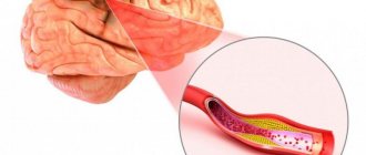
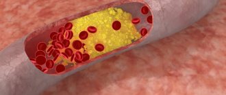
Atherosclerosis
In atherosclerosis, fats are deposited on the inner walls of blood vessels, forming plaques. If treatment is not started in time, hypoxia of brain tissue may develop.
When conducting an examination, three stages of the disease can be identified:
- There are a few inclusions of cholesterol on the walls of blood vessels.
- Cholesterol becomes more and more, platelets are added to it, plaques are formed, narrowing the lumen of blood vessels.
- Calcium ions settle on the plaques.
In the third stage, a person often experiences severe headaches, cognitive dysfunction and memory impairment are possible.
Stroke
This disease is an acute disorder of cerebral circulation, in which an area of the brain ceases to be supplied with blood, resulting in tissue necrosis. There are several types of pathology:
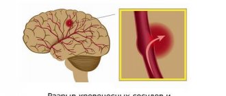
- Hemorrhagic. In this type of stroke, an artery in the brain ruptures.
- Ischemic. A blockage of the artery lumen with a thrombus occurs.
- Lacunar. When blood vessels become blocked, cysts can form.
- Cardioembolic. The cause of blockage of blood vessels is cardiac emboli.
If symptoms of a stroke appear, an urgent MRI is necessary, which will show the extent of damage to surrounding tissues. According to statistics, only 10% of people can fully recover from a stroke; 90% of patients develop serious complications. It is timely diagnosis that will help significantly facilitate rehabilitation after illness.
MRI of the cerebral arteries allows one to accurately determine the type of stroke and the location of circulatory disorders.
If the examination was done in the first hours after a stroke, the lesions will be clearly reflected in the images and doctors will be able to begin emergency treatment to resolve the blood clot. If a stroke is detected after some time, the edema resolves, the sulci of the cerebral cortex and the lateral ventricles expand.
Hypertension
Hypertension is a disease of the cardiovascular system, which is characterized by increased blood pressure caused by impaired vascular regulation and cardiac pathologies. In the cerebral arteries, MRI reveals indirect signs of hypertensive angiopathy.
Arteriovenous malformation
Malformation occurs when several vessels of different diameters become entwined together. One artery feeds this node, and a vein drains blood. MRI requires finding these vessels. During the study, a contrast agent may occasionally be used to help determine the location of the malformation.
Content
- MRI of head vessels
- Angiography, vasography, vascular MRI - what's the difference?
- What pathologies will an MRI of the head vessels reveal?
- Indications for MRI of head vessels
- What does MRI of head vessels show?
- Contraindications to MRI of head and neck vessels
- Description of diseases diagnosed using arteriography
- Aneurysm
- Arteriovenous malformation
- Stenosis and blockage of cerebral vessels
- MRI signs of pathologies MRI signs of arteriovenous malformation
- MRI - signs of narrowing and blockage of blood vessels
- Signs of an aneurysm
- Signs of vasculitis
What does MRI of head vessels show?
An MRI study of cerebral vessels will detect a violation of the blood supply in a certain area of the area being studied. To study in more detail the smallest changes in blood microcirculation, the study is carried out using a contrast agent (often gadolinium), which is injected into the patient intravenously.
MRI diagnostics allows you to timely detect the consequences of strokes and degenerative processes occurring in the tissues and arteries of the brain. This research method also makes it possible to diagnose Alzheimer's disease, multiple sclerosis, and demyelination (selective damage to the myelin sheath).
When describing the images obtained, the terms “filling defect” or “wall protrusion” are often used.
When an intraluminal thrombus was discovered during diagnosis, the contours of the aneurysm on tomograms appear as an irregularity with sharp, jagged edges. The defect itself appears to be either partial or poorly functioning. The signal from the thrombosed area of the aneurysm disappears on tomograms or has a chaotic periodicity.
Description of diseases diagnosed using arteriography
Arteriography can only be prescribed by a specialist. Patient complaints of dizziness, migraine and tinnitus are not indications for a contrast study. With such symptoms, the patient should consult a doctor to determine the need for a procedure.
With the help of a contrast MRI study you can:
- diagnose the development of a cerebral aneurysm;
- determine blockage or degree of narrowing of blood vessels (stenosis);
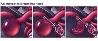
- check the location of vascular clips;
- determine the presence of arteriovenous malformation;
- diagnose vasculitis.
Each of the above diseases should be considered separately.
Indications for MRI of head vessels
- TBI (traumatic brain injury) or other physical impact of a traumatic nature.
- Atherosclerosis (accumulation of cholesterol in the walls of blood vessels, which causes their narrowing and blockage).
- Dilation of the walls of blood vessels and pathological forms of inflammation occurring in them.
- Compression of the great vessels.
- Aortic dissection, dystrophic changes in its tissues.
- Congenital and acquired heart defects.
- Painful reaction at the junction of the neck and skull, intensifying when turning/tilting the head.
- Headaches (usually pulsating) of unknown etiology.
- Pathological narrowing of arteries and veins.
- Strokes and heart attacks.
- Tumor processes (malignant and benign), cysts.
- Suspicion of metastasis.
- Weakness, fatigue, dark spots before the eyes, dizziness.
- Deviations from the norm in blood pressure readings.
MRI image of a child's brain vessels
What pathologies will an MRI of the head vessels reveal?
- Cerebrovascular accidents caused by a variety of etiological factors.
- Complete or partial obstruction of blood through the vessels and veins in the area under study.
- Blockage of arteries with blood clots.
MRI examination also allows:
- Determine the stability of the spinal column if its incorrect position interferes with blood circulation.
- Assess the permeability of blood through the tissue structures of the brain.
- Detect impaired metabolism.
- Obtain a layer-by-layer detailed image of vessels, arteries, veins and capillaries in the area under study.
- Assess the condition of the cerebral cortex.