Diagnosis of diseases of the central nervous system (CNS) using magnetic resonance imaging makes it possible to identify and differentiate pathological processes in the early stages of development.
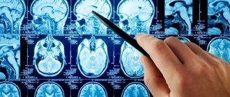
A series of MRI images for suspected stroke
An induction field is used to visualize the state of the cerebral substance and cerebral vessels. Under the influence of a directed electromagnetic pulse, the hydrogen atoms in water molecules change position, then returning to their place. The resonance of charged particles is detected using sensitive sensors and processed using complex software algorithms.
Diagnosis of strokes using MRI is an effective way to determine pathological changes in the condition of intracranial vessels and assess the consequences of cerebrovascular accidents. Magnetic resonance imaging of the head is prescribed to clarify the diagnosis if the examination is insufficiently informative and in the case of an initial visit to a specialist. A referral for the procedure can be given by a neurologist, neurosurgeon, oncologist, or traumatologist.
What is a stroke, its types
A stroke is a disorder of cerebral circulation leading to brain damage.
The pathology is widespread. In the Russian Federation alone, there are 3 cases of stroke per 1000 inhabitants. In the post-mortem extract, it is listed as the cause of death in 23.5% of people.
Even if patients do not die after suffering a vascular accident, more than 80% of them remain disabled. Often neurological disorders are so severe that the patient is unable to care for himself. Stroke is the third leading cause of death.
There are 2 types of stroke: ischemic and hemorrhagic. The mechanism of their development and the features of treatment have nothing to do with each other. There is also a special type of hemorrhagic vascular damage - subarachnoid hemorrhage.
Ischemic
Ischemic stroke is a cerebral circulatory disorder accompanied by an acute onset. Pathology develops due to disruption or complete cessation of blood supply to the brain. This leads to softening of its tissues and infarction of the affected area. It is cerebral vascular ischemia that is one of the main causes of death worldwide. Such a stroke occurs 6 times more often than hemorrhagic lesions.
It can be of 2 types:
- Thrombotic . Develops due to blockage of blood vessels in the brain by a blood clot.
- Embolic . Occurs when blood vessels located far from the brain become blocked. The most common source of embolism is the heart muscle (cardioembolic stroke).
In 80% of cases, the pathological focus is localized in the middle cerebral artery. Other vessels account for the remaining 20%.
Reasons that can provoke ischemic damage to cerebral arteries and veins:
- Myocardial infarction.
- High or low blood pressure.
- Atrial fibrillation.
- Diabetes.
- Lipid metabolism disorders.
Risk factors include: old age, hereditary predisposition to vascular accidents, as well as lifestyle features.
Symptoms of ischemic stroke do not increase as quickly as symptoms of hemorrhagic brain damage.
Its manifestations:
- Drowsiness, dazedness.
- Brief fainting.
- Headache, dizziness.
- Nausea and vomiting.
- Pain in the eyes that gets worse with movement.
- Cramps.
- Sweating, hot flashes, dry mouth.
Depending on which part of the brain is affected, the neurological manifestations of ischemia differ. The lower and upper extremities are affected to a greater or lesser extent, paresis of the tongue and face is observed, and visual and/or auditory function deteriorates.
Hemorrhagic
Hemorrhagic stroke is bleeding into the cranial cavity. The most common cause of blood vessel rupture is high blood pressure.
Other provoking factors include:
- Aneurysm.
- Malformation of cerebral vessels.
- Vasculitis.
- Systemic connective tissue diseases.
- Taking certain medications.
- Amyloid angiopathy.
The onset of the pathology is acute; most often the manifestation occurs against the background of high blood pressure. A person experiences severe headaches, dizziness, accompanied by vomiting or nausea. This state is quickly replaced by stupor, loss of consciousness, and even the development of coma. Convulsions are possible.
Neurological symptoms manifest themselves in the form of memory loss, deterioration of sensitivity and speech function. One side of the body, which is on the opposite side of the lesion, loses the ability to function normally. This applies not only to the muscles of the torso, but also to the face.
A stroke with blood escaping into the ventricles of the brain is difficult to endure. The victim develops symptoms of meningitis and has seizures. He quickly loses consciousness.
The next 3 weeks after a stroke are considered the most difficult. At this time, cerebral edema progresses. It is he who is the main cause of death of patients. Starting from the fourth week, in surviving people, the symptoms of the lesion begin to reverse. From this time on, the severity of brain damage can be assessed. They are used to determine what degree of disability to assign to the victim.
Subarachnoid hemorrhage
Subarachnoid hemorrhage is understood as a condition that develops as a result of a breakthrough of blood vessels into the subarachnoid space of the brain. This pathology is a type of hemorrhagic stroke.
The subarachnoid space contains cerebrospinal fluid, the volume of which increases due to blood flow. The patient's intracranial pressure increases and meningitis of aseptic nature develops. The situation is aggravated by the reaction of cerebral vessels. They spasm, which leads to ischemia of the affected areas. The patient develops an ischemic stroke or transient ischemic attack.
The following reasons lead to hemorrhage into the subarachnoid space::
- Traumatic brain injuries with damage to the integrity of blood vessels.
- Aneurysm rupture.
- Dissection of the carotid or vertebral artery.
- Myxoma of the heart.
- A brain tumor.
- Amyloidosis.
- Diseases associated with blood clotting disorders.
- Uncontrolled use of anticoagulants.
The pathology manifests itself as a severe headache. Possible loss of consciousness. In parallel, symptoms of meningitis develop, with neck stiffness, vomiting, and photophobia. A distinctive sign is an increase in body temperature. In severe cases, there is a disorder of respiratory function and cardiac activity. With prolonged fainting and coma, one may suspect that blood has entered the ventricles of the brain. This occurs during its massive outpouring and threatens with serious consequences.
What does an MRI show after a stroke?
Tomograms visualize the state of intracranial vessels, white and gray cerebral matter, and cerebral cortex. The pathogenesis of the disease determines what an MRI shows for a stroke of one type or another.
As a result of scanning, the doctor receives photographs of thin sections of the scanned area; the layer thickness is from 1 mm. Layer-by-layer images show the area under study in three mutually perpendicular projections (lateral, central, transverse). If it is necessary to clarify the location of cerebral structures and determine the interaction of the pathological focus and healthy tissue, a 3D model of the brain is built.
Intracerebral hemorrhage on MRI
In the acute period, magnetic resonance imaging helps to identify the causes of stroke. Based on tomograms, the doctor evaluates the nature of the blood supply to the brain, the condition of the walls, tone, and lumen of the intracranial arteries.
With the development of a focus of necrosis, the location, size, and degree of damage to the cerebral substance are determined.
MRI after a stroke is prescribed during the rehabilitation period. The examination helps to monitor the processes of restoration of damaged areas and timely diagnose the development of long-term consequences.
Signs and symptoms of stroke
A stroke manifests unexpectedly for a person, although sometimes it is preceded by certain symptoms. If you interpret them correctly, you can avoid a terrible vascular catastrophe.
Warning signs of an impending stroke include:
- Prolonged headaches. They do not have a clear localization. It is not possible to cope with them with the help of analgesics.
- Dizziness. It occurs at rest and can intensify when performing any actions.
- Ringing in the ears.
- Sudden attack of atrial fibrillation.
- Difficulty swallowing food.
- Memory impairment.
- Numbness of arms and legs.
- Loss of coordination.
- Insomnia.
- Increased fatigue.
- Decreased overall performance.
- Rapid heartbeat and constant thirst.
The listed signs may have varying intensities. You should not ignore them; you should consult a doctor.
Symptoms of ischemic stroke increase slowly. With hemorrhagic brain damage, the clinical picture unfolds rapidly.
You can suspect a brain catastrophe based on the following symptoms::
- General cerebral symptoms . The patient experiences unbearable headaches. Nausea ends with vomiting. Consciousness is impaired, and both stupefaction and coma may occur.
- Focal symptoms . They directly depend on where exactly the lesion is localized. The patient may have decreased or completely lost muscle strength on one side of the body. Half of the face is paralyzed, causing it to become distorted. The corner of the mouth lowers, the nasolabial fold smoothes out. On the same side, the sensitivity of the arms and legs decreases. The victim’s speech deteriorates and he has difficulty oriented in space.
- Epileptiform symptoms . Sometimes a stroke provokes an epileptic attack. The patient loses consciousness, has convulsions, and foam appears at the mouth. The pupil does not react to the light beam; on the side of the lesion it is dilated. The eyes move right and left.
- Other symptoms . The patient's breathing quickens and the depth of inspiration decreases. A significant decrease in blood pressure and increased heart rate are possible. Often a stroke is accompanied by uncontrolled urination and defecation.
When the first signs of a stroke appear, you should not hesitate to call an ambulance.
Signs of stroke on MRI
Using magnetic resonance imaging, acute cerebrovascular accident can be detected after the first clinical symptoms appear.
A stroke on MRI of the brain has the following characteristic signs:
- thrombosis, embolism, blood vessel aneurysm;
- cerebral edema;
- necrosis of the cerebral substance (hyperintense lesion on T2-weighted images);
- displacement of the affected brain structures towards healthy tissue;
- formation of liquefaction cysts.
In the acute period (first 6 hours), diffusion-weighted (DW) MRI is performed. The resulting images reflect pathological changes in the brain matter 5 minutes after the onset of signs of stroke. DWI images help determine the area of the lesion, show swelling of the tissue around the lesion, and displacement of cerebral structures.
The acute period (1-7 days) is characterized by the appearance of clearly limited bright areas on MRI in T2-weighted mode. On T1WI, foci of necrosis have a hypointense signal.
Brain on MRI with different scanning modes
Over the next two weeks, the area of edema in the photographs decreases, and the affected area is clearly visible. The early rehabilitation period is characterized by the appearance of a cyst formed in place of necrotic tissue. Later, the formation of alternative neural connections is observed, which allows the restoration of lost brain functions.
Differential signs of stroke on MRI make it possible to clarify the nature of the disease in the first hours, when the use of thrombolysis and regeneration of areas affected by ischemia are possible.
Diagnostic methods
It is important to quickly distinguish a stroke from other diseases that can lead to the development of similar symptoms. It is almost impossible to do this on your own, as well as to determine the type of vascular accident.
The main difference between an ischemic stroke is a gradual increase in symptoms that do not lead to loss of consciousness. With hemorrhagic hemorrhage, the patient passes out quickly. However, stroke does not always have a classic course. The disease may begin and progress atypically.
Diagnosis begins with examination of the patient. The doctor collects anamnesis and determines the presence of chronic diseases. Most often, you can get information not from the victim himself, but from his relatives. The doctor performs an ECG, determines the heart rate, takes a blood test, and measures blood pressure.
It is possible to make the correct diagnosis and obtain maximum information about the patient’s condition thanks to instrumental diagnostic methods. The best option is a CT scan of the brain. Performing an MRI is difficult because the procedure takes a long time. It takes about an hour. It is impossible to spend this amount of time diagnosing an acute stroke.
Computed tomography allows you to clarify the type of pathology, where it is concentrated, to understand how badly the brain is damaged, whether the ventricles are affected, etc. The main problem is that it is not always possible to perform a CT scan in the shortest possible time. In this case, doctors have to focus on the symptoms of the disease.
To determine the source of the stroke, the method of diffusion-weighted tomography (DWI) is used. The information will be received within a few minutes.
Other examination methods include:
- Lumbar puncture.
- Cerebral angiography.
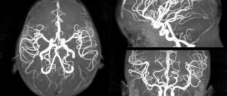
- Magnetic resonance angiography. It is performed without the introduction of a contrast agent.
- Doppler ultrasound.
Once the diagnosis is made, the doctor will immediately begin treatment.
Diagnosis of strokes using MRI
The MR scan picture for stroke depends on the type of process. Various pathogenesis leads to the appearance of additional signs of stroke on MRI images. Magnetic resonance imaging is more informative in relation to cerebral infarction, the diagnosis of which can be difficult due to the absence of a clear clinical picture at an early stage.
Ischemic stroke
The disease is characterized by the appearance of foci of necrosis caused by impaired blood supply to a certain area of the brain. In the acute stage, thickening of the convolutions of the cerebral cortex is observed, the boundary between gray and white matter is erased. During the recovery period, the infarction zone is defined as an area of cystic or glial degeneration.
Lacunar stroke, which is a type of ischemia, is characterized by the appearance of small (up to 20 mm) foci and hemorrhages in the area of deep structures of the brain. The cause is thrombosis of a small intracranial artery, which caused the formation of a cyst (lacuna).
Intracranial hemorrhage on MRI (arrow indicates hematoma)
Hemorrhagic stroke
Hemorrhages are poorly detected on magnetic resonance imaging. In hemorrhagic stroke, MRI shows hematomas as outlined lesions with signal enhancement (T1-weighted).
Parenchymal hemorrhage is characterized by the accumulation of blood directly in the brain tissue. Perifocal edema appears as an area of reduced density with a rounded shape.
Subarachnoid hemorrhages
Foci of necrosis occur under the arachnoid membrane of the brain. On MRI, the picture of subarachnoid hemorrhage is similar to the changes characteristic of hemorrhagic stroke. The images visualize the affected area and allow you to determine the cause of the disease.
Intracerebral hemorrhage requires follow-up scans. As the hematoma increases, compression of the surrounding structures may increase and perifocal edema may spread.
Acute hypertensive encephalopathy
MRI of the brain shows an increase in the signal on T2 VI, characteristic of a stroke, localized mainly in the parieto-occipital regions, pons, and cerebellum. There are no signs of diffusion on DW images.
Depending on the MRI picture taken after a stroke, it is determined what nature, ischemic or hemorrhagic, the pathological process has. Information about the causes of the disease is necessary to choose the most effective treatment method.
Who is at risk
There are people who need to be especially wary of developing a stroke, as they are at risk.
Among them:
- Persons with hypertension.
- Patients with diabetes.
- Men and women over 65 years of age.
- People with abdominal obesity.
- Persons with a hereditary predisposition to vascular pathologies.
- Patients who have previously had a stroke or heart attack.
- Patients with diagnosed atherosclerosis.
- Women over 35 years of age taking oral contraceptives.
- Smokers.
- People suffering from heart rhythm disturbances.
- People with high cholesterol levels.
Most often, patients with the listed diagnoses are registered at the dispensary. Special mention should be made of people living in a state of chronic stress. Emotional stress negatively affects all systems of the body and can cause a stroke.
How to provide first aid for a stroke
There is a clear algorithm for providing first aid to a person suffering from a stroke :
- Call a medical team . To do this, you need to dial 103 from a landline phone. If you happen to have a smartphone at hand, then the call is made to the single number 112. The doctor must immediately be informed that the person is unwell and there is a suspicion of a stroke.
- The victim must be laid on a flat surface so that his head is higher than his body. They take off his glasses and remove his lenses. If possible, you need to help him get removable dentures.
- If there is no consciousness, then you need to open the patient’s mouth slightly and turn his head to the side . This is done to prevent aspiration of vomit. It is imperative to listen to the patient’s breathing.
- For better access to fresh air, it is recommended to open a window or vent..
- Before the arrival of the medical team, it is necessary to prepare documents, if any..
Doctors need to be informed about the person’s illnesses, as well as what medications he is taking. It is prohibited to give the victim any medications. Medication correction should be carried out by emergency physicians. You should not try to give water or food to a person. This may make the situation worse.
If the patient falls and has an epileptic attack, there is no need to unclench his teeth or try to hold him. It is necessary to protect the victim from injury. To do this, place a soft object, such as a pillow, under his head. If a stroke with an epileptic attack happened on the street, then you can use a jacket or other suitable thing. Foam flowing from the mouth is wiped away with a cloth. The head should be elevated at all times.
There is no need to try to bring a person to his senses with ammonia. Until the end of the attack, it should not be moved from place to place.
If breathing stops, resuscitation measures must be started immediately. To do this, perform a heart massage and breathe mouth to mouth or mouth to nose.
Treatment and rehabilitation
The patient receives treatment in a hospital. All patients with suspected stroke are hospitalized on an emergency basis. The optimal period for providing medical care is the first 3 hours after a brain accident has occurred. The person is placed in the intensive care unit of a neurological hospital. After the acute period has been overcome, he is transferred to the early rehabilitation unit.
Until the diagnosis is established, basic therapy is carried out. The patient’s blood pressure is adjusted, the heart rate is normalized, and the required blood pH level is maintained. To reduce cerebral edema, diuretics and corticosteroids are prescribed. Craniotomy is possible to reduce the degree of compression. If necessary, the patient is connected to an artificial respiration apparatus.
Be sure to direct efforts to eliminate the symptoms of stroke and alleviate the patient’s condition. He is prescribed medications to lower body temperature, anticonvulsants, and antiemetics. Medicines that have a neuroprotective effect are used.
Pathogenetic therapy is based on the type of stroke. In case of ischemic brain damage, it is necessary to restore nutrition to the affected area as quickly as possible. To do this, the patient is prescribed drugs that resolve blood clots. It is possible to remove them mechanically. When thrombolysis fails, the patient is prescribed Acetylsalicylic acid and vasoactive drugs.
In case of a stroke, it is extremely important to provide timely treatment to the damaged areas of the brain. A course of use of the drug accelerates the process of recovery of brain cells after a stroke, even in cases of impaired blood circulation or hypoxia. This allows for rapid restoration of memory, thinking, speech, swallowing reflex and restoration of other functions of daily activities. Gliatilin has a positive effect on the transmission of nerve impulses, protects brain cells from repeated damage, which prevents the risk of recurrent stroke.
The drug is well tolerated by patients; it is contraindicated for use by pregnant, lactating women and people with hypersensitivity to choline alfoscerate.
Courses will need to be taken regularly. You definitely need to do physical therapy, undergo physical therapy, and visit a massage therapist. After a stroke, many patients have to restore motor skills over a long period of time and learn to care for themselves independently.
Relatives and friends should provide support to the patient and not leave him alone with the problem. Psychologists are involved in the work. Sessions with a speech therapist are often required.
Is it possible to do an MRI after a stroke?
Magnetic resonance imaging does not have a negative impact on the patient's health. If you suspect the development of necrosis of brain tissue, a scan will help determine the nature and intensity of the pathological process.
Signs of acute cerebrovascular accident on MRI
The disease must be differentiated from lesions of cerebral structures caused by:
- poisoning;
- hypoglycemia;
- growth of tumors;
- epilepsy attack, etc.
MRI for cerebral stroke is highly effective during the recovery period. In the acute period, scanning is used if an ischemic lesion is suspected, characterized by the patient's disorientation without loss of consciousness and the absence of severe headache. MRI allows you to clarify the localization of the pathological focus and study in detail the damaged areas of the brain.
Possible consequences, complications
The main danger of a stroke is death. If a person survives, the disease will still make itself felt with certain complications.
Early consequences include:
- Brain swelling.
- Coma.
- Pneumonia.
- Paralysis. It can be partial or complete. Most often, one half of the body is affected.
- Repeated stroke.
- Bedsores.
- Mental disorders. They can manifest themselves in moodiness, irritability, aggression, and anxiety. Sometimes dementia develops.
- Sleep disorders.
- Myocardial infarction, gastric ulcer. These disorders develop against the background of increased levels of stress hormones.
After an ischemic stroke, death occurs in 15-25% of cases. Hemorrhagic damage to the blood vessels of the brain leads to the death of 50-60% of patients. The cause of death is precisely severe complications, for example, pneumonia or acute heart failure. The first 3 months after a stroke are considered the most dangerous.
Arms recover worse in patients than legs. A person's future health is determined by the severity of brain damage, the speed of medical care, his age and the presence of chronic diseases.
Long-term consequences include:
- Formation of blood clots in various parts of the body.
- Depression.
- Speech problems.
- Memory loss.
- Deterioration of intellectual abilities.
After a stroke, you have to deal with the consequences for many months. Sometimes a person never manages to fully recover. For rehabilitation to be as successful as possible, you must strictly follow all the doctor’s instructions.
Stroke is a serious pathology because it affects the brain. Therefore, even the slightest suspicion of a developing vascular accident is a reason to urgently seek medical help.
Brain after stroke
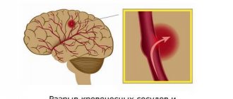
A brain stroke (see photo 3) provokes pathological changes that occur due to vascular problems, for example, arterial thrombosis, which abruptly stops the access of oxygen to brain cells. 20% of the oxygen entering the body goes to ensure an active brain - all the processes occurring in the brain are so costly.
As a result of a disruption in the supply of oxygen to neurons, vital cellular processes are disrupted and they become unable to carry out their conduction function. Five minutes of ischemia is enough for the brain after a stroke to lead to irreversible changes in local areas. Even a micro-stroke does not go away without leaving a trace, although the severity of complications is certainly much less.
A brain stroke provokes disruption of basic processes - speech, motor activity, and thinking. Brain cells responsible for certain processes gradually lose their functions, and after some time necrosis occurs - cell death. Depending on how many cells are affected, the severity of the stroke manifests itself. The location of the lesion gives visual manifestations of pathology.
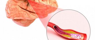
What does a hemorrhagic stroke look like?
Hemorrhagic stroke (see photo 4) occurs when a patient has a cerebral hemorrhage due to vascular problems. Brain disorders provoke atherosclerosis, thrombosis and other pathologies of cerebral vessels. In parallel with hemorrhage, cerebral edema develops, aggravating the pathology.
Between 50 and 80% of such strokes are fatal. Determining a hemorrhagic stroke on an MRI is very simple - the area of hemorrhage and swelling will become visible as a result of the scan.
It is noteworthy that hemorrhagic stroke is much less likely to provide warning signs of disorders. People may not feel sick at all, but threatening symptoms increase in a matter of seconds. The headache of a hemorrhagic stroke is much stronger. Rarely, there may be an ocular stroke - with intracerebral hemorrhage, damage to the small vessels of the eyes is noticeable.
How does ischemic stroke manifest?
Ischemic stroke (see photo 5), as a rule, has a short period of manifestation of pathological symptoms. With this type of brain damage, the symptoms increase slowly, there is practically no headache, and if there is any, it is weak and aching. Against the background of such signs, pressure almost never increases, as happens with a hemorrhagic stroke.
After a stroke, patients may experience vomiting and involuntary urination. The first signs of a stroke in a woman are usually more difficult to bear than in a man and can be triggered by pregnancy, hormonal surges, and women’s lower resistance to stress. Ischemic stroke of the brain can cause disability, but patients remain alive.
Diagnostic methods
It is important to quickly distinguish a stroke from other diseases that can lead to the development of similar symptoms. It is almost impossible to do this on your own, as well as to determine the type of vascular accident.
The main difference between an ischemic stroke is a gradual increase in symptoms that do not lead to loss of consciousness. With hemorrhagic hemorrhage, the patient passes out quickly. However, stroke does not always have a classic course. The disease may begin and progress atypically.
Diagnosis begins with examination of the patient. The doctor collects anamnesis and determines the presence of chronic diseases. Most often, you can get information not from the victim himself, but from his relatives. The doctor performs an ECG, determines the heart rate, takes a blood test, and measures blood pressure.
It is possible to make the correct diagnosis and obtain maximum information about the patient’s condition thanks to instrumental diagnostic methods. The best option is a CT scan of the brain. Performing an MRI is difficult because the procedure takes a long time. It takes about an hour. It is impossible to spend this amount of time diagnosing an acute stroke.
Computed tomography allows you to clarify the type of pathology, where it is concentrated, to understand how badly the brain is damaged, whether the ventricles are affected, etc. The main problem is that it is not always possible to perform a CT scan in the shortest possible time. In this case, doctors have to focus on the symptoms of the disease.
To determine the source of the stroke, the method of diffusion-weighted tomography (DWI) is used. The information will be received within a few minutes.
Other examination methods include:
- Lumbar puncture.
- Cerebral angiography.
- Magnetic resonance angiography. It is performed without the introduction of a contrast agent.
- Doppler ultrasound.
Once the diagnosis is made, the doctor will immediately begin treatment.
Who is at risk
There are people who need to be especially wary of developing a stroke, as they are at risk.
Among them:
- Persons with hypertension.
- Patients with diabetes.
- Men and women over 65 years of age.
- People with abdominal obesity.
- Persons with a hereditary predisposition to vascular pathologies.
- Patients who have previously had a stroke or heart attack.
- Patients with diagnosed atherosclerosis.
- Women over 35 years of age taking oral contraceptives.
- Smokers.
- People suffering from heart rhythm disturbances.
- People with high cholesterol levels.
Most often, patients with the listed diagnoses are registered at the dispensary. Special mention should be made of people living in a state of chronic stress. Emotional stress negatively affects all systems of the body and can cause a stroke.
How to provide first aid for a stroke
There is a clear algorithm for providing first aid to a person suffering from a stroke :
- Call a medical team . To do this, you need to dial 103 from a landline phone. If you happen to have a smartphone at hand, then the call is made to the single number 112. The doctor must immediately be informed that the person is unwell and there is a suspicion of a stroke.
- The victim must be laid on a flat surface so that his head is higher than his body. They take off his glasses and remove his lenses. If possible, you need to help him get removable dentures.
- If there is no consciousness, then you need to open the patient’s mouth slightly and turn his head to the side . This is done to prevent aspiration of vomit. It is imperative to listen to the patient’s breathing.
- For better access to fresh air, it is recommended to open a window or vent..
- Before the arrival of the medical team, it is necessary to prepare documents, if any..
Doctors need to be informed about the person’s illnesses, as well as what medications he is taking. It is prohibited to give the victim any medications. Medication correction should be carried out by emergency physicians. You should not try to give water or food to a person. This may make the situation worse.
If the patient falls and has an epileptic attack, there is no need to unclench his teeth or try to hold him. It is necessary to protect the victim from injury. To do this, place a soft object, such as a pillow, under his head. If a stroke with an epileptic attack happened on the street, then you can use a jacket or other suitable thing. Foam flowing from the mouth is wiped away with a cloth. The head should be elevated at all times.
There is no need to try to bring a person to his senses with ammonia. Until the end of the attack, it should not be moved from place to place.
If breathing stops, resuscitation measures must be started immediately. To do this, perform a heart massage and breathe mouth to mouth or mouth to nose.
Treatment and rehabilitation
The patient receives treatment in a hospital. All patients with suspected stroke are hospitalized on an emergency basis. The optimal period for providing medical care is the first 3 hours after a brain accident has occurred. The person is placed in the intensive care unit of a neurological hospital. After the acute period has been overcome, he is transferred to the early rehabilitation unit.
Until the diagnosis is established, basic therapy is carried out. The patient’s blood pressure is adjusted, the heart rate is normalized, and the required blood pH level is maintained. To reduce cerebral edema, diuretics and corticosteroids are prescribed. Craniotomy is possible to reduce the degree of compression. If necessary, the patient is connected to an artificial respiration apparatus.
Be sure to direct efforts to eliminate the symptoms of stroke and alleviate the patient’s condition. He is prescribed medications to lower body temperature, anticonvulsants, and antiemetics. Medicines that have a neuroprotective effect are used.
Pathogenetic therapy is based on the type of stroke. In case of ischemic brain damage, it is necessary to restore nutrition to the affected area as quickly as possible. To do this, the patient is prescribed drugs that resolve blood clots. It is possible to remove them mechanically. When thrombolysis fails, the patient is prescribed Acetylsalicylic acid and vasoactive drugs.
If a patient develops a hemorrhagic stroke, it is important to stop the bleeding. To do this, the patient is prescribed drugs that thicken the blood, for example, Vikasol. It is possible to perform an operation to remove the resulting hematoma. It is aspirated using special equipment, or through open access by performing craniotomy.
Relatives and friends should provide support to the patient and not leave him alone with the problem. Psychologists are involved in the work. Sessions with a speech therapist are often required.
What does a stroke look like on an MRI of the brain?
Monochrome images allow you to identify altered areas that differ in color from healthy tissue. Foci of ischemia and hemorrhage have clear boundaries, making it possible to clarify the extent of damage to brain structures.
Cerebral infarction on MRI
Tomograms visualize pathologies of intracranial blood vessels, helping to determine the causes of stroke development. To increase the information content, contrast magnetic resonance scanning is used.
Whether an MRI is necessary for a stroke is decided by the doctor, taking into account the clinical picture of the disease. The method is highly effective. But since the session lasts from 15 to 30 minutes, patients in serious condition undergo an alternative CT study.
The Magnit Clinic offers an MRI scan of the brain if a stroke is suspected and during the recovery period. A modern closed tomograph from the German company Siemens creates a field of 1.5 Tesla, which allows you to obtain high-resolution photos. You can sign up for the procedure by calling +7 (812) 407-32-31 or on the clinic’s website.