Brain puncture is a multi-purpose medical manipulation, with the help of which one or more therapeutic and diagnostic purposes are pursued. A puncture (injection) is any penetration with a needle or trocar into the cavity of a vein, another vessel, or an organ, to obtain material for research and diagnosis, optimize functions and remove obstacles, and perform operations.
Modern techniques make it possible to combine operational and diagnostic goals, achieving them simultaneously.
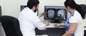
Taking fluid for analysis does not exclude the use of other diagnostic methods. Modern technologies allow parallel ultrasound to determine the location of the dislocation, for example, a cyst. This combination can successfully remove the tumor.
There is no need to be afraid of a puncture - it is not only a diagnostic method, but also a treatment method that was used before, but in an indirect form.
What is a cerebral puncture?
Penetration into the cranium at the location of parts of the brain is carried out less frequently than other manipulations in places that are less dangerous and threaten negative consequences.
Although any puncture can cause complications if it is carried out unprofessionally, affects some important segments or becomes a source of infection. Each invasive procedure has features specific to a particular department, developed techniques and precautions. Brain puncture (cerebral puncture) is a collective name for a therapeutic or diagnostic procedure carried out according to indications at a strictly defined destination:
- in the lower parts of the temporal or frontal lobes;
- over the tympanic space or mastoid process;
- ventricular, in the region of the lateral ventricles;
- within the central nervous system, to obtain a sample to study the spinal cord and brain simultaneously.
To carry out the procedure, a special needle and scalpel are used, the trepanation window is cut out with a special cutter, and bone bleeding is stopped by rubbing wax or electrocoagulation. To regulate the flow of cerebrospinal fluid there is a special device - a mandrin. In most cases, the procedure is performed under local anesthesia, in compliance with all necessary conditions of sterility and preparation of a sterile surgical field.
However, just in case, a large operating room is being prepared, which in rare cases can be used to perform open brain surgery. This scenario is possible when surgical complications arise - damage to a vessel, air entering the cavity, or insertion of a needle to an unexpected height.
Although sometimes the reason for further surgical tactics is an insufficiently studied pathology located directly in the brain (abscess, abscess, cyst, neoplasm).
BIOPSY AND BONE MARROW ASPIRATION
How the examination is performed . Bone marrow puncture (aspiration) in patients is usually done from the posterior iliac crest of the pelvis with the patient lying on his stomach. The area where the procedure is performed is disinfected. The puncture is performed with a special puncture needle. To conduct the study, imported disposable needles are used. In a bone marrow biopsy, a large needle is used to take a sample of the hard part. Typically, when a bone marrow biopsy is performed, it is done at the same time as aspiration. When puncturing the posterior crest of the iliac bone of the pelvis, local anesthesia is performed with novocaine or lidocaine, as well as for trepanobiopsy. If you are allergic to these medications, be sure to tell your doctor before the procedure! Do not confuse a bone marrow puncture from the ilium with a spinal puncture of the spinal canal, during which cerebrospinal fluid is taken for analysis. These are completely different procedures!
Preparing for the examination . Bone marrow collection is often performed on an outpatient basis and usually does not require special preparation. Taking bone marrow is often painless and the procedure requires little time: the biopsy method usually takes about 20 minutes, the aspiration method - 5-10 minutes.
Before the procedure . For most people, local anesthesia is all that is needed to make the examination comfortable. It is possible to conduct research under anesthesia.
After the procedure . After the bone marrow is collected, a sterile dressing is applied to the area. You can then go home and return to your daily activities.
Wound care . The dressing in the area where the bone marrow is collected should remain dry for 24 hours.
Bone marrow analysis . A bone marrow analysis (myelogram calculation and trepanobiopsy assessment) is carried out by a laboratory diagnostics doctor and a pathologist who issues an appropriate conclusion. The conclusion of a bone marrow analysis is needed by your hematologist or oncologist in order to make the correct diagnosis. Test results are prepared within several days. Check with your doctor when you can get them. In some cases, it may be necessary to perform multiple tests over time.
Risks and complications when performing the examination . Complications from bone marrow aspiration or biopsy are rare, but some patients may experience bleeding from the site where the bone marrow is collected.
What is it done for - diagnostic and therapeutic purposes
Obtaining cerebrospinal fluid to determine treatment tactics, analysis and diagnostic prognosis is carried out with the goal of achieving a certain result, and before prescribing a puncture, the tasks are strictly delimited. However, there are situations when a cerebral puncture for the purpose of research, collection of cerebrospinal fluid, as material for diagnostics, turns into shunting or removal of excess fluid to reduce pressure inside the skull.
Ventricular puncture (penetration into the lateral ventricles of the brain) helps doctors achieve several goals:
- performing diagnostics by obtaining important biological fluid for research;
- measuring intracranial pressure or conducting studies with a radiopaque substance;
- operations performed using a special device - a ventriculoscope, or shunting in the cerebrospinal fluid system;
- reducing intracranial pressure by removing spinal fluid if the natural outflow system does not work.
Well-established techniques and precautions allow operations to be performed as needed, using only local anesthesia. The methods and routes of penetration have been developed over many years of practice, and the data obtained in most cases helps to carry out more effective treatment based on objective information.
How to take a brain puncture
The operation is carried out under local anesthesia with strict adherence to all the rules of sanitary processing, first the incision, and then cutting the bone with a special tool, after which cerebrospinal fluid begins to flow through the hole, which is taken to alleviate the patient’s condition and conduct tests.
The surgical field is limited to sterile tissue, and the outflow of biological fluid is strictly controlled, as well as possible bleeding when a burr hole appears.
Precautions and Rules
It is necessary to take into account all indications and contraindications, possible obstacles to the operation. Thorough sanitization at each stage, preparation of spare instruments, a large operating room, careful monitoring of the patient’s condition at each stage.
Mandrin and other instruments must be thoroughly disinfected
The manipulation is carried out with the patient positioned on his back, with his head bowed to his chest. The neurosurgeon determines the incision line by touch.
There is a method of penetration through the orbit (the so-called Dogliotti method), and there is another method - according to Germanovich, who developed penetration through the temporal bone from below.
When is a lumbar puncture necessary and why not?
lumbar puncture
Lumbar puncture is performed both for diagnostic purposes and for therapy, but always with the consent of the patient, except in cases where the latter, due to his serious condition, cannot contact the staff.
For diagnosis, a spinal puncture is performed if it is necessary to examine the composition of the cerebrospinal fluid, determine the presence of microorganisms, fluid pressure and patency of the subarachnoid space.
Therapeutic puncture is needed to evacuate excess cerebrospinal fluid or introduce antibiotics and chemotherapy drugs into the intrathecal space in case of neuroinfection or oncopathology.
The reasons for lumbar puncture are mandatory and relative, when the decision is made by the doctor based on a specific clinical situation. Absolute indications include:
- Neuroinfections - meningitis, syphilitic lesions, brucellosis, encephalitis, arachnoiditis;
- Malignant tumors of the brain and its membranes, leukemia, when CT or MRI cannot make an accurate diagnosis;
- The need to clarify the causes of liquorrhea with the introduction of contrast or special dyes;
- Subarachnoid hemorrhage in cases where non-invasive diagnosis is not possible;
- Hydrocephalus and intracranial hypertension - to remove excess fluid;
- Diseases requiring the administration of antibiotics and antitumor agents directly under the membranes of the brain.
Among the relative ones are pathology of the nervous system with demyelination (multiple sclerosis, for example), polyneuropathy, sepsis, unidentified fever in young children, rheumatic and autoimmune diseases (lupus erythematosus), paraneoplastic syndrome. A special place is occupied by lumbar puncture in anesthesiology, where it serves as a method of delivering an anesthetic to the nerve roots to provide fairly deep anesthesia while maintaining the patient’s consciousness.
If there is reason to suspect a neuroinfection, then the cerebrospinal fluid obtained by puncture of the intrathecal space will be examined by bacteriologists, who will establish the nature of the microflora and its sensitivity to antibacterial agents. Targeted treatment significantly increases the patient's chances of recovery.
With hydrocephalus, the only way to remove excess fluid from the subarachnoid spaces and the ventricular system is puncture, and often patients feel relief almost immediately as soon as cerebrospinal fluid begins to flow through the needle.
If tumor cells are detected in the resulting liquid, the doctor has the opportunity to accurately determine the nature of the growing tumor, its sensitivity to cytostatics, and subsequent repeated punctures can become a way to administer drugs directly to the area of tumor growth.
Lumbar puncture may not be performed on all patients. If there is a risk of harm to health or danger to life, then the manipulation will have to be abandoned. Thus, the following are considered contraindications for puncture:
- Cerebral edema with risk or signs of herniation of brain stem structures or cerebellum;
- High intracranial hypertension, when removal of fluid can provoke dislocation and wedging of the brain stem;
- Malignant neoplasms and other space-occupying processes in the cranial cavity, intracerebral abscesses;
- Occlusive hydrocephalus;
- Suspicion of dislocation of stem structures.
The conditions listed above are fraught with the descent of the stem structures to the foramen magnum with their wedging, compression of vital nerve centers, coma and death of the patient. The wider the needle and the more fluid removed, the higher the risk of life-threatening complications. If the puncture cannot be delayed, then the minimum possible volume of cerebrospinal fluid is removed, but if wedging occurs, a certain amount of liquid is reintroduced.
If the patient has suffered a severe traumatic brain injury, massive blood loss, has extensive injuries, or is in a state of shock, it is dangerous to perform a lumbar puncture.
Other obstacles to the procedure may include:
- Inflammatory pustular, eczematous skin changes at the point of the planned puncture;
- Pathology of hemostasis with increased bleeding;
- Taking anticoagulants and antiplatelet agents;
- Aneurysm of cerebral vessels with rupture and bleeding;
- Pregnancy.
These contraindications are considered relative, increasing the risk of complications, but in cases where puncture is vitally necessary, they can be neglected if maximum caution is observed.
Important Invega (paliperidone)
Doctor's competence
Taking biological fluid for analysis or to alleviate the patient’s condition is considered a surgical intervention of a high degree of complexity. The procedure is the responsibility of the neurosurgeon and anesthesiologist. The first one must certainly have extensive practical experience in order to transfer the process from puncture to open brain surgery in case of complications.
No one is immune from errors or complications during the operation. The result may be bleeding and hematoma, damage to the brain substance itself or its vessels, displacement of brain structures or rapid swelling.


