MRI of the vessels of the brain and neck is prescribed to patients to identify existing pathological processes. Tomography is rightfully considered the most highly accurate type of examination. Diagnosis of soft tissue disease through other measures is very problematic.
Thus, ultrasound examination is considered a low-informative procedure, which is used for this purpose in rare cases. For example, when for one reason or another it is not possible to undergo an MRI of the vessels and arteries of the neck or brain, and it is advisable to perform a computed tomography scan only to identify tumors. In addition, CT scanning is associated with radiation exposure to the patient’s body, so scanning cannot be called completely safe and cannot be carried out without compelling reasons.
What is MRI of the brain based on?
The diagnostic effect of brain MRI is based on nuclear magnetic resonance. In response to the powerful radiation created by the generator, the hydrogen atomic nuclei contained in the tissues line up along the electromagnetic field lines and begin to vibrate. Each atom becomes like a spinning mini-spinning top, emitting energy waves.
Different structures emit different amounts of energy - some give it out more intensely, while others less so. The difference is recorded by a device that takes pictures (slices) in different projections.
To do this, the patient is placed inside a tomograph in which generators maintain a high-frequency electromagnetic field. Special radio emitters generate pulses, and coils record the energy sent by vibrating atoms.
The resulting slice images are combined into a three-dimensional matrix using a special computer program, in which dark or light unhealthy areas are visualized against a gray background.
Advantages of magnetic resonance imaging over other methods
An MRI examination gives results much more accurately than x-rays, echoencephalography (EchoEG), ultrasound and other diagnostic options. It allows you to obtain maximum data about existing tumors, diseases, post-traumatic and post-stroke changes. Unlike CT and X-rays, in this case the body is not irradiated.
Only soft tissues are visualized in the finished images. The bones of the skull are not visible, so they do not interfere with analysis and decoding.
The contrast agent used in MRI diagnostics is much less likely to cause allergic reactions compared to X-ray contrast agents used for radiography.
How does the procedure work?
The patient removes all metal jewelry and takes out removable dentures containing metal.
The patient is placed on a movable table and secured with special belts. This measure is necessary because you will have to lie inside the tomograph, remaining motionless, for a long time.
A device equipped with wires that transmit and receive radio signals is placed on the head. The equipment is quite noisy and tires with constant clicks and whistles. Therefore, the patient’s ears are protected with earplugs. After this, the table slides into the device, and the specialist sits down at the computer, in which the transmitted data is analyzed and processed.
The technique takes pictures, the quality of which depends on the characteristics of a particular MRI scanner. The thinner the visual slices the equipment makes, the more accurate the final images will be. The duration of diagnosis is 20-30 minutes, and when contrast is used - up to an hour.
After MRI diagnostics, you can immediately return to your normal life. No side effects subsequently or during the MRI examination occur, with the exception of an extremely rare allergy to gadolinium salts.
Finished photographs are handed out printed or recorded on a magnetic medium - disk or flash card. It can be sent to email with SMS notification.
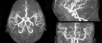
How is MRI of the vessels of the brain and neck performed?
Diagnosis is carried out using a special device - a magnetic resonance imaging scanner, which looks like a large tunnel from the outside. Before the scan begins, the patient is explained how to behave correctly inside the device, namely:
- It is prohibited to bring any gadgets with you;
- it is unacceptable to make any, even the most insignificant movements;
- You cannot make movements with your mouth (for example, try to speak).
Of course, in exceptional cases, when the patient feels significant changes in well-being during an MRI of the vessels and arteries of the neck or brain (which happens extremely rarely and is most likely caused by fear of undergoing diagnostics), he will always be able to contact the medical staff using a speaker and report changes in your health. If there are compelling reasons, the doctor may decide to interrupt the procedure.
The patient is also informed that there is no need to be afraid of the characteristic sounds of a working tomograph - this is a completely natural phenomenon. The doctor will ask how you are feeling and suggest taking sedatives if necessary.
If MRI of brain vessels needs to be performed using contrast, the doctor will definitely ask about the presence of allergic manifestations to medications.
The scanning takes place without the use of radiation exposure, which means it is completely harmless to the patient. Due to the fairly short examination time, patients in most cases tolerate it well, because the likelihood of panic attacks during an MRI of the vessels and arteries of the neck or brain is minimized.
Types of MRI of the brain
- Standard - done without the introduction of contrasting solutions, but at the same time provides a sufficient amount of information.
- With contrast, before which drugs containing gadolinium salts are injected into the vein - gadopentetic and gadoteric acids, Omniscan, Magnevist, etc. These solutions penetrate into the bloodstream and, once in the rays of the MRI scanner, highlight the resulting “picture”. At the same time, the changed areas become better visible, which simplifies decoding. The technique is most often used to detect vascular abnormalities, multiple sclerosis and tumor formations. The dose of contrast agent is selected individually, taking into account weight.
- MR angiography – is performed to assess the condition of blood vessels in atherosclerosis, aneurysms, blood clots and pre-stroke conditions. It is done with gadolinium contrast to show blood flow problems in detail.
- MR imaging of the pituitary gland, an appendage that is an endocrine gland. The pituitary gland secretes hormones responsible for reproductive function, tissue metabolism and the regulation of human growth. An examination is prescribed if an adenoma is suspected - a benign tumor that causes migraine-like pain, hormonal imbalances, gigantism, infertility, obesity and sexual dysfunction. The same method is used to identify malignant pituitary formations that have similar symptoms and are accompanied by a pronounced deterioration in health.
Indications for general anesthesia for MRI
Intravenous or inhalational sedation is only necessary for patients who are unable to hold their body still for long periods of time. Main indications for general anesthesia:
- Claustrophobia is the fear of closed spaces. Such patients, while inside the device, experience panic, which negatively affects their health and makes MRI diagnostics impossible.
- Mental disorders accompanied by unpredictability of behavior and high excitability.
- Uncontrollable involuntary head movements (swaying, shaking, tics).
- Epilepsy and other types of convulsive readiness and seizures - anesthesia is given only intravenously due to the risk of provoking a seizure attack.
- Early childhood. Small children cannot lie still for a long time in an MRI scanner, so light mask anesthesia is indicated for them.
- Severe pain syndrome, in which prolonged stay in one position causes discomfort, cramps, pain and spasms.
Limitations for MRI of brain and neck vessels
It is not always possible to conduct tomography for people with mental illness, patients with uncontrolled movements of the limbs, as well as people with a fear of being in a confined space. Often, scanning of such patients is carried out in a state of medicated sleep, since otherwise it is impossible to carry out a diagnosis.
It is especially difficult to perform MRI of cerebral vessels in patients on mechanical ventilation. Not every medical institution has specialists who can take on such responsibility.
Very often, tomography is performed under anesthesia and in young children. It is very difficult for children to remain immobilized throughout the entire examination period, and under anesthesia, the scanning is very calm for both them and the doctors. Another advantage of using anesthesia is that young patients will not experience any stress and will not have negative memories.
Indications for magnetic resonance imaging of the brain
- Neoplasms or their metastases. Diagnosis is prescribed for persistent migraine-like pain, sudden loss of vision and hearing, auditory, olfactory and visual hallucinations, attacks of confusion, sudden reading and writing disorders, often accompanying cancer pathologies.
- Epilepsy and other diseases manifested by fainting, confusion and convulsions.
- Suspicion of single or multiple cystic cavities filled with fluid, bloody or other contents.
- Possible presence of parasites (cysticercus and echinococci) carried through the vascular bed with the bloodstream into the head.
- Inflammations – meningitis, encephalitis, arachnoiditis, myelitis. Lesions caused by infections - measles, herpes, tuberculosis, toxoplasmosis, tick-borne encephalitis.
- Rehabilitation after stroke, traumatic brain injury and surgery. Using magnetic resonance diagnostics, the doctor evaluates the effectiveness of the treatment and predicts long-term results.
- The likelihood of developing multiple sclerosis, Alzheimer's disease and other degenerative processes.
- Children are examined for congenital pathologies and hydrocephalus.
With all these diseases, life and health directly depend on a timely diagnosis. Therefore, at the slightest suspicion of brain disorders in yourself or your child, you need to come to the clinic and be examined.
What does the scan reveal?
It is advisable to conduct MRI of the vessels and arteries of the neck to identify many diseases and pathological processes, which include:
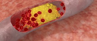
- cerebrovascular accident due to the presence of atherosclerotic plaques on the inner walls of blood vessels;
- received injuries to the head and neck;
- brain infections of any etiology;
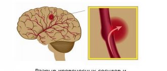
- life-threatening conditions due to brain damage (for example, cerebral infarction);
- any neoplasms;
- pathological structure of the vascular system of the brain;
- multiple sclerosis.
What the results show
MRI studies, especially those performed with contrast, reveal numerous pathological processes. In the sections, compactions, cystic cavities, and hematomas (collections of blood) are visible in detail. Scars, parasites and their cysts, foci of degeneration, sclerosis and inflammation are identified.
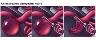
Vascular changes are diagnosed, manifested by impaired patency, narrowing or dilation of blood vessels, the appearance of aneurysms (protrusion of walls) and thrombosis.
The degree of tissue damage in traumatic brain injuries, hemorrhagic and ischemic strokes is determined. The affected areas look lighter and are visible even if they are small in size and have sparse neurological symptoms.
Congenital defects are determined - underdevelopment and hypertrophy of the organ, small and incorrectly located gyri, cysts, holoprosencephaly - lack of division into hemispheres. Hydrocephalus is detected - accumulation of fluid in the ventricles, which with this anomaly are greatly enlarged.
Pathological areas and neoplasms appear as dark or lightish spots of various sizes and shapes, standing out against a grayish background. Oncological seals, especially malignant ones, have blurred, uneven edges and surrounding areas of necrosis.
MR diagnostics is recommended periodically for everyone who has been treated for oncological pathologies of any location. It detects metastases, which usually accompany cancer recurrence.
Contraindications
- Installed pacemakers and other electronic devices that are disrupted by the surrounding electromagnetic field.
- Fixed dentures with metal elements present in the mouth, crowns containing metal, braces and other orthodontic structures. The metal they contain is heated by a magnet and deteriorates, damaging surrounding tissue.
- Tattoos applied to the skin, made with metal-containing ink. Due to the heat caused by the electromagnets, burns may occur in these areas. In the absence of information about the pigment. used when applying a tattoo, it is better not to risk it and do a CT scan, ultrasound or x-ray. Examination is also prohibited for any metal piercing that cannot be removed.
- MRI examinations with contrast are not performed during pregnancy or if contrast agents are intolerant. Such an examination is not prescribed for severe kidney pathologies that make it difficult to eliminate gadolinium.
Magnetic resonance imaging is a safe and highly informative procedure that detects pathologies in the early stages. Therefore, in case of migraine-like phenomena, coordination problems, a sharp decrease in hearing and vision, fainting, or progressive memory deterioration, you must definitely go to the clinic and be examined. The price of MRI diagnostics is low and quite affordable for Muscovites and residents of Moscow Region.
Get an MRI of the brain >>>
Contraindications for examination
Despite the fact that MRI of the head and neck vessels is an absolutely safe diagnosis, there are still some contraindications for its use. If we look at it as a whole, they are not at all different from those contraindications that apply to scanning other parts of the body.
First of all, MRI of cerebral vessels is unacceptable for people with metal objects in their body. Any implants, stimulators, prostheses act as contraindications. The exception is products made of titanium. After the doctor is aware of the presence or absence of such objects in the body, the patient will be allowed to undergo the examination or will be refused. In this case, the patient must provide the specialist with appropriate documentary evidence of his words. The type of material from which a particular implant is made is recorded in a special certificate issued after the surgical intervention.
Another contraindication to MRI of the vessels of the head and neck is scanning using contrast for women carrying a child. The chemical substance, despite its relative safety for an adult, can cause irreparable harm to a developing fetus, which is why such a ban is due.
The use of contrast is unacceptable regardless of the duration of pregnancy, as well as during the lactation period. At the same time, MRI of the vessels and arteries of the neck without contrast enhancement is quite acceptable for pregnant women in the 2nd and 3rd trimesters, when the fetus has developed all vital organs and systems.