1.What is MR angiography
MR angiography (magnetic resonance angiography) is a technique that allows you to examine the blood vessels of the brain. The result of such diagnostics is a three-dimensional image of the entire vascular system of the area under study. These data provide the doctor with information not only about the anatomical features, but also about the functional viability of the vessels. The result of MR angiography of the cerebral arteries is an accurate diagnosis or confirmation of the suspected disease, which serves as the basis for a further treatment plan. MR angiography is also used to evaluate the effectiveness of already used therapy or surgical assistance.
MR angiography is a safe study that even excludes arterial puncture. Diagnosis can be done with or without contrast agent. At the same time, the information content and accuracy of such a survey is higher than that of many other methods. MR angiography of the cerebral arteries is a radiation diagnostic method, but the radiation parameters of the tomograph are completely safe. The procedure is carried out using a magnetic resonance tomograph with a field strength of 0.5 Tesla.
A must read! Help with treatment and hospitalization!
MRI angiography of vessels: which vessels can be examined?
Vascular MR angiography allows you to examine such areas of the human vascular system as:
- cerebral vessels
- neck vessels
- spinal vessels
- thoracic aorta, small arteries and veins
- abdominal aorta, small arteries and veins
- coronary vessels of the heart
- iliac arteries and veins
- vessels of the upper and lower extremities.
Initial appointment with a NEUROLOGIST
ONLY 1800 rubles!
(more about prices below)
2. Indications and contraindications for MR angiography of cerebral arteries
The main indications for studying the vascular system of the brain are related to head injuries and elucidation of the causes, localization and prevalence of ischemic stroke.
This study is also indicated for chronic headaches, when it is necessary to exclude or confirm vegetative-vascular dystonia as the cause of this condition. If there is a decrease in hearing and vision, the cause can also be identified during an examination of the vessels of the head.
In addition, any disturbances in the blood supply to the brain, suspicion of possible vascular disorders, or suspected arterial insufficiency may be an indication for MR angiography of the cerebral arteries. The diagnosis assumed based on the results of other types of diagnostics is most accurately confirmed during MR angiography.
Magnetic resonance imaging is safe and has a narrow list of contraindications. The main restrictions are related to the presence of metal implants in the body, about which the doctor must be warned by the patient.
In addition, it is better to postpone any MR examinations in pregnant patients.
Renal failure is also a relative contraindication. The advisability of performing MR angiography in such patients is clarified after studying the anamnesis and assessing the degree of risk.
Visit our Neurology page
Alternatives to CT scan of the brain
Alternative diagnostic methods include primarily MRI. But it is not recommended if the patient has any metal elements in the body: a pacemaker, vascular clips, etc. X-ray angiography is also sometimes used.
If you want to know where is the best place to get a CT scan of the brain, contact us - we will sign you up for a study at a clinic where they conduct the study using the latest equipment and guarantee high-quality diagnostic results.
3. How is the procedure of MR angiography of the cerebral arteries performed?
When going for diagnostics using an MRI scanner, you need to expect that the procedure will take a significant amount of time - from 30 minutes to 1.5 hours.
Special preparation for the study is not required, however, the doctor must warn the patient about the need to remove all metal objects, including dentures, and also asks to inform about the presence of implants in the body, their location, volume, and the material from which they are made.
The patient is placed horizontally on the surface of the tomograph, after which the head is fixed with a special helmet. For the accuracy of the diagnosis, any head movements must be excluded.
If the patient is prone to claustrophobia, he will be asked to take sedatives.
The operation of the tomograph is accompanied by rather sharp sounds, so special protective headphones are put on the ears of the person being examined. The scanning unit makes rotational movements around the patient's head and transmits a three-dimensional image to the monitor. The doctor deciphers the data received and draws conclusions about the state of the patient’s vascular system of the brain.
The results of MRI diagnostics are given to the patient in the form of a report, which may contain a printed scan of the image or a disk with files.
About our clinic Chistye Prudy metro station Medintercom page!
Advantages of brain MRA
is that it can be performed simultaneously with MRI and does not take much time. MRA of the neck arteries is extremely important for osteochondrosis and episodes of cerebrovascular accidents. MRA is an accurate, safe, quick and painless technique for diagnosing vascular pathologies. There is only one contraindication for MRA, as for any magnetic resonance imaging study: an artificial heart pacemaker. The presence of metal objects in the body is not a contraindication. In St. Petersburg, one of the leading specialists in the field of radiation diagnostics is Professor A.V. Kholin. MRI in St. Petersburg is performed by him in different clinics. In case of complex vascular pathologies, the entire arsenal of radiation methods for studying blood vessels can be used.
MR venography gives an idea of the condition of the venous sinuses and the outflow of blood through the main veins.
MR venography. Reconstruction in the sagittal plane, Color processing of the image. Norm.
MR venography of the brain. Reconstruction in the transverse plane. Normal dural sinuses and great veins.
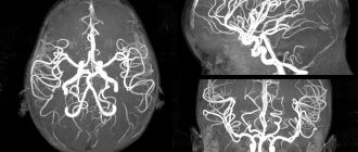
MRA is used not only to study cerebral vessels, but also to study peripheral arteries
MRI of St. Petersburg allows you to choose the location for vascular examination. However, Professor A.V. Kholin’s research has its own distinctive features.
4.What results does MR angiography of the cerebral arteries give?
The volumetric 3D image obtained by the doctor on the tomograph monitor makes it possible to comprehensively study the anatomy and functions of the vascular system of the brain. In this case, aneurysms, vascular occlusions, vascular malformations, places of stenosis and atherosclerotic changes are clearly visible.
Many chronic conditions - headaches of unknown origin, tinnitus, dizziness - may have vascular disorders as their root cause. Often, only through MR angiography can a clear connection be established between a particular manifestation and a specific organic or functional abnormality and adequate treatment, therapeutic or surgical, be prescribed.
The following diseases are most often diagnosed during MR angiography of cerebral arteries:
- aneurysm;
- vascular wall dissection;
- vascular stenosis;
- vasculitis;
- atherosclerosis.
Advantages of CT scan of brain vessels
CT not only displays the vascular network in three dimensions, but also allows you to study in detail any large or small vessels. This research method is considered the most useful and effective, since it makes it possible to diagnose almost any dangerous disease at an early stage.
During CT angiography, you can examine the structure of the vascular wall and the tissue that surrounds the veins and arteries. This provides valuable information necessary, for example, for the early detection of hemorrhagic stroke. In the images, you can see the size of the hematoma, study the degree of damage to the brain centers and nuclei, and also assess the level of compression of the brain matter.
There is no pain during the examination.
MR angiography of cerebral vessels
During an MRI study of cerebral vessels, a three-dimensional image is obtained and an assessment is made of the state of the blood supply to the brain and biological processes in the brain tissue.
The technique is used in the practice of neurologists, neurosurgeons, and vascular surgeons for accurate diagnosis and selection of targeted treatment. The study can be performed with or without contrast.
MR angiography of cerebral vessels allows:
- study the condition and structure of the vessels supplying blood to the brain;
- detect abnormalities in vascular development, pathological branches;
- measure the width of the lumen of an artery or vein and determine the degree of its narrowing (stenosis);
- identify aneurysms (thinning of vascular walls in a certain area with the risk of vessel rupture);
- determine the speed of blood flow;
- detect a stroke and determine its nature (ischemic or hemorrhagic);
- diagnose sinus thrombosis in the brain;
- detect a brain tumor;
- identify post-traumatic hematoma.
Indications for MR angiography of cerebral vessels:
- frequent attacks of headaches with impaired hearing and vision, with dizziness;
- previous head trauma;
- ischemic cerebral vascular disease;
- suffered a stroke;
- cardiopsychoneurosis.
MR angiography of neck vessels
MR angiography of the arterial and venous vessels of the neck is an informative research method that allows you to visualize the vessels and assess the state of the circulatory system that supplies the brain. Visualization of the carotid arteries allows you to diagnose the cause of insufficient blood supply to the brain, detect arteriovenous shunts, and vascular anomalies.
Indications for MR angiography of neck vessels
- patient complaints of headaches with blurred vision, dizziness; memory impairment; migraine attacks; insomnia;
- presence of neurological symptoms during examination;
- the neurologist suspects vertebrobasilar insufficiency (due to narrowing of the lumen of the vertebral and carotid arteries).
Imaging of the deep jugular veins may reveal thrombosis. Using vascular tomography, this pathology is diagnosed, which is inaccessible for diagnosis by other methods.
Contraindications to MR angiography of brain and neck vessels
Contraindications include:
- the patient has a pacemaker, electronic implant, artificial heart valves, insulin pump, etc.;
- allergy to contrast agent;
- mental disorder, epilepsy;
- renal and hepatic failure;
- the presence of metal bodies in the body (staples on blood vessels, bullets, dental implants, braces, etc.);
- pregnancy (as a last resort, examination after the 1st trimester is allowed, but without the use of contrast).
- claustrophobia.
Metal implants will not cause harm to the patient, but they can significantly distort images and cause incorrect diagnosis. The most modern MRI scanners allow you to change the device settings and prevent distortion of the study results. The possibility of performing MR angiography can be decided by a doctor who has been warned about the presence of metal in the patient’s body.
When breastfeeding a child, the mother expresses milk on the eve of the procedure to feed the baby over the next 24 hours (until the contrast agent is removed from the body), or feeds the child with formula milk.
What can be revealed during an MRA examination?
During this diagnostic procedure, the following diseases and pathologies of cerebral vessels can be visualized:
- aneurysm defects (pathological protrusion of the vascular wall in a separate area);
- dissection of the walls of blood vessels;
- deformation of blood vessels (pathological expansion or narrowing);
- abnormalities in the development of blood vessels (asymmetry of their branching, excessive looping of the vascular network, etc.);
- identify the condition of the ring of vascular hemispheres;
- determine the condition of the arteries in the cerebral cortex;
- see the functioning of the cranial veins.
MRA of the brain
Magnetic resonance angiography of the brain is a safe, modern and highly informative diagnostic technique that allows you to assess anatomical disorders and detect functional changes in the brain. It reflects the condition of the blood vessels of the cervical spine and brain.
The content of the article:
- Indications for magnetic resonance angiography of the brain
Thanks to this technique, specialists will be able to confirm or refute the diagnosis; its use is also relevant when planning angioplasty and stenting (a type of vascular surgery).
Angiography makes it possible to see vessels on a magnetic resonance imaging scanner at any location. During it, the doctor takes many pictures, which then undergo careful computer processing, due to which they turn into a sufficiently contrasting and clear two-dimensional or three-dimensional image. The main advantages of MRA of the brain are harmlessness and absolute painlessness.
Magnetic resonance angiography is a non-invasive technique, since there is no surgical impact on the body. Basically, it is performed without the use of a contrast agent. Also, during the study there is no ionizing radiation or radiation exposure. The procedure itself takes no more than 40 minutes, and the patient just needs to lie down and not move.
Afterwards, the tomograph displays an image of the vascular bed, informing about the spatial location and relationships of all vascular structures. The conclusion describes not only the above-mentioned features of the state of the brain, but also pathologies, if any were detected.
MRA of the brain is prescribed for microstrokes, strokes, traumatic brain injuries, congenital heart defects, vegetative-vascular dystonia and intracranial pressure disorders.
Magnetic resonance angiography is important in diagnosing the following diseases:
- aneurysm dissection;
- local expansion of the walls of blood vessels;
- vascular stenosis;
- vasculitis (inflammatory processes in the vascular walls);
- atherosclerosis of the arteries.
Any MRA also has its contraindications; angiography cannot be used if the patient has pacemakers and hemostatic clips on the vessels of the brain. The presence of electronic, ferromagnetic or metal implants also prohibits diagnostics. There is also a certain relative contraindication that does not recommend this study for pregnant women in the first trimester.