Indications
CT is prescribed to detect inflammatory processes, brain tumors, hemorrhages and the consequences of traumatic brain injury (TBI).
In addition, computed tomography is one of the methods for monitoring the effectiveness of treatment and rehabilitation for various diseases - the bones of the skull, membranes and structures of the brain, blood vessels and paranasal sinuses are clearly visible in the images.
A computed tomography scan of the brain may be prescribed by the attending physician (therapist) or a neurologist in the following cases:
- suspected swelling, inflammation, hemorrhage after TBI;
- acute circulatory disorder (stroke);
- abscess and cysts of the brain;
- infectious diseases (encephalitis, meningitis);
- oncopathology (tumors, metastases);
- abnormalities in the development of brain structures and blood vessels;
- pathologies of blood vessels (thrombosis, aneurysm);
- hydrocephalus (“dropsy” of the brain - excessive accumulation of cerebrospinal fluid (CSF) in the ventricular system of the brain);
- convulsions, visual disturbances, dizziness, decreased sensitivity, confusion - neurological symptoms for no apparent reason;
- causeless headaches for 2-3 months or more;
- preoperative examination;
- individual contraindications to MRI (presence of a pacemaker, insulin pump, metal prostheses).
Contraindications to CT scan of the brain
For CT diagnostics, X-ray radiation is used, albeit in a minimal dosage. Therefore, CT scanning is not recommended for pregnant women (risk of developing birth defects in the child).
Note: CT scanning with contrast is not contraindicated for nursing mothers if after the procedure the baby is weaned from the breast for 2 days (the contrast agent enters the secretions of the mammary glands).
Also, contrast-enhanced CT is not advisable for individuals with severe renal impairment. When the drug is excreted in the urine, side effects may develop in the form of intoxication (poisoning).
Acute pain syndrome and pathological inability to remain still (hyperkinesis) can become a serious obstacle to the study, since you cannot move during the procedure.
Contrast agents should not be administered to people who are allergic to iodine.
It is advisable to avoid any kind of radiation, including minimal radiation, for patients with multiple myeloma and those suffering from endocrine pathology.
For psychologically unstable individuals, as well as for patients with claustrophobia, CT is either not prescribed at all or is performed in a state of light medicated sleep.
And for obese people (weight over 120-130 kg), a CT scan is simply impossible, since their body volumes may exceed the diameter of the diagnostic chamber.
Pros of CT
Screening tomography has special advantages in comparison with other brain diagnostic methods. The main advantages are:
- non-invasiveness - the procedure is painless due to the absence of the need to use surgical or other medical instruments (the patient’s discomfort can arise exclusively when using a contrast reagent and only when it is administered intravenously through a catheter);
- high accuracy of data - layer-by-layer examination of the head helps to identify changes in the structure of its parts in the early stages of development, and the use of an additional amplifier further increases the information content;
- speed - the study takes place as quickly as possible, since it usually lasts about 60 minutes (after an hour the patient receives the results);
- minimal radiation exposure - the dosage of X-rays with CT is much lower than with radiography, so the study is allowed to be performed more often
Additional advantages of diagnostic tomography:
- the ability to obtain information about bones and soft structures simultaneously;
- reducing the amount of contrast agent, which reduces the risk of complications;
- allowed for patients with metal implants in the body, which is mainly a contraindication to MRI.
Another advantage of the technique is the possibility of conducting diagnostics for people who are afraid of being in confined spaces. To do this, they use open-type devices that do not close completely, which allows the patient to feel comfortable throughout the entire examination period. The disadvantages include the inability to conduct research during pregnancy, preparation for the procedure with contrast, as well as exposure to radiation on the body, albeit in a minimal amount.
Methodology
The duration of the procedure ranges from 2-3 minutes to half an hour, depending on the purpose of the study. Before the examination begins, the patient must remove all jewelry and metal objects.
The patient is positioned on a movable couch in the chamber, his head is secured with a special fixing device (it is required to remain completely still). The table with the patient moves inside the tomograph. During scanning, the outer part of the device will rotate around its axis, and the couch under the subject will move slightly in the horizontal plane. The tomograph makes a slight noise during operation, which does not cause much discomfort to the patient.
During the procedure, the medical staff is in an adjacent room and observes the process through glass. At the same time, a two-way connection is maintained with the patient: the doctor can inquire about his well-being, and he, in turn, can report any changes in his condition.
When a contrast agent is injected, you may experience a metallic taste in your mouth and a feeling of warmth or cold spreading through your veins. This is a normal reaction of the body to the administration of the drug.
It is not normal for the patient to experience nausea, dizziness, headache or stomach discomfort. He should immediately report these symptoms to his doctor. This may be a side effect of the injected contrast agent.
Computed tomography for children
CT scanning can be performed on children from 3 years of age (for emergency indications), if they can lie still for 15-20 minutes. If this is not possible, then the child is given a light anesthesia. Otherwise, the procedure for examining children is no different from adult tomography.
Complications
There are no serious complications after computed tomography, except for allergic reactions to the contrast agent. But this may not happen if the medical staff of the institution collects the history of the patient’s life and illness correctly and as completely as possible.
MRI of the brain and CT: what is common?
- The results of the examination of patients are a series of layer-by-layer images. They are formed when a computer processor processes data from a tomograph. They are given to the visitor on disk or printed out.
- It involves scanning a person in a supine position. In some cases, when examining certain areas of the body, the person being diagnosed may be asked to turn on their side.
- They last quite a long time (sometimes up to an hour), while urging the patient to remain still.
- The appearance of the installations is the same.
- In both methods, a contrast agent is administered.
Brain CT results
Decoding the results and preparing a conclusion takes from 1 to 1.5 hours. Also, many clinics practice sending results to the patient’s email address. The patient is given photographs and/or a CD with a recording of a three-dimensional image with detailed descriptions of them.
The patient is fine if:
- the bones of the skull, the brain and its vessels are of normal size;
- no foreign inclusions, tumors, hematomas, etc. were found;
- no symptoms of bleeding or fluid accumulation;
- the integrity of the bone tissue is not compromised.
Any deviation from the norm is considered a sign of a particular disease. Therefore, with the received conclusion, the patient is immediately sent to the doctor who prescribed the examination.
Computed tomography of the brain
Computed tomography, or CT, is a diagnostic method based on the use of x-rays. X-rays produced in the X-ray tube of the tomograph affect the area of the patient's body being examined from different angles. The device’s sensors receive layer-by-layer scanning data, which is immediately processed by a computer and “lined up” into an overall picture. Thus, the doctor sees a detailed image of the brain and can make a diagnosis based on this. Unlike radiography, CT is much more informative and allows you to examine in detail any part of the organ. Computed tomography differs from magnetic resonance imaging in the lower cost of the study and the absence of some contraindications: the presence of metal prostheses, implants and a pacemaker in the patient is not an obstacle to CT.
Indications for performing a CT scan of the brain
- prolonged and/or severe headaches;
- paralysis of limbs;
- dizziness, fainting;
- disturbance of consciousness;
- deterioration of vision and hearing perception;
- nausea, vomiting;
- newly developed convulsive syndrome;
- repeated convulsive syndrome in combination with recent trauma, fever, headache, cancer history;
- recent trauma accompanied by vomiting, fainting, bleeding disorders and/or penetrating skull trauma.
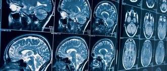
CT scan of the skull and brain allows for an informative comprehensive assessment of the condition of the vault, base of the skull, paranasal sinuses, temporal bones, as well as soft tissue formations, arteries, veins, brain sinuses, and cavity structures. This X-ray technique is capable of informatively and quickly diagnosing a whole range of brain disorders. CT is used to detect:
- localization of foreign bodies;
- accumulations of pus in the epidural and subdural spaces;
- arachnoiditis;
- small focal hemorrhages;
- the nature and localization of tissue damage to the eyeball, orbit, inner ear, auditory nerve;
- violations of the integrity of the bone tissue of the skull;
- hydrocephalus;
- abscesses;
- brain cysts;
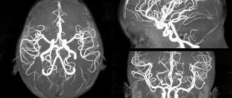
- pathologies of the blood vessels of the brain;
- neoplasms of various nature;
- areas of bruises and other brain damage, meningeal hematomas;
- ischemic and hemorrhagic strokes;
- meningitis and its complications;
- for fractures of the base of the skull - leakage of cerebrospinal fluid;
- cerebral edema and assessment of its degree;
- encephalitis and other disorders.
When it comes to diagnosing tumors, CT is used as an additional method after MRI. In addition, this technique is used if the patient has absolute restrictions on MRI. Brain CT is also widely used in preparing a patient for surgery, developing tactics for further therapy, and monitoring its effectiveness. Native CT of head tissue is used as a screening method to search for a possible disorder. When the pathology is non-oncological in nature, CT is not inferior in information content to MRI, except for cases of minor changes in the white matter and pituitary microadenomas. CT scan of the brain without contrast is mainly performed to detect all of the above pathologies, with the exception of neoplasms, metastases and vascular disorders. In particular, native research is carried out in the acute phase of a stroke; also, using this technique, it is possible to assess the degree of atrophy of the brain substance and determine the type of hydrocephalus. We note the importance of non-contrast CT in identifying the consequences of a head injury, since the images visualize disorders already after 6 hours after the incident, while another alternative research method - MRI - reveals such consequences only after 3 days. In case of injury, CT allows you to obtain the most complete data on the morphological state of the brain and skull, perform dynamic monitoring and draw conclusions about the consequences of the injury.
Alternative methods - MRI
Unlike magnetic resonance imaging (MRI), which is most effective in studying the physical state of the brain, CT studies the chemical structure of tissues. That is, CT allows you to determine their X-ray density, which usually changes with various diseases.
When examining the brain, MRI perfectly visualizes soft tissues, which is important for diffuse and focal lesions of brain structures and pathologies of the spinal cord. But on an MRI, the bones of the skull are practically not visible - a CT scan of the brain in this regard provides much more information. Therefore, in some cases, both studies are prescribed simultaneously, as complementary to each other and giving the most complete picture of the disease.
Difference between CT and MRI of the brain
- They differ in operating principles. With computer diagnostics, the body is scanned with X-rays, and with magnetic resonance imaging, it is placed under a magnetic field.
- On a CT scan, a specialist monitors the physical parameters of the brain, and on an MRI, he looks at the chemical components of brain tissue.
- During a CT scan of the head, only this part of the patient's body is pushed into the scanner. For MRI, the entire person's brain is placed in the machine.
- On a computed tomography scan, the cranial bones are clearly visible. With MRI, the focus of imaging is on the soft tissue of the brain.
- The head is examined on MRI if there is a suspicion of oncology, genetic diseases of neurons, inflammation processes in the nerves or membranes of the brain.
- A CT scan checks to see if a person is having a stroke.


