The brain is a complex and poorly understood human organ that is difficult to diagnose. However, the brain is the control center responsible for the operation of all systems in the body. Therefore, any pathological processes in it affect the physical and mental health of a person.
MRI is one of the methods for studying the brain to determine negative conditions in it. Diagnostics is prescribed for adults and children and is a safe examination method.
What it is
The method refers to a non-invasive examination using radio waves and a magnetic field. During the scanning process, the doctor receives an image showing parts of the brain. Diagnosis is carried out without radiography. The study reveals disturbances in the functioning of the nervous and vascular systems, aneurysms, and tumors.
Also, based on the results of the examination, the level of activity of the cerebral cortex is determined. When using a contrast agent, tissues are better visualized, which helps to more accurately identify pathologies.
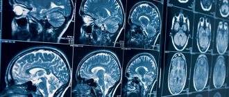
Decoding MRI of the brain
For diagnosticians at DiMagnit MC, 15-20 minutes are enough to analyze the received images.
The center’s specialist doctor will inform the patient about the results obtained and advise how to proceed further. This consultation is free.
Upon completion of the diagnosis, the patient is given a disk with the image and a conclusion, which are included in the cost of the study. If necessary, the survey results can be duplicated onto the visitor’s flash card for an additional fee.
Interpretation of MRI scans
Advantages of the method
Advantages of brain tomography:
- the patient does not experience pain during diagnosis;
- During the examination, no foreign substances are introduced into the human body;
- no ionizing radiation;
- clear images help to accurately recognize pathology and prescribe treatment;
- as prescribed by a specialist, an examination of the head and cervical/upper spine is carried out to assess the functional activity of the brain - based on the results, a safe method of surgical intervention is prescribed;
- areas of the brain hidden by the bones of the skull are examined - other methods do not diagnose these areas;
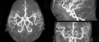
- MRI helps to obtain a complete picture of the brain and vascular system even without the use of a contrast agent;
- tumors and other pathologies are detected in the early stages of development.
Make an appointment now!
Why is an examination prescribed?
MRI diagnostics helps to recognize pathological changes in the soft tissues of the lining of the brain, inflammatory reactions, and disorders of the central nervous system. Also, with the help of an examination, the conditions of the parts of the head are studied: the visual and occipital lobes, the pituitary gland, the cerebellum, areas of thinking and memory, and the ventricles of the brain.
Before the procedure, the patient undergoes tests, the results of which determine the type of diagnosis. For example, tests showed that the patient had an excess level of the hormone prolactin. The doctor writes a referral for examination of the cerebellum.
Examination of the brain in a magnetic field helps determine the presence of:
- Multiple sclerosis - lesions of the myelin sheath of nerve fibers. The diagnosis determines the stage of the disease, the level of damage, after which the doctor prescribes therapy.
- Benign and malignant tumors. Brain scanning is used to identify the affected area, monitor the development of the tumor, as well as the treatment process.
- Disorders in the cerebral cortex - Parkinson's disease, Alzheimer's disease. MRI determines the density of white and gray matter, the presence of pathologies in the subcortex and cortex of the head.
- Ischemic strokes, heart attacks. The images determine the area and density of ischemic lesions, the presence of cortical necrosis, the appearance of edema, and the stage of development of the disease.
- Brain injuries are damage to blood vessels and tissues due to trauma. Diagnostics reveals the first signs of VSD.
- Mental disorders of endogenous and exogenous types. The method helps to identify hereditary pathologies, changes after traumatic brain injury, viral infection, and poisoning with toxic substances. MRI diagnostics determines schizophrenia.
For children, head tomography is prescribed for the following pathologies:
- head injury, concussion after injury, intrauterine infectious processes;
- hypoxia, ischemia and other disruptions in brain development;
- epileptic seizures, cerebral hemorrhage;
- the first stages of multiple sclerosis;
- dangerous diseases such as pituitary adenoma, brain tumors, cysts and suspicions of them;
- a sharp deterioration in vision and hearing, malfunction of the inner ear;
- high intracranial pressure.
Diagnostics helps to study the state of the brain and its parts, identify the causes of headaches in children, and prevent the development of autism.
What diseases will an MRI of the brain show?
Magnetic resonance scanning is used to identify the following brain pathologies:
- tumors of the brain and meninges;
- demyelinating and degenerative diseases (Parkinson's disease, Alzheimer's disease, multiple sclerosis);
- cysts, including pituitary cysts;
- empty sella syndrome;
- acute pathologies of cerebral vessels (hemorrhagic and ischemic strokes, thrombosis, aneurysms);
- complicated inflammatory processes (meningitis, sinusitis and others).
Degenerative brain changes on MRI scans
The final diagnosis is established by the doctor who gave the referral for the study. Usually a neurologist deals with brain diseases, but if a tumor is suspected, a referral can be made by an oncologist.
The radiologist conducting the study does not make a final diagnosis, but analyzes the images obtained and describes the detected abnormalities and formations. After the examination, the images are sent to the attending physician, who confirms or removes the initial diagnosis.
Differences between MRI and CT scan of the brain
Magnetic resonance imaging of the head differs from other diagnostic methods in the following:
- the examination is performed in 3-4 projections, which provides more information for identifying diseases and choosing treatment methods;
- pathologies are detected in the early stages of development - ischemic changes are detected 2-3 hours after a stroke;
- even minimal changes in the brain that cause multiple sclerosis are determined;
- parts of the brain that are not diagnosed by computed tomography (CT) are studied - the brain stem, the cerebellum.
What does a brain MRI show?
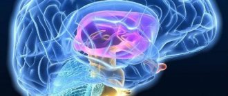
Magnetic tomography is one of the most highly accurate visual diagnostic methods. Using magnetic scanning, you can obtain detailed and detailed images of the organ in three projections. Even small structures such as the pituitary gland are clearly visible in the images taken. Magnetic resonance scanning makes it possible to see the structural features of the brain, which means carefully studying the structure of the nervous substance of the cerebral cortex for abnormalities, to see hemorrhages and inflammation of the meninges.
When is a diagnosis prescribed?
A brain examination is prescribed for the following ailments:
- developmental anomalies and diseases of cerebral vessels;
- head injuries and bruises, cerebral hemorrhages;
- sharp deterioration of vision and hearing;
- tumor development;
- infectious reactions in the central nervous system - meningitis, HIV infections, abscesses;
- pituitary adenoma, epilepsy;
- pathologies of the base of the skull;
- multiple sclerosis, sinusitis, neurodegenerative diseases;
- aneurysms, thrombosis and other anomalies in the vessels of the head.
Diagnosis is also carried out after surgery. Prescribed for frequent migraines and fainting, tinnitus, sudden deterioration of memory and attention, impaired coordination of movements, and mental disorders.
Indications and contraindications for MRI with contrast
The main indications for MRI with contrast are:
- tumors (detection);
- differential diagnosis of neoplasms;
- pituitary adenoma;
- postoperative diagnosis of tumor metastases;
- multiple sclerosis (activity or passivity);
- postoperative control (stability of the intervertebral disc).
Contraindications to MRI with contrast:
- pregnancy and breastfeeding;
- allergy to contrast agent;
- renal failure.
Contraindications
Head tomography is not prescribed if the patient has absolute contraindications, but is allowed at the discretion of the doctor in case of relative contraindications. Diagnostics is prohibited if the following conditions exist:
- the patient has prosthetic heart valves, pacemakers or neurostimulators;
- presence of insulin pumps, inner and middle ear prostheses, cochlear implant;
- the patient has an Ilizarov apparatus;
- the presence of metal implants, ferromagnetic particles, fragments in the body.
Relative indications include:
- tremor, the patient’s inability to hold his breath for a long time during examination;
- presence of dental braces, dentures, stents, vena cava filters;
- heart failure, clip instead of gallbladder;
- pregnancy;
- claustrophobia;
- severe pain while standing still;
- coronary artery bypass grafting.
MRI of the brain - contraindications
Despite all the advantages of MRI examination, this type of diagnosis is not indicated for everyone. There are two categories of reasons that prevent an MRI scan.
Absolute contraindications
Brain tomography is not allowed if:
- the presence of metal objects in the body (clips, prostheses made of ferromagnetic metal, bullet fragments, etc.);
- weight category from 130 kg and waist circumference over 150 cm;
- implanted pacemaker, neurostimulator, cochlear implant;
- in the first trimester of pregnancy;
- children under five years of age.
Undesirable moments
There are a number of situations that create problems for prescribing MRI diagnostics or complicate its implementation:
- complicated cardiovascular diseases;
- mental disorders in the acute stage;
- claustrophobia;
- tattoos with metal in dye;
The presence of implants made of non-ferromagnetic materials does not interfere with magnetic resonance examination, provided that documents for the prosthesis are available, three months have passed since its installation and the implant is outside the area of interest of medical specialists.
Preparatory stage
The doctor, based on the test results, determines whether to perform a tomography with or without the introduction of a contrast agent. If the first option is chosen, then the patient does not take food or liquid 5 hours before the diagnosis. Before the procedure, you must remove all jewelry and accessories, watches and objects with metal elements.
During a consultation with a doctor, the patient reports the presence of chronic diseases, allergies to medications, claustrophobia, and pregnancy.
When preparing a child for a head tomography, it is not recommended to give him anything to drink or eat 3 hours before the diagnosis. If an MRI is performed with the administration of contrast, the examination is performed on an empty stomach. Before the procedure, the child should be shown to an allergist to check for an allergic reaction to the injected substance.
Diagnostic features
If a brain tomography is performed with the introduction of a contrast agent, the examination takes 1-1.5 hours.
Stages:
- the patient removes objects and clothing with metal elements;
- lies on the tomograph table on his back;
- the doctor administers a contrast agent intravenously;
- if there is tremors or inability to lie still, the patient takes sedatives;
- legs and arms are secured with belts, bolsters are placed under the neck;
- the table is pushed into the tomograph capsule - the doctor monitors the procedure from a special room with equipment;
- during the diagnosis, the patient hears slight noises of the tomograph operating - the procedure is safe and painless, a slight sensation is felt in the area of the injection of the substance;
- head diagnostics lasts 15 minutes - the patient is recommended to remain in a stationary position to obtain accurate scan results.
Features of examination of children
Brain tomography in children is carried out under the drug n8a8p00ko70z0o-7m9, -6v5 Propofol is administered. Anesthesia helps keep the child still during diagnosis. Children over 5 years old are given a sedative and given psychological adjustment and support before the tomography.
During the examination, the baby is shown cartoons and toys. Some clinics use open tomographs, when only the child's head is placed in the capsule, and the parents stand nearby and hold his hand.
Before diagnosis, it is recommended to take the child to the toilet, remove electronic items and jewelry with metal inserts from clothes and body. Next, the child is dressed in special clothes, “introduced” to the tomograph and allowed to listen to how it works. This helps to calm the baby before the procedure and obtain his consent to the examination.
Side effects
After tomography, the patient may experience nausea, dizziness, vomiting, weakness, and disorientation in space. This is a normal reaction. It occurs in people with increased sensitivity, when there are metal objects on the patient’s body, and when the rules for preparing for an MRI are not followed. Unpleasant sensations go away on their own. However, if side effects do not disappear for a long time, it is recommended to consult a doctor.
If the rules are followed, the procedure takes place without pain or side effects. The images help doctors accurately determine the cause of the disease and prescribe effective treatment.