MRI of the pituitary gland with contrast is a magnetic resonance examination of the most important endocrine gland of the human body, due to its small size, the use of contrast agents is required. Contrast agents for MRI contain nanoparticles of gadolinium, a metal that can increase the sensitivity and information content of the method. Once in the blood, gadolinium is evenly distributed throughout all tissues of the human body. Inflammatory processes, tumors and other anomalies, on the contrary, lead to local accumulation of contrast, which is why this area stands out in the images, facilitating diagnosis.
Where is the pituitary gland located?
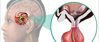
The pituitary gland is the most important organ of the endocrine system (gland), which produces a number of biologically active substances (hormones) that regulate the balance of energy, nutrients and minerals, cell division, sexual functions and reproduction, metabolism and many other aspects of the normal functioning of the human body. This is hard to believe, especially considering the small size of the pituitary gland - only 13 mm in length, 5-6 mm in thickness (the size of a pea or bean). The mass of the pituitary gland does not exceed 0.5 grams.
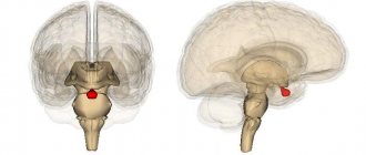
The pituitary gland is located at the base of the brain, inside the skull, in an area called the sella turcica (due to the shape of the bones, which actually resemble a saddle). The only method for diagnosing pituitary gland diseases that allows obtaining detailed and high-quality images is magnetic resonance imaging. MRI of the pituitary gland is always performed with contrast - this allows to increase the accuracy of diagnosis and the information content of the images, which is important, given the miniature size of the organ.
How is the pituitary gland examined in Moscow?
To increase the information content of the study, doctors may prescribe contrast, for example, to clarify the location and size of the formation, the intensity of blood circulation, and the condition of the blood vessels. Preparation for an MRI of the pituitary gland does not include any special actions; the patient simply comes to the appointment at the appointed time. For children 2-4 years old, it is recommended to have a CT scan instead of magnetic resonance imaging - it is faster, and it will be easier for the baby not to move for a short time than 20-40 minutes in the machine.
Pre-procedure testing may include an allergy test (if contrast is used). How is an MRI of the pituitary gland done? You remove metal objects and lie down on the machine table. The doctors will do the rest for you, and within 30 minutes you will receive the results of the study.
Indications and contraindications for MRI of the pituitary gland with contrast
Indications for the use of contrast-enhanced MRI of the pituitary gland:
- pituitary tumors/cysts of various origins;
- metastases to the pituitary gland;
- empty sella syndrome;
- hypofunction of the pituitary gland (decrease in the levels of pituitary hormones - prolactin, somatotropin, TSH, LH, FSH, etc.);
- hyperfunction of the pituitary gland (increased levels of pituitary hormones);
- apoplexy (hemorrhage) in the pituitary gland, pituitary necrosis (Sheehan syndrome);
- Itsenko-Cushing's disease;
- other endocrine disorders associated with the functional activity of the pituitary gland.
Symptoms in which an MRI of the pituitary gland is recommended:
- headache;
- decreased visual acuity (loss of visual fields, double vision, distortion of visible objects);
- excessively short or high stature inappropriate for age, delayed physical development;
- constant thirst with increased fluid consumption and frequent urination;
- infertility, both male and female;
- impotence in men, irregularities or absence of the menstrual cycle in women;
- obesity.
Indications for MRI of the pituitary gland
Magnetic resonance imaging better visualizes all brain structures, including the pituitary gland. Thanks to a step-by-step section of 3 mm, the radiologist can detect even small pathologies and formations. The peculiarity of this study is safety for the body, since, unlike CT, the patient is not exposed to x-ray radiation during the scanning process.
Prescribed in the following cases:
- any morphological disorders of the pituitary gland (developmental anomalies, hemorrhage into the pituitary gland, adenomas, hormonally inactive tumors, etc.);
- increase in hormonal levels, in particular prolactin ;
- suspected Cushing's syndrome;
- disruptions in the menstrual cycle;
- erectile dysfunction;
- vision problems;
- regular headaches;
- control of the dynamics (observation) of previously identified pathologies in the pituitary gland.
Disorders of the pituitary gland affect human growth, which can lead to the development of dwarfism or gigantism. Because benign tumors are more common, magnetic resonance imaging is used to monitor their development or after treatment.
Briefly about
Address: Moscow, metro st. 1905, no. 7, building 1
Working hours: seven days a week, from 7 am to 2 am
Equipment: Powerful Philips 1.5 Tesla
Specialists: All employees have at least 5 years of experience
Contraindications to contrast-enhanced MRI of the pituitary gland
MRI of the pituitary gland with contrast is contraindicated:
- patients who have metal foreign bodies in their bodies (iron filings in the eyes, steel clips on blood vessels, orthopedic structures, etc.) – the metal heats up and can move from its place under the influence of magnetic fields;
- patients with pacemakers, defibrillators, neurostimulators, cochlear implants, insulin pumps - magnetic fields can damage complex electronics;
- pregnant women in the first 13 weeks of pregnancy - MRI is not recommended in the first trimester of pregnancy without good reason;
- children under 5 years of age - MRI in children under 5 years of age is performed in specialized hospitals under anesthesia and medical supervision;
- patients with severe claustrophobia - people with a fear of closed spaces often cannot remain calm and still during an MRI examination.
In addition, MRI diagnostics may be hampered by excess weight (more than 130 kg) and circumference (more than 150 cm in girth) of the body. Patients with such data do not physically fit in the magnetic resonance imaging machine.
How is magnetic resonance diagnostics performed?
The method of performing an MRI of the pituitary gland with or without contrast is quite simple. The tomograph is located in a separate room. At the same time, the patient does not remain for a single minute without close attention, because the medical staff monitors the progress of the scan from the next room. Communication with the subject is maintained through special speakers.
Before the scan begins, the doctor conducts an explanatory conversation with the patient, asks about possible contraindications, explains to him the rules of behavior during the examination, and inquires about his well-being. If the patient experiences a feeling of fear, the doctor may suggest taking a sedative.
After this, the patient is escorted to the room with the tomograph, he changes into comfortable clothes. If the MRI is performed with contrast, the substance is administered to the patient intravenously. Then the subject is placed on a special table, the body is fastened with straps, and then placed inside the tomograph. The examination time can vary from 30 to 50 minutes - it all depends on whether an MRI of the pituitary gland is performed without contrast or with its use, as well as on the required number of images.
As soon as the scanning is completed, the patient will be able to go into the corridor to wait for interpretation of the images and a report from the radiologist. There is no need to stay in the clinic or change your usual routine, the only exception is that if the MRI was performed using contrast, a woman in the lactation period will have to stop breastfeeding for 2 days.
If the patient does not have enough free time, he can ask the medical staff to send the images and conclusion to him by email.
Preparing for MRI of the pituitary gland with contrast
Magnetic resonance imaging of the pituitary gland with contrast agents is performed without any preliminary preparation. The study can be carried out at any time convenient for the patient. The main condition for safe MRI is the absence of foreign metal objects on the patient’s body. Magnetic fields generated during the examination can lead to heating of jewelry, piercings, and metal clothing fittings, with the risk of burns. It is strictly prohibited to carry mobile phones, electronic gadgets, credit and other magnetic cards, storage media, external batteries, etc. Otherwise, injury or burns may occur. The patient can leave anything unnecessary in the storage room.
Contrast drugs very rarely cause side effects - the most common of them is a mild allergy in the form of a small rash and itching on the skin. Severe allergic reactions to contrast are rare and also serve as a contraindication for its administration. In such cases, the study is performed without contrast.
How does a contrast-enhanced MRI of the pituitary gland work?
Magnetic resonance imaging is performed on an outpatient basis, using special equipment - an MRI scanner. The research setup is a large cylindrical block with a tunnel equipped with a special bed on which the patient lies during the examination. A lattice structure resembling a helmet is installed around the patient’s head - this is an additional coil that increases the quality and clarity of images of the brain and pituitary gland.
Before the examination, the patient is asked to wear special headphones - they protect the ears from the noise that occurs during the operation of the tomograph. Headphones can transmit music, which helps the patient relieve stress, and also serve to communicate with the operator. To make an emergency call to the operator, the patient can press a pear-shaped button, which is in his hands at all times during the examination.
The duration of an MRI of the pituitary gland is approximately 30 minutes. In the first half, the study is performed without contrast. Then, 15 minutes after the start of the procedure, the nurse administers an intravenous contrast agent, after which the study is repeated with contrast enhancement.
All that is required of the patient during diagnosis is to lie down, remaining calm and still. MRI is an absolutely safe and painless procedure that is not accompanied by any unpleasant sensations. Slight discomfort may occur in people prone to anxiety and panic attacks due to the limited space of the tunnel.
After completing the examination, you must wait for the images to be decrypted (usually this takes a maximum of 15-30 minutes). The images themselves are recorded free of charge on a CD and sent to the patient along with a written report from the radiologist. If desired, you can record pictures on a flash drive or print them on film.
Interpretation of the results of MRI of the pituitary gland with contrast
MRI results of the pituitary gland - a series of thin layer-by-layer images of the sella turcica area. Since the pituitary gland is the size of a pea, each section has a minimum thickness of 1 mm. The radiologist carefully examines each image, looking for deviations from the norm. Normal MRI picture of the pituitary gland: dimensions do not exceed 1-1.3 cm, absence of cysts, tumors and other heterogeneous inclusions, the anterior and posterior lobes of the pituitary gland are clearly distinguishable. When administered, the contrast is evenly distributed throughout the pituitary gland. In addition, the condition of the tissues surrounding the pituitary gland - nerve fibers, blood vessels and other anatomical structures - is described.
Local accumulation of contrast is the main sign of the presence of a pituitary tumor. Most often, doctors deal with so-called pituitary adenomas - these are hormonally active tumors that secrete pituitary hormones. An excess of certain hormones leads to endocrine diseases with corresponding symptoms.
Adenomas in the photographs look like formations in the pituitary tissue of arbitrary size, intensively accumulating a contrast agent. Moreover, in the absence of contrast, the tumor may not be visible - very small tumors or microadenomas can hardly be detected without contrast. This is why MRI of the pituitary gland is almost always performed with contrast enhancement.