Clinical picture of osteomyelitis of the tooth socket
The clinical picture of osteomyelitis of the tooth socket is characterized by complaints of acute throbbing pain both in the area of the socket and in the area of neighboring teeth. General symptoms such as weakness, hyperthermia, headache, chills, impaired performance and sleep are also observed. Swelling of the perimandibular soft tissues develops, the submandibular lymph nodes are enlarged, dense, painful on palpation.
With osteomyelitis of the socket of only one molar of the lower jaw, the inflammatory process can spread to the area of the masseter or medial pterygoid muscle, which, in turn, can cause difficulty opening the mouth. When examining the oral cavity, the doctor may detect a dirty gray coating on the bottom and walls of the socket. You can also smell a specific smell. Percussion of adjacent teeth is painful.
The mucous membrane in the area of the transitional fold is hyperemic and swollen. Palpation of the alveolar process from the buccal and oral sides is sharply painful both in the area of the socket and adjacent teeth.
The acute phase of inflammation lasts about 6–8 days, sometimes 10 days. Then the inflammatory phenomena decrease, the process becomes chronic. The general condition improves, body temperature decreases. Swelling and hyperemia of the mucous membranes also decrease, and then pain on palpation of the alveolar process, swelling of facial tissues and submandibular lymphadenitis disappear. After 12–15 days, the tooth socket is filled with loose, pathological granulation tissue, sometimes bulges out of the socket, and pus can be released when pressure is applied.
On the x-ray we see fuzzy blurred contours of the compact lamina of the alveoli; osteoporosis and bone destruction in the alveolar region are pronounced. In rare cases, after 20–25 days from the beginning of the acute period, it is possible to identify small sequesters.
Causes of limited osteomyelitis of the socket
1. as a complication of alveolitis. 2. as a result of decreased immunity
Treatment of socket osteomyelitis
In the acute stage of the disease, therapy begins with revision of the hole. Under conduction or infiltration anesthesia, the tooth socket is cleaned of remnants of a blood clot, food and pathological tissue. Then the well is washed from a syringe with a weak antiseptic solution or a biologically active drug. The following drugs are used: staphylococcal and streptococcal bacteriophage, proteolytic enzymes, lysozyme. After this, a bandage with Alvogyl is applied to the wound.
To reduce inflammation and pain, dissection of the infiltrated area of the mucous membrane and periosteum is performed. An incision is made along the transitional fold 1.5–2 cm long, as well as an incision on the inside of the alveolar process. Drug therapy is prescribed: antibiotics, sulfa and antihistamines, analgesics, physical therapy (UHF, ultrasound, helium-neon laser). To increase specific immunological reactivity, phagocytosis stimulants are prescribed: pentoxyl, methyluracil, milife, lemongrass.
After relief of acute inflammation, treatment with multivitamins and stimulants of nonspecific resistance of the body is continued (methyluracil 0.5 g or pentoxyl 0.2 g 3-4 times a day, sodium nucleinate 0.2 g 3 times a day, milife 0.2 G). Concomitant ultrasound or laser therapy of the inflammation focus is necessary. Approximately 20 days from the onset of the acute inflammatory process, if the wound has not healed and sequesters are found on the radiograph, pathological granulations and small sequesters are removed with a surgical spoon, the walls and bottom of the hole are scraped out. The wound is washed with an antiseptic solution, dried and loosely packed with gauze soaked in iodoform liquid.
Dressings (treating the hole with an antiseptic solution and changing the iodoform gauze in it) are done every 2-3 days until young granulation tissue forms on the walls and bottom of the hole.
Prevention of socket osteomyelitis is the same as for alveolitis.
- Before the intervention, professional oral hygiene is necessary
- the doctor must inspect the socket and perform hemostasis by compression
- if more than two teeth are removed, apply sutures
- After tooth extraction, it is necessary to give clear recommendations to the patient
If, nevertheless, the appearance of alveolitis could not be avoided, and the patient complained of pain, it is necessary to stop the process as quickly as possible. Therefore, the patient must be informed about possible complications and motivated not to delay visiting the dentist if he experiences pain.
Prognosis of post-traumatic neuropathy
Important! It is impossible to classify the extent and prognosis of sensory nerve injury based on clinical findings early after injury. Thus, in order to assess the actual outcome of nerve damage, it is necessary to re-interview, examine and test the patient after 2 to 3 weeks of drug treatment.
Prognosis for recovery after injury to the inferior alveolar nerve during implantation:
Full recovery – 50%
Partial recovery – 44%
No signs of recovery – 6%
Trigeminal neuritis
Today, traumatic neuritis of the trigeminal nerve is considered one of the most common causes of pain syndromes in the maxillofacial area.
As a complication, it occurs during the removal of permanent molars of the lower jaw, due to damage to the lower alveolar nerve in the mandibular canal. The apices of the roots of the lower molars are in close proximity to the mandibular canal and in some cases may be located in the canal itself. Sometimes, due to chronic apical periodontitis, the bone between the root apex and the wall of the mandibular canal is resorbed. When a tooth is loosened by an elevator, the lower alveolar nerve can be injured, which will lead to partial or complete disruption of the functions of the third branch of the trigeminal nerve. The result is pain in the jaw, numbness of the lower lip and chin, decreased or absent sensitivity of the gums, decreased electrical excitability of the dental pulp on the affected side. Usually all these phenomena gradually disappear after a few weeks.
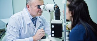
Electroodontometry
Electroodontometry is the most effective method for assessing the functional state of the trigeminal nerve when it is damaged. EDI is based on a study of the reaction of the teeth of the lower jaw to electrical stimulation. The method is performed on all teeth of the lower jaw, with preserved pulp both in the affected area and on the opposite healthy side.
S.N. scale Fedotova (1997) to assess the severity of damage to the inferior alveolar nerve based on electrical odontometry data:
- mild degree - reaction of teeth with preserved pulp on the side of nerve damage within 20-40 μA;
- moderate severity – reaction of teeth to currents from 40 to 100 µA;
- severe degree - complete loss of pain sensitivity, reaction of teeth to currents above 100 μA
The use of EDI to diagnose traumatic injuries of the inferior alveolar nerve is impossible if:
- lower jaw teeth endodontically treated
- lower jaw teeth are covered with orthopedic structures
- metal elements of splinting structures are fixed on the teeth
- missing teeth
Locations for measuring electrical excitability of facial skin
Assessment of the area of paresthesia in traumatic neuritis of the inferior alveolar nerve
The zone of paresthesia is identified - impaired sensitivity of the skin based on a tactile test, photographed, followed by an assessment of the area of the paresthesia zone: points are drawn on the border of areas of normal sensitivity of the skin, the red border of the lips and the paresthesia zone, which are then connected by a continuous line. Zones of hyper-, hypo- and anesthesia were marked with different colors.
I - vertical lines:
- midline,
- a line passing through the outer edge of the philtrum,
- a line passing through the outer edge of the wing of the nose,
- pupil line;
II - horizontal lines:
- lip line,
- a line running along the lower edge of the red border of the lips,
- the border line between the chin and lower lip,
- a line drawn along the most protruding part of the chin,
- line of the border of the chin and submental areas.
Schematic representation of the areas for measuring facial skin paresthesia.
Each of the 12 resulting squares was assigned a score depending on the nature of the sensitivity disorder:
0-sensitivity is not impaired;
1-skin hyperesthesia
2-hypoesthesia of the skin;
3-anesthesia of the skin.
Next, the sum of points was calculated and divided by 12 (quadrants).
With the results:
3.0-2.1 – severe sensitivity impairment was diagnosed;
2.0–1.1 – moderate severity;
less than 1.0 – mild severity of the pathology being studied
Causes of trigeminal neuralgia
Symptoms of trigeminal neuralgia occur against the background of compression of the trigeminal nerve root by the cerebellar artery or vein in the area of its entry into the brain stem (the artery or vein compresses the nerve or wraps around the root), resulting in damage to the sheath of nerve fibers.
This pathology develops most often during inflammatory processes in the nasal mucosa, periodontal tissue or other organs, which lead to a narrowing of the lumen of the canal through which the nerve passes.
At CELT you can get advice from a neurologist.
- Initial consultation – 4,000
- Repeated consultation – 2,500
Make an appointment
In addition, trigeminal neuralgia can develop due to:
- hypothermia of nerve fibers with subsequent spasms of facial muscles;
- disruptions in the functioning of the endocrine system;
- compression due to growth of tumors;
- pathologies of neurogenic and vascular nature;
- herpes virus;
- atherosclerosis of intracerebral arteries.
Treatment of neuritis
Treatment of neuritis in traumatic injuries must be timely and wait-and-see tactics are unacceptable. For mild damage to the inferior alveolar nerve, decongestant therapy (prednisolone, veroshpirone) is sufficient. In case of moderate severity of damage, drugs that improve the conductivity of the nerve trunk (neuromedin) are added to decongestant therapy. In case of severe damage to the nerve trunk, in the absence of positive dynamics in the restoration of sensitivity within 4 months, the patient should be referred for a consultation with a neurosurgeon in order to resolve the issue of the possibility of restoring the anatomical integrity of the nerve trunk.
Various reflexotherapy methods are widely used in the complex treatment of diseases of the peripheral nervous system. In the complex treatment of diseases of the peripheral nervous system, reflexology methods such as electroacupuncture and transcutaneous electrical neurostimulation are widely used.
Prevention of neuritis
Prevention of neuritis of the inferior alveolar nerve is the correct technique for removing teeth, correct diagnosis and correct reading of the radiograph, and a gentle technique for dislocating the roots of teeth in the lower jaw with an elevator.
Features of manifestations of iatrogenic damage to the trigeminal nerve
- Painful discomfort, changes in sensations, numbness (anesthesia).
- Functional consequences - Patients who experience pain from touch or cold often have difficulty with daily functions: kissing, communicating, speaking, eating and drinking, etc. https://www.ncbi.nlm.nih.gov/pubmed/22677874
- Psychological consequences - patients develop various anxiety, fear, anger, post-traumatic stress disorder. Psychological disorders can be aggravated in cases where informed consent for implantation, endodontic treatment, orthognathic surgery, etc., which specifically indicates possible nerve damage, has not been signed before medical intervention.
Rice. 6. Schematic representation of the relationship of the branches of the inferior alveolar nerve with the formed implant bed. Pain during drilling is an important diagnostic criterion for the proximity of the nerve.
Abscess after tooth extraction
An abscess after tooth extraction is a long-term complication of a tooth extraction operation that occurs as a result of contamination of the wound surface with microorganisms. When teeth are removed, not only the tissues surrounding the tooth can be injured, but also the mucous membranes of the mouth and cheeks. A fresh wound is a favorable environment and entry point for microorganisms. Therefore, under appropriate circumstances, an abscess may form in the soft tissues or in the hole, which over time and without treatment can “spill” with the formation of phlegmon in several areas.
Causes of abscess
The cause of an abscess may be failure to comply with the rules of asepsis and antisepsis directly during the tooth extraction operation. As a result of incorrect actions by the doctor, infection occurs in the tooth socket and subsequent suppuration. Also, infection of the socket and soft tissues can occur due to the fault of the patient himself if he does not follow all the dentist’s recommendations. The patient must strictly follow the prescribed procedures, otherwise repeated surgery cannot be avoided.
Prevention and treatment of tooth abscess
Prevention of the appearance of an abscess after tooth extraction is basic regular oral hygiene and following all the recommendations of the dentist. Also, preventive measures include visiting the dentist if you experience pain within a few days after tooth extraction. Treatment of an abscess consists of opening and cleaning the abscess cavity, removing purulent formations and prescribing anti-inflammatory therapy. Sometimes the abscess opens on its own, and the pain disappears. But this does not mean that the process has been stopped, so further treatment by a dentist is necessary. Otherwise, the infection may spread to contact areas and intensify the process.