- What does the biopsy show?
- Is it possible to do without a biopsy?
- Types and methods of biopsy
- Does a biopsy hurt?
- Do I need special preparation for a biopsy?
- Is a biopsy safe? What are the consequences and complications?
A biopsy is a diagnostic procedure performed to obtain a tissue sample (biopsy) from a “suspicious” location, such as a tumor or polyp. A biopsy is necessary to confirm the diagnosis of cancer.
What does the biopsy show?
All cells of the body have a characteristic structure, depending on what tissue they belong to. As a malignant tumor develops, the structure of the cells is disrupted, and these changes can be seen under a microscope.
A doctor examining samples of tissue or cells obtained through a biopsy can tell definitively whether a patient has cancer. While other tests suggest cancer with varying degrees of probability, a biopsy helps to establish an accurate diagnosis.
What can you see
With the help of a biopsy we can solve a very wide range of problems:
- Obtain complete information about the characteristics of the pathology;
- Determine the nature of the tumor;
- Confirmation or refutation of a diagnosis made earlier;
- Diagnostics for the purpose of differentiation;
- Determine the stage of pathology;
- Eliminate the source of pathology;
- Evaluation of treatment effectiveness, etc.
Prostate biopsy
Types and methods of biopsy
A doctor can obtain a biopsy, a tissue sample for examination, in a variety of ways. Depending on this, there are several types of biopsy:
- razor;
- puncture;
- trephine biopsy;
- incisional;
- excision.
Imprint smears, scrapings, shave biopsy
Sometimes it is enough to obtain only a few cells for a biopsy. For example, for early detection of cervical cancer, a smear-imprint is performed from the mucous membrane of the cervix. The material obtained in this way is quite enough to perform laboratory research.
You can also make smears of nipple discharge if breast cancer is suspected.
During a shave biopsy, the doctor cuts off a layer of a certain thickness from the surface of an area of skin using a sharp instrument. A bleeding surface is left on which a pressure bandage is applied.
Needle biopsy
The name of the method comes from the Latin word punctio - “injection”. In turn, puncture biopsy is divided into types: fine-needle, thick-needle (trephine biopsy), aspiration.
Fine needle biopsy
This type of needle biopsy is used when it is necessary to obtain a small number of cells. The doctor inserts a thin needle into the suspicious area and removes a small amount of tissue.
Core needle biopsy
This type of biopsy is optimal in many cases, since it does not require an incision, and at the same time allows you to obtain a fairly large amount of tissue. Core needle biopsy is often used for suspected breast, liver, prostate and a number of other tumors.
Trephine biopsy is used to take samples of skin and bone marrow. The doctor uses a special instrument that resembles a needle, only thicker, in the form of a hollow cylinder with sharp edges. It is immersed in the desired place, and as a result it is filled with a column of fabric.
Aspiration biopsy
In an aspiration biopsy, tissue is removed using a vacuum aspirator—a special cylinder that creates negative pressure. It is connected to a needle. During the procedure, the doctor may obtain several fragments of suspicious tissue at once.
Aspiration biopsy is often used in gynecological practice.
Scan-guided biopsy
Sometimes a suspicious formation is almost impossible to palpate through the skin due to its small size, but can be detected during radiography, ultrasound, or MRI. In this case, the biopsy is performed under the guidance of an x-ray or other image, which helps the doctor guide the needle and control the position of its tip.
During a stereotactic biopsy, images in at least two planes are used, which helps to accurately determine the position of the suspicious lesion and the needle in three-dimensional space. Biopsy under scanning control can be fine-needle, thick-needle, or aspiration.
Biopsy during surgery
During surgery, your doctor may remove part of the tumor (incisional biopsy) or all of it (excisional biopsy). This allows you to obtain the maximum amount of tissue for research. But this type of biopsy has a drawback: the diagnosis is established after the patient has been operated on.
If the surgeon removes the entire studied formation or organ during a biopsy, the procedure is also a therapeutic measure. If the formation (for example, a polyp) turns out to be benign, after its removal a complete cure occurs.
Biopsy during endoscopy
When examining some organs, such as the gastrointestinal tract, an endoscope is used - a thin tube with a video camera and a light source at the end. Through it, you can insert special endoscopic forceps or a needle to take a biopsy from the esophagus, stomach or intestines. This type of biopsy is also called targeted biopsy.
If a tissue sample from the colon is needed, an endoscope is inserted through the anus, a procedure called fibrocolonoscopy or sigmoidoscopy (depending on which part of the colon needs to be examined). If material needs to be obtained from the stomach, esophagus, duodenum, the endoscope is inserted through the mouth, and the study is called fibrogastroduodenoscopy (FGDS).
A biopsy can also be performed during bronchoscopy, cystoscopy (endoscopic examination of the bladder) and other types of endoscopy.
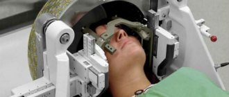
Stereotactic brain biopsy
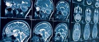
A stereotactic brain biopsy is a procedure that allows a neurosurgeon to diagnose brain damage. The procedure is performed in an operating room, usually under general anesthesia, and involves removing a small piece of tissue, most often brain matter, but sometimes samples are taken from blood vessels or the dura mater (the outer membrane covering the brain). In most cases, the neurosurgeon uses stereotactic equipment to localize the biopsy site. This equipment allows neurosurgeons to map the brain in a three-dimensional coordinate system and select appropriate target coordinates for biopsy needle direction.
Primary brain tumors affect almost 30,000 people each year, and metastatic tumors affect almost 200,000 people. The most common primary brain tumors include glioma and meningioma. Magnetic resonance imaging (MRI) provides information about the location, size, and relationship of the tumor to surrounding structures. MRI is sometimes supplemented with magnetic resonance spectroscopy (MRS) to provide information about the chemical composition of a tumor. In addition, diffusion/perfusion-weighted imaging provides information about the flow of blood and water through the tumor.
However, the most informative method for making a diagnosis is to obtain a tissue sample. The decision about whether to perform a biopsy or attempt to completely remove the tumor takes into account numerous factors and is carefully weighed by the neurosurgeon, often in consultation with other neuro-oncology colleagues. If it is determined that biopsy is the optimal course of action for further treatment, the safest and most accurate route to access the tumor is chosen. The same principle applies to metastatic brain tumors where the primary malignancy is unknown or in a situation where the neurosurgeon suspects an infectious process and there is a need for a tissue sample to confirm the diagnosis.
About stereotactic surgery
Stereotaxis is a process by which neurosurgeons use MRI or CT scans, targeted algorithms and a computer workstation to pinpoint the location of a tumor or other lesion within the brain. Previously, this had to be done by placing a metal frame on the patient's head, but this method has now been largely replaced by the stereotactic system, which uses small fiducial markers, about the size of a coin, that gently adhere to various parts of the scalp to provide orientation. Systems that use stereotaxis to simplify neurosurgical procedures are known as stereotactic navigation systems; they are also referred to as frameless stereotactic neuronavigation systems.
There are several frameless stereotactic neuronavigation systems available for use in neurosurgical procedures. They are manufactured by various biomedical engineering companies and go by different names. They all have comparable accuracy and efficiency and use essentially the same principles to complete the task.
Technique for performing stereotactic brain biopsy
Once the patient falls asleep in the operating room, his head is fixed in a certain position, and the fiducials on the scalp are recorded by cameras in a computerized navigation system. A minimal amount of hair is shaved and a small incision is made. The incision area is thoroughly cleaned and sterilely draped. A small hole is made in the skull and the covering of the brain (dura mater) is exposed. A stereotactic biopsy needle, long, soft-nosed, and blunt-tipped, is guided into the target using a neuronavigation system to obtain biopsy samples.
The samples are examined in real time, frozen section analysis is performed by a pathologist, and additional samples are then taken for other pathology studies. It takes 3-4 days to receive final results. Once the incision is closed, a clean, dry dressing is applied, which is usually removed after two to three days. The procedure is usually performed in a day hospital and requires an overnight stay. Some patients undergo it on an outpatient basis. The sutures are removed after 10-14 days.
Indications for stereotactic brain biopsy
The most common diseases that can be diagnosed by biopsy are tumors, infections (abscess), inflammation (encephalitis), demyelinating diseases (multiple sclerosis) or neurodegenerative diseases (Alzheimer's disease). A biopsy can help identify lesions that do not require surgical treatment or identify patients who for some reason are contraindicated for surgical treatment so that they can decide on another type of therapy.
Benefits of Stereotactic Brain Biopsy
The stereotactic biopsy needle reaches the deepest recesses of the brain, allowing neurosurgeons to obtain a sample and make an accurate diagnosis in the safest manner possible. The procedure is carried out in a day hospital setting.
Risks of Stereotactic Brain Biopsy
Risks associated with stereotactic biopsy include intracranial hemorrhage (approximately 1%), infection (1%), or failure to obtain tissue for diagnosis (1%), which may require a repeat biopsy. However, for the most part, it is a safe procedure performed by neurosurgeons with careful planning and great care, which provides valuable information for further treatment. Modern software and equipment provide a high level of accuracy with minimal risk to surrounding brain tissue.
Recovery and follow-up after stereotactic brain biopsy
After a brain biopsy, patients are usually observed for a short time before being sent home, although some patients may be kept in hospital overnight.
Sometimes patients experience minimal discomfort around the incision site. Most patients can return to their normal activities the next day. The neurosurgeon, usually in collaboration with colleagues in radiation medicine and oncology, and sometimes with infectious disease specialists, monitors subsequent care and treatment based on the results of the biopsy. Return back to the clinic Diagnostics Treatment Our doctors Useful information
Do I need special preparation for a biopsy?
Usually no special preparation is required. The clinic requires you to sign a written consent to perform medical procedures (biopsy). The doctor will tell you what the procedure is, how it will be performed, what the risks are, and answer your questions.
If necessary, local anesthesia is administered before the biopsy using an injection or spray. Sometimes medicated sleep or general anesthesia is used. In this case, you will be asked not to drink or eat for a certain time before the procedure.
WHAT IS THE SURGICAL INTERVENTION?
It is possible to perform an open biopsy using craniotomy, as when removing a tumor. In this case, a window is opened in the skull bone by removing a fragment, which is subsequently replaced in its original place. However, this approach is not always practiced and is limited to superficial lesions in prominent areas of the brain or when samples need to be taken from the cranial bone or meninges.
If possible, partial resection of the lesion is performed and a sample of this tissue is taken for analysis. Minimally invasive biopsy methods are chosen: for diffuse damage to brain tissue without clear boundaries for small and deep lesions for significant lesions in a very visible area of the brain with a high risk of serious neurological consequences To do this, a trepanation or a single small hole is made as an entry point into the skull, through which a cannula is inserted and directed to specific, pre-planned coordinates.
The classic method, which is still used in many centers and for other types of operations, is stereotaxy. Stereotaxy involves obtaining target coordinates by performing a CT scan of the skull immediately before entering the operating room. A metal frame is attached to the patient's skull to serve as a reference for obtaining coordinates, which will later be attached to another component on the operating table.
Because this procedure is not particularly pleasant for the patient, has higher logistical requirements, and takes longer to prepare, an alternative technique has been developed. The cannula is inserted into specific coordinates in the brain using neuronavigation technology (frameless stereotactic brain tumor biopsy), which is used at the Instituto Clavel. This method provides maximum accuracy and safety.
During neuronavigation biopsy of brain tumors, the patient's head must be in a fixed position relative to the landmark of the navigation device. An accurate, high-quality MRI of the patient's skull is loaded onto the console of this device and a target is determined to hit.
The neuronavigator combines the downloaded image with the marked target and shows on the screen the position of the biopsy cannula in relation to the target, allowing neurosurgeons to reach it safely. The path is selected in the image in such a way as to avoid risk areas inside the brain and reach the desired depth.
Each time biopsy samples are taken from brain tumors, a fresh sample is sent to the laboratory to be analyzed and assessed for viability and the presence of abnormal tissue before sending additional samples for final examination and wound closure.
Is a biopsy safe? What are the consequences and complications?
This depends on the type of biopsy. If it is performed during surgery, then the risks are due to the surgery itself. During a puncture biopsy, the needle may enter a vessel or neighboring organs (for example, the gallbladder during a liver biopsy), bleeding, infection, and pain for some time after the procedure. If the biopsy is performed by an experienced specialist in a well-equipped clinic, there are virtually no risks.
At Euroonko you can perform various types of biopsies. We employ highly qualified doctors and use modern equipment.
Book a consultation 24 hours a day
+7+7+78
Types of diagnostics
There are these types of biopsies:
- Stereotactic brain biopsy. This type of tissue collection is used for hard-to-reach areas of the brain. Typically, stereotactic biopsy is performed using a special installation - a stereotactic system, equipped with a coordinating frame and the possibility of three-dimensional orientation. The structure of the device allows you to choose the safest trajectory and entry point. To increase accuracy, stereotactic biopsy is performed under the guidance of magnetic resonance or computed tomography.
- Needle biopsy. A hole is drilled in the skull through which a puncture needle is inserted to excise the tissue.
- Open biopsy.
It is performed during brain surgery.