Dysarthria in children.
Dysarthria in children is one of the types of speech disorders that occurs due to damage to the central nervous system. The most obvious disorders of this disease are changes in intonation, tempo and rhythm of speech, as well as a violation of voice formation and a disorder of articulation of sounds.
Children who suffer from dysarthria have great difficulty chewing and swallowing. They sometimes have difficulty buttoning clothes, hopping on one leg, or cutting with scissors. It is also difficult for them to write, so they study in specialized schools. Very often there are contradictions between neurologists and speech therapists regarding dysarthria. If a neurologist does not see obvious disturbances in the function of the cranial nerves, he cannot call the speech disorder dysarthria. This question is almost a stumbling block between neurologists and speech therapists. This is due to the fact that a neurologist, after making a diagnosis of dysarthria, is obliged to carry out serious therapy for the treatment of brainstem disorders, although such disorders (excluding dysarthria) do not seem to be noticeable. The medulla oblongata, as well as the cervical spinal cord, often experiences hypoxia during childbirth. This leads to a sharp decrease in motor units in the nerve nuclei responsible for articulation. During a neurological examination, the child adequately performs all tests, but cannot cope properly with articulation, because it is necessary to perform complex and fast movements that are beyond the strength of weakened muscles.
What treatment does neurology offer?
A mandatory component of treatment is therapy of the underlying disease. For hypertension, antihypertensive drugs are prescribed; for atherosclerosis, drugs are prescribed to lower cholesterol levels and prevent the formation of blood clots. For neuroinfections, antimicrobial therapy is indicated. For tumors, surgery is performed and radiation therapy is used.
For all variants of pseudobulbar syndrome, medications are useful to activate neurometabolism (nootropics) and improve microcirculation in the brain.
Treatment for children includes sessions with a speech therapist to correct phonation disorders and work with a speech pathologist. Additionally, exercise therapy, physiotherapy, and breathing exercises are performed. Neurology considers the use of stem cells to be a promising method of therapy.
Causes of dysarthria
1. Organic damage to the central nervous system as a result of the influence of various unfavorable factors on the developing brain of a child in the prenatal and early periods of development. Most often, these are intrauterine lesions that are the result of acute, chronic infections, oxygen deficiency (hypoxia), intoxication, toxicosis of pregnancy and a number of other factors that create conditions for the occurrence of birth trauma. In a significant number of such cases, asphyxia occurs during childbirth and the child is born premature. 2. The cause of dysarthria may be Rh factor incompatibility. 3. Dysarthria occurs somewhat less frequently under the influence of infectious diseases of the nervous system in the first years of a child’s life. Dysarthria is often observed in children suffering from cerebral palsy (CP). According to E.M. Mastyukova, dysarthria with cerebral palsy manifests itself in 65-85% of cases. Classification of clinical forms of dysarthria Classification of clinical forms of dysarthria is based on identifying different locations of brain damage. Children with various forms of dysarthria differ from each other in specific defects in sound pronunciation, voice, and articulatory motor skills, require different speech therapy techniques and can be corrected to varying degrees.
Forms of dysarthria
Reasons for appearance
The causes of the syndrome are associated with disruption of the brain. It is based on the phenomenon of multiple small hemorrhages in both hemispheres.
The following reasons can lead to the syndrome:
- degenerative changes in the brain caused by diseases (dementia, Alzheimer's disease, Pick's disease and others);
- infectious diseases affecting the brain (encephalitis, meningitis, cerebral syphilis);
- cerebral atherosclerosis;
- two or more previous cerebrovascular accidents.
Bulbar dysarthria
Bulbar dysarthria (from the Latin bulbus - a bulb, the shape of which is the medulla oblongata) occurs when there is a disease (inflammation) or tumor of the medulla oblongata. In this case, the nuclei of the motor cranial nerves located there (glossopharyngeal, vagus and sublingual, sometimes trigeminal and facial) are destroyed. Characteristic is paralysis or paresis of the muscles of the pharynx, larynx, tongue, and soft palate. A child with a similar defect has difficulty swallowing solid and liquid food and has difficulty chewing. Insufficient mobility of the vocal folds and soft palate leads to specific voice disorders: it becomes weak and nasal. Voiced sounds are not realized in speech. Paresis of the muscles of the soft palate leads to the free passage of exhaled air through the nose, and all sounds acquire a pronounced nasal (nasal) tone. In children with the described form of dysarthria, atrophy of the muscles of the tongue and pharynx is observed, and muscle tone also decreases (atonia). The paretic state of the tongue muscles causes numerous distortions in sound pronunciation. Speech is slurred, extremely unclear, slow. The face of a child with tabloid dysarthria is amicable.
Symptoms
Most often, the syndrome occurs suddenly in the patient. In some cases, it develops gradually. The elements of the clinical picture look like this:
- The voice changes. The patient begins to speak “in his nose”, the voice becomes more hoarse;
- intonation expressiveness disappears. Speech is disrupted: consonant sounds fall out of it;
- difficulties with chewing and swallowing food. Some of the food constantly accumulates on the teeth, gets stuck between them, on the palate; liquid food may leak through the nose;
- violation of facial expressions. Facial expressions may not correspond to the actual feelings and emotions experienced;
- paresis of facial muscles, usually bilateral. The facial muscles lose the ability to move, the face becomes like a frozen mask;
- strange behavior. When asked to open his eyes, a person opens his mouth. When touched to the corner of the mouth, upper lip, or showing teeth, he may begin to cry or laugh. This is the so-called forced crying and laughter;
- oral automatism - voluntary contraction of the circular muscles of the mouth. The tendon reflex of the upper jaw increases;
- symptoms of hemiparesis may be present (impaired motor activity in a certain part of the body);
- possible manifestations of extrapyramidal disorder (slowing movements with increased muscle tone);
- disruption of the brain and nervous system leads to a decrease in the patient’s intelligence.
Subcortical dysarthria
Subcortical dysarthria occurs when the subcortical nodes of the brain are damaged. A characteristic manifestation of subcortical dysarthria is a violation of muscle tone and the presence of hyperkinesis. Hyperkinesis is violent involuntary movements (in this case in the area of articulatory and facial muscles) that are not controlled by the child. These movements can be observed at rest, but usually intensify during speech. The changing nature of muscle tone (from normal to increased) and the presence of hyperkinesis cause peculiar disturbances in phonation and articulation. A child can correctly pronounce individual sounds, words, short phrases (especially in a game, in a conversation with loved ones or in a state of emotional comfort) and after a moment he is unable to utter a single sound. An articulatory spasm occurs, the tongue becomes tense, and the voice is interrupted. Sometimes involuntary screams are observed, and guttural (pharyngeal) sounds “break through.” Children may pronounce words and phrases excessively quickly or, conversely, monotonously, with long pauses between words. Speech intelligibility suffers due to unsmooth switching of articulatory movements when pronouncing sounds, as well as due to disturbances in the timbre and strength of the voice. A characteristic sign of subcortical dysarthria is a violation of the prosodic aspect of speech - tempo, rhythm and intonation. The combination of impaired articulatory motor skills with disorders of voice formation and speech breathing leads to specific defects in the sound aspect of speech, which manifest themselves variably depending on the child’s condition, and are reflected mainly in the communicative function of speech. Sometimes with subcortical dysarthria in children, hearing loss is observed, complicating a speech defect.
Pathogenesis
Normally, every person in the brain has so-called “old” parts that work without his control. The syndrome appears when failures occur in the regulation of the movement of impulses from the nerve nuclei. Loss of impulses is possible with brain pathology. In this case, the kernels begin to work in emergency mode, causing the following processes:
- Change in articulation. There is a violation of the pronunciation of many sounds.
- Problems with swallowing due to the development of paralysis of the soft palate.
- Activation of oral automatism reflexes, which should normally be present in newborns.
- The appearance of an involuntary grimace.
- Impaired phonation and decreased mobility of the vocal cords.
Often, deviations in the functioning of the nervous system are caused not only by damage to the nuclei, but also by changes in the brain stem and cerebellum. In newborns, the disorder is usually bilateral, which increases the risk of cerebral palsy.
Cortical dysarthria
Cortical dysarthria is very difficult to isolate and recognize. With this form, voluntary motor skills of the articulatory apparatus are impaired. In its manifestations in the sphere of sound pronunciation, cortical dysarthria resembles motor alalia, since, first of all, the pronunciation of words with a complex sound-syllable structure is impaired. In children, the dynamics of switching from one sound to another, from one articulatory posture to another, is difficult. Children are able to clearly pronounce isolated sounds, but in the speech stream the sounds are distorted and substitutions occur. Combinations of consonant sounds are especially difficult. At an accelerated pace, hesitations appear, reminiscent of stuttering. However, unlike children with motor alalia, children with this form of dysarthria do not experience disturbances in the development of the lexico-grammatical aspect of speech. Cortical dysarthria should also be distinguished from dyslalia. Children have difficulty reproducing articulatory posture, and it is difficult for them to move from one sound to another. During correction, attention is drawn to the fact that defective sounds are quickly corrected in isolated utterances, but are difficult to automate in speech.
Treatment
First of all, differential diagnosis is necessary to exclude bulbar syndrome, parkinsonism, nephritis, and neuroses.
There is no universal pill or injection that cures this disease. Each patient requires an individual approach. The doctor will select the necessary medications taking into account the condition of each individual patient.
It is important to fight the disease that has become the root cause of pseudobulbar syndrome. Appropriate treatment is selected for each patient. At the same time, the syndrome itself is treated.
A whole list of medications can be used to relieve symptoms and restore muscle function:
- To improve the transmission of nerve impulses, blood supply to the brain (Cerebrolysin, Aminalon);
- drugs to strengthen the walls of blood vessels;
- metabolic agents that improve metabolic processes in brain tissue;
- nootropic drugs (nootropil, pantogam);
- drugs for the breakdown of acetylcholine (prozerin, oxazil).
In addition to medications, the patient is prescribed physiotherapy, massage, physical therapy, breathing exercises, and a rehabilitation course to restore speech. In severe cases, physiotherapy and massage are combined with medications and acupuncture.
Modern medicine also suggests using stem cells for treatment. The principle of their action is associated with the replacement of affected cells with healthy ones. This is an expensive and complex procedure, which is resorted to in rare cases.
Pseudobulbar dysarthria
Pseudobulbar dysarthria is the most common form of childhood dysarthria. Pseudobulbar dysarthria is a consequence of organic brain damage suffered in early childhood, during childbirth or in the prenatal period as a result of encephalitis, birth injuries, tumors, intoxication, etc. The child experiences pseudobulbar paralysis or paresis caused by damage to the pathways coming from the cerebral cortex to the nuclei of the glossopharyngeal, vagus and hypoglossal nerves. According to the clinical manifestations of disorders in the area of facial and articulatory muscles, it is close to bulbar. However, the possibilities of correction and full mastery of the sound-pronunciation side of speech with pseudobulbar dysarthria are much higher. As a result of pseudobulbar palsy, the child's general and speech motor skills are impaired. The baby sucks poorly, chokes, chokes, and swallows poorly. Saliva flows from the mouth, facial muscles are disturbed. The degree of impairment of speech or articulatory motor skills may vary. Conventionally, there are three degrees of pseudobulbar dysarthria: mild, moderate, severe. 1. A mild degree of pseudobulbar dysarthria is characterized by the absence of gross disturbances in the motor skills of the articulatory apparatus. Articulation difficulties lie in slow, insufficiently precise movements of the tongue and lips. Disorders of chewing and swallowing are revealed faintly, with occasional choking. The pronunciation of such children is impaired due to insufficiently clear articulatory motor skills, speech is somewhat slow, and blurring is typical when pronouncing sounds. More often, the pronunciation of sounds that are difficult to articulate suffers: zh, sh, r, ts, ch. Voiced sounds are pronounced with insufficient participation of the voice. Soft sounds are difficult to pronounce and require adding to the main articulation the raising of the middle part of the back of the tongue to the hard palate. Pronunciation deficiencies have an adverse effect on phonemic development. Most children with mild dysarthria experience some difficulty in auditory processing. When writing, they encounter specific errors in replacing sounds (t-d, t-ts, etc.). There is almost no violation of the structure of the word: the same applies to grammatical structure and vocabulary. Some uniqueness can only be revealed through a very careful examination of children, and it is not typical. So, the main defect in children suffering from mild pseudobulbar dysarthria is a violation of the phonetic aspect of speech. Children with a similar disorder, who have normal hearing and good mental development, attend speech therapy classes at the regional children's clinic, and at school age - a speech therapy center at a comprehensive school. Parents can play a significant role in eliminating this defect. 2. Children with moderate dysarthria make up the largest group. They are characterized by amicity: lack of movement of the facial muscles. The child cannot puff out his cheeks, stretch out his lips, or close them tightly. Tongue movements are limited. The child cannot lift the tip of his tongue up, turn it to the right, left, or hold it in this position. Switching from one movement to another is a significant difficulty. The soft palate is often inactive, and the voice has a nasal tone. Characterized by profuse salivation. The acts of chewing and swallowing are difficult. The consequence of dysfunction of the articulatory apparatus is a severe pronunciation defect. The speech of such children is usually very slurred, slurred, and quiet. The articulation of vowels, usually pronounced with a strong nasal exhalation, is characteristic due to the inactivity of the lips and tongue. The sounds “a” and “u” are not clear enough, the sounds “i” and “s” are usually mixed. Of the consonants, p, t, m, n, k, x are most often preserved. The sounds ch and ts, r and l are pronounced approximately, like a nasal exhalation with an unpleasant “squelching” sound. The exhaled mouth stream is felt very weakly. More often, voiced consonants are replaced by voiceless ones. Often sounds at the end of words and in combinations of consonants are omitted. As a result, the speech of children suffering from pseudobulbar dysarthria is so incomprehensible that they prefer to remain silent. Along with the usually late development of speech (at the age of 5-6 years), this circumstance sharply limits the child’s experience of verbal communication. Children with such a disorder cannot study successfully in a comprehensive school. The most favorable conditions for their education and upbringing are created in special schools for children with severe speech impairments, where these students receive an individual approach. 3. A severe degree of pseudobulbar dysarthria - anarthria - is characterized by deep muscle damage and complete inactivity of the speech apparatus. The face of a child suffering from anarthria is mask-like, the lower jaw droops, and the mouth is constantly open. The tongue lies motionless on the floor of the oral cavity, lip movements are sharply limited. The acts of chewing and swallowing are difficult. Speech is completely absent, sometimes there are individual inarticulate sounds. Children with anarthria with good mental development can also study in special schools for children with severe speech impairments, where, thanks to special speech therapy methods, they successfully master writing skills and a curriculum in general education subjects. A characteristic feature of all children with pseudobulbar dysarthria is that with distorted pronunciation of the sounds that make up a word, they usually retain the rhythmic contour of the word, i.e., the number of syllables and stress. As a rule, they know the pronunciation of two- and three-syllable words; four-syllable words are often reproduced reflectively. It is difficult for a child to pronounce consonant clusters: in this case, one consonant is dropped (squirrel - “beka”) or both (snake - “iya”). Due to the motor difficulty of switching from one syllable to another, there are cases of likening syllables (dishes - “posyusya”, scissors - “noses”). Impaired motor skills of the articulatory apparatus leads to improper development of the perception of speech sounds. Deviations in auditory perception caused by insufficient articulatory experience and the lack of a clear kinesthetic image of sound lead to noticeable difficulties in mastering sound analysis. Depending on the degree of speech motor impairment, variously expressed difficulties in sound analysis are observed. Most special tests that reveal the level of sound analysis are not available to dysarthric children. They cannot correctly select pictures whose names begin with a given sound, come up with a word containing a certain sound, or analyze the sound composition of a word. For example, a twelve-year-old child who has studied for three years in a public school, answering the question what sounds in the words of the regiment, cat, names p, a, k, a; k, a, t, a. When completing the task of selecting pictures whose names contain the sound b, the boy puts aside a jar, a drum, a pillow, a scarf, a saw, and a squirrel. Children with better preserved pronunciation make fewer mistakes; for example, they select the following pictures based on the sound “s”: bag, wasp, plane, ball. For children suffering from anarthria, such forms of sound analysis are not available.
Pseudobulbar palsy (supranuclear bulbar palsy)
- a syndrome characterized by paralysis of muscles innervated by the V,VII, IX, X, XII cranial nerves, as a result of bilateral damage to the corticonuclear pathways to the nuclei of these nerves.
In this case, bulbar functions suffer, mainly articulation, phonation, swallowing and chewing (dysarthria, dysphonia, dysphagia). But unlike bulbar palsy, muscle atrophy is not observed and reflexes of oral automatism take place: increased proboscis reflex; Astvatsaturov’s naso-labial reflex appears; Oppenheim reflex (sucking movements in response to line irritation of the lips), distant-oral and some other similar reflexes, as well as pathological laughter and crying. The study of the pharyngeal reflex is less informative. The main causes of pseudobulbar palsy:
1. Vascular diseases affecting both hemispheres (lacunar state in hypertension, atherosclerosis; vasculitis). 2. Perinatal pathology and birth trauma, including. 3. Congenital bilateral periaqueductal syndrome. 4. Traumatic brain injury. 5. Episodic pseudobulbar palsy in epileptiform opercular syndrome in children. 6. Degenerative diseases affecting the pyramidal and extrapyramidal systems: ALS, primary lateral sclerosis, familial spastic paraplegia (rarely), OPCA, Pick's disease, Creutzfeldt-Jakob disease, progressive supranuclear palsy, Parkinson's disease, multiple system atrophy, other extrapyramidal diseases. 7. Demyelinating diseases. 8. Consequences of encephalitis or meningitis. 9. Multiple or diffuse (glioma) neoplasm. 10. Hypoxic (anoxic) encephalopathy (“disease of the revitalized brain”). 11. Other reasons.
Vascular diseases
Vascular diseases affecting both hemispheres are the most common cause of pseudobulbar palsy. Repeated ischemic disorders of cerebral circulation, usually in people over 50 years of age, with hypertension, atherosclerosis, vasculitis, systemic diseases, heart and blood diseases, multiple lacunar cerebral infarctions, etc., as a rule, lead to a picture of pseudobulbar palsy. The latter can sometimes develop with a single stroke, apparently due to decompensation of latent cerebrovascular insufficiency in the other hemisphere. With vascular pseudobulbar palsy, the latter may be accompanied by hemiparesis, tetraparesis, or bilateral pyramidal insufficiency without paresis. A vascular disease of the brain is detected, usually confirmed by an MRI.
Perinatal pathology and birth trauma
Due to perinatal hypoxia or asphyxia, as well as birth trauma, various forms of cerebral palsy (CP) can develop with the development of spastic-paretic (diplegic, hemiplegic, tetraplegic), dyskinetic (mainly dystonic), ataxic and mixed syndromes, including with a picture of pseudobulbar palsy. In addition to periventricular leukomalacia, these children often have unilateral hemorrhagic infarction. More than half of these children show symptoms of mental retardation; approximately one third develop epileptic seizures. The medical history usually contains indications of perinatal pathology, delayed psychomotor development, and the neurological status reveals residual symptoms of perinatal encephalopathy. The differential diagnosis of cerebral palsy includes some degenerative and hereditary metabolic disorders (glutaric aciduria type I; arginase deficiency; dopa-responsive dystonia; hyperekplexia (with rigidity); Lesch-Nyhan disease), as well as progressive hydrocephalus, subdural hematoma. MRI detects certain abnormalities in the brain in almost 93% of patients with cerebral palsy.
Congenital bilateral aqueduct syndrome
This defect occurs in pediatric neurological practice. It leads (like congenital bilateral sclerosis of the hippocampus) to a pronounced impairment of speech development, which sometimes even imitates childhood autism and a picture of pseudobulbar palsy (mainly with speech disorders and dysphagia). Mental retardation and epileptic seizures are observed in approximately 85% of cases. MRI reveals malformation of the perisylvian gyri. Severe traumatic brain injury (TBI) Severe traumatic brain injury in adults and children often leads to various types of pyramidal syndrome (spastic mono-, hemi-, tri- and tetraparesis or plegia) and pseudobulbar disorders with severe speech and swallowing disorders. The association with a history of trauma leaves no room for diagnostic doubt.
Epilepsy
Episodic pseudobulbar palsy
has been described in epileptiform opercular syndrome in children (paroxysmal oral apraxia, dysarthria and drooling), observed in the slow phase of night sleep. The diagnosis is confirmed by epileptic discharges in the EEG during a nocturnal attack.
Degenerative diseases
Many degenerative diseases involving the pyramidal and extrapyramidal systems may be accompanied by pseudobulbar syndrome. Such diseases include amyotrophic lateral sclerosis, progressive supranuclear palsy (these forms are the most common cause of pseudobulbar syndrome), primary lateral sclerosis, familial spastic paraplegia (rarely leads to severe pseudobulbar syndrome), Pick's disease, Creutzfeldt-Jakob disease, Parkinson's disease, secondary parkinsonism, multiple system atrophy, less often - other extrapyramidal diseases.
Demyelinating diseases
Demyelinating diseases often involve the corticobulbar tracts on both sides, leading to pseudobulbar syndrome (multiple sclerosis, post-infectious and post-vaccination encephalomyelitis, progressive multifocal leukoencephalopathy, subacute sclerosing panencephalitis, AIDS-dementia complex, adrenoleukodystrophy).
This group (“myelin diseases”) includes metabolic diseases of myelin
(Pelizius-Merzbacher disease, Alexander disease, metachromatic leukodystrophy, globoid leukodystrophy).
Consequences of encephalitis and meningitis
Encephalitis, meningitis and meningoencephalitis, along with other neurological syndromes, may also include pseudobulbar syndrome in their manifestations. Symptoms of an underlying infectious brain lesion are always identified.
Multiple or diffuse glioma
Some variants of brainstem glioma manifest variable clinical symptoms depending on its location within the caudal, middle (pons) or oral parts of the brainstem.
Most often, this tumor begins in childhood (in 80% of cases before the age of 21) with symptoms of involvement of one or more cranial nerves (usually VI and VII on one side), progressive hemiparesis or paraparesis, and ataxia. Sometimes conduction symptoms precede damage to the cranial nerves. Headaches, vomiting, and swelling in the fundus occur. Pseudobulbar syndrome develops. Differential diagnosis with pontine form of multiple sclerosis
, vascular malformation (usually cavernous hemangioma) and brainstem encephalitis. MRI provides significant assistance in differential diagnosis. It is important to distinguish between focal and diffuse forms of glioma (astrocytoma).
Hypoxic (anoxic) encephalopathy
Hypoxic encephalopathy with serious neurological complications is typical for patients who have survived resuscitation measures after asphyxia, clinical death, prolonged coma, etc. The consequences of severe hypoxia, in addition to prolonged coma in the acute period, include several clinical variants, including dementia with (or without) extrapyramidal syndromes, cerebellar ataxia, myoclonic syndromes, and Korsakoff's amnestic syndrome. Delayed postanoxic encephalopathy with a poor outcome is considered separately. Sometimes there are patients with hypoxic encephalopathy in whom persistent residual effects consist of predominant hypokinesia of bulbar functions (hypokinetic dysarthria and dysphagia) against the background of minimally expressed or completely regressing general hypokinesia and hypomimia (this variant of pseudobulbar disorders is called “extrapyramidal pseudobulbar syndrome” or “pseudopseudobulbar syndrome” "). These patients do not have any abnormalities in the limbs and trunk, but are disabled due to the above-mentioned manifestations of a peculiar pseudobulbar syndrome.
Other causes of pseudobulbar syndrome
Sometimes pseudobulbar syndrome manifests itself as a component of more extensive neurological syndromes.
For example, pseudobulbar syndrome in the picture of central pontine myelinolysis (malignant neoplasm, liver failure, sepsis, alcoholism, chronic renal failure, lymphoma, cachexia, severe dehydration and electrolyte disturbances, hemorrhagic pancreatitis, pellagra) and the overlapping locked-in syndrome
(occlusion basilar artery, traumatic brain injury, viral encephalitis, post-vaccination encephalitis, tumor, hemorrhage, central pontine myelinolysis).
Central pontine myelinolysis is a rare and potentially fatal syndrome that manifests itself with the rapid development of tetraplegia (due to medical illness or Wernicke encephalopathy), and pseudobulbar palsy due to demyelination of the central parts of the pons, which is visible on MRI and in turn can lead to “locked-in” syndrome. person." “Locked-in man” syndrome ("isolation" syndrome, de-efferentation syndrome) is a condition in which selective supranuclear motor de-efferentation leads to paralysis of all four limbs and caudal parts of the cranial innervation without impairment of consciousness. The syndrome is manifested by tetraplegia, mutism (aphonia and anarthria of pseudobulbar origin) and the inability to swallow while conscious; in this case, the possibility of communication is limited only by vertical movements of the eyes and eyelids. CT or MRI reveals destruction of the medioventral part of the pons.
Treatment of dysarthria
Due to the fact that dysarthria is not an independent disease and can be observed with various manifestations of nervous system disorders, the content and order of all treatment measures will be established after the doctor makes a clinical diagnosis, taking into account the age and condition of the child. Treatment of dysarthria in children is carried out comprehensively. Depending on the stage of the disease, the following measures are used: speech therapy correction; drug treatment; massage; breathing exercises; Exercise therapy. Medicines are prescribed by a neuropsychiatrist. There are no drugs for the disease as such. The doctor prescribes medications that only remove the symptoms of the disease and alleviate the general condition of the patient.
TREATMENT METHODS
Since erased dysarthria is a neurological problem, then, accordingly, you will need the help of a neurologist. Although the speech therapist also takes an active part in the treatment process. Neurological tests are carried out, the results of which provide grounds for further research into the anamnesis.
To accurately diagnose the disease, you will need to undergo the following examinations:
- MRI or echo examination of the brain;
- electroneurography;
- electromyography;
- electroencephalography;
- transchannel magnetic stimulation.
Magnetic resonance imaging of the brain makes it possible to detect damage to its structure, the presence of neoplasms or an inflammatory process. Other studies help determine the extent of the lesion in order to select the correct and most effective treatment method.
Properly selected individual therapy can achieve positive dynamics in the development of the disease.
The method of treating pseudobulbar dysarthria is determined by a neurologist or psychoneurologist together with a speech therapist. Depending on the type and stage of the disease, the following methods are used:
- correction using speech therapy techniques;
- drug therapy;
- course of massage sessions;
- exercise therapy;
- breathing exercises.
Manifestations of dysarthria, even if they do not disappear completely, become controlled and practically unnoticeable. The action of pharmacological drugs is aimed at alleviating the symptoms and general condition of the patient.
What is pseudobulbar syndrome
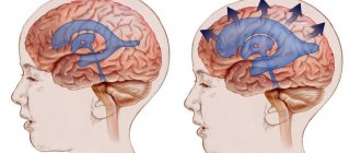
The essence of this disease comes down to the appearance of multiple large and small foci of hemorrhage, which lead to damage in both hemispheres of the fibers connecting the motor nuclei of the cerebral cortex with the brain stem.
This type of lesion can develop due to repeated strokes. But there are cases when pseudobulbar syndrome (PS) makes itself felt even without previous cases of hemorrhage.
With such a problem, as a rule, bulbar functions begin to suffer. We are talking about swallowing, chewing, articulation and phonation. Violation of such functions leads to pathologies such as dysphagia, dysphonia, and dysarthria. The main difference between this syndrome and the bulbar syndrome is that there is no development of muscle atrophy and reflexes of oral automatism are observed:
- increased proboscis reflex;
- Oppenheim reflex;
— Astvatsaturov’s nasolabial reflex;
- distant-arial and other similar reflexes.
Symptoms of PBA
The most common symptom of pseudobulbar lesions is mood-inappropriate behavior. Laughing hysterically at a funeral. Uncontrollable crying when someone tells a joke. These are classic signs of this condition. In some cases, the symptoms are less contradictory and simply exaggerated. Persons living with APD are generally aware of the inadequacy of responses, but lack voluntary control over their behavior. Symptoms may be intermittent or severe and debilitating, negatively impacting quality of life.
Characteristics of PBA symptoms include:
- Outbreaks occurring several times a day.
- Explosions lasting from several seconds to several minutes.
- A sudden and unpredictable attack - some people compare it to a seizure.
Due to the inability to control inappropriate emotional outbursts associated with pseudopulbar can affect, if you live with this condition, you may withdraw socially from your relationships and find yourself unable to carry out daily activities related to work, daily life and social activities.
Some people mistakenly associate the symptoms of this disorder with clinical depression. The difference between them lies in the similarity of mood. If you live with depression, your uncontrollable crying is most likely related to sadness. With the pseudopulbar effect, as discussed, your uncontrollable crying may be triggered by a funny story. The duration of symptoms is another key distinguishing feature. Symptoms of PBA are short-lived and unpredictable. Symptoms of depression tend to last longer and are consistent with mood. That is, he cries because he feels depressed. Often, however, depression and PBA coexist.
We suggest you read: Why you have no luck in your personal life with men: 5 main reasons
Living with Pseudobulbar Affect
There is no cure, but there are options. Talking to other people about your condition will not only help them understand, but it will also help you when you are suffering from an episode in front of them. Monitor for other comorbidities that occur with this disease, including clinical depression. When you feel the episode starting, straighten your posture to stop the full-blown PBA reaction. Finally, focus your mind on the opposite mood you are experiencing. While you can't control your behavior, you can nip it in the bud before it even starts. And just remember that if you have an episode, it will be over in a few seconds or minutes. You can live a good life with pseudopulbar - just being open, honest and aware of your condition is the first step in treatment alone.
How to influence the condition in newly born children
If pseudobulbar syndrome has been diagnosed in newborns, treatment will involve an integrated approach. First of all, this is a massage of the orbicularis oris muscle, feeding through a tube and electrophoresis with proserin on the cervical spine.
Speaking about the first signs of recovery, it is worth noting that they include the appearance of reflexes of the newborn, which were previously absent, stabilization of the neurological status and positive changes in the deviations recorded earlier. Also, with successful treatment, there should be an increase in motor activity against the background of physical inactivity or an increase in muscle tone in the case of severe hypotension. In children with long gestational ages, a meaningful reaction to contact and emotional tone improves.