X-ray of the skull: when is it needed and how is it performed?
Radiography is a quick and reliable way to determine pathologies that are located within dense tissues.
The advantages of a skull x-ray are as follows:
- high information content of images taken in various projections;
- high efficiency;
- non-invasiveness and relatively simple technology for performing the procedure;
- accessibility - today head x-rays
can be taken in almost any clinic; - low radiation exposure.
What does a lung x-ray show?
X-ray of the lungs allows you to identify the presence and determine the nature of pathologies of the lungs, heart, spine and lymph nodes. The study is prescribed for a general assessment of the health of the respiratory system or to clarify the diagnosis for diseases:
- pneumonia;
- emphysema;
- sarcoidosis;
- tuberculosis;
- pleurisy;
- malignant neoplasms;
- bronchitis.
The procedure allows you to detect dangerous pathologies at an early stage, determine their location and area of distribution. They usually appear in the image as light spots called shadows. They are classified by density, size and shape. Neoplasms or an abscess in the lungs provide a shadow, and tissue compaction indicates the development of an inflammatory process. Opacities outside the lungs may be a sign of an aortic aneurysm, tumors of the esophagus, or the spine.
The doctor may refer the patient for an x-ray of the lungs several times to assess the dynamics of treatment or in its absence. This approach is justified by the fact that the harm from an untreated disease is much greater than from the radiation received.
The radiation indicator during an X-ray of the lungs ranges from 0.03-0.3 mSv per procedure, so even when taking pictures in several projections, its total dose will not harm health. A person receives approximately the same amount of radiation in two weeks in normal life.
When is a skull x-ray prescribed?
Survey X-ray of the skull
in different settings can be prescribed to patients who are concerned about:
- cephalgia, or, in other words, headache, of varying localization and intensity;
- trembling in the limbs;
- the appearance of a veil before the eyes or darkness;
- nosebleeds;
- painful chewing of food;
- decreased visual and hearing acuity;
- cases of fainting for no apparent reason;
- the appearance of asymmetry of the facial bones.
X-rays of the head are also indicated for mechanical injuries - bruises, blows, falls from a height, and so on.
Various specialists can prescribe an X-ray examination of the skull: a neurologist, a surgeon, an oncologist, an ophthalmologist and others.
What does the procedure show?
X-ray images clearly visualize:
- cheek bones;
- lower jaw bones;
- bony pyramid of the nose;
- sphenoid bone;
- eye sockets;
- temporomandibular joints;
- mastoid processes of the temporal bones.
If a more accurate and detailed diagnosis of the condition of the bone tissues of the skull is necessary, targeted images are taken, which can show the following pathological conditions:
- formed calcifications - can provoke pathological development of the cranial bones;
- partial calcification of tumors;
- hemorrhages and hematomas;
- fluid in the paranasal sinuses;
- fractures of the skull bones.
Using the X-ray method, it is possible to identify congenital pathologies of the skull, as well as increased intracranial pressure. The latter may be indicated by so-called impressions - marks similar to fingerprints that are located on the inside of the bone tissue.
Advantages of skull x-ray over other methods
X-ray of the skull is the most accurate study, and this is confirmed by its following advantages:
- affordable price;
- obtaining a complete clinical picture of the condition of the bone tissues of the skull;
- minimum amount of time to get results;
- the therapeutic technique carried out in dynamics.
Medical will provide the opportunity to consult with a specialist in the field of the disease. All CT scans are reviewed by a professional radiologist. Appropriate treatment is prescribed taking into account the characteristics of the body.
Preparation for the procedure
Special preparation x-ray of the head
does not require. Before the procedure, the patient must remove all metal jewelry, glasses, and, if possible, dentures. If the dentures are fixed or a metal implant is installed, the radiologist must be warned about this in advance. Then, depending on the configuration of the X-ray machine, the patient takes a lying, sitting or standing position.
A lead vest or apron is placed on the patient's body to prevent exposure below the neck level. The head is secured with special clamps, since the first condition for obtaining a high-quality image is immobility.
Features of skull x-ray
The principle of operation of skull radiography is based on the ability of X-rays to penetrate the tissues of the body. Depending on their density, structure, and chemical composition, the penetration of the rays changes and a black and white image of the internal structures is formed. In analog devices this happened on a special film, in modern digital devices the data is transferred to a computer, and you can view it directly on the monitor.
X-ray is the method that is most informative regarding dense tissues. In photographs they appear in light shades, and soft fabrics appear in dark shades.
There are two types of research:
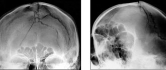
- Survey radiography of the skull is a general photograph of the head to assess the picture as a whole.
- A targeted x-ray of the skull bones is a photograph of one of the parts of the head in order to examine in detail the pathology detected on a general x-ray.
Technique
To take an X-ray, the patient must stand up, sit near the X-ray machine, or lie down on its work table. It is important to remain still and not breathe while filming. If you need to take a picture in several projections, the doctor will tell you how to change the position.
X-ray of the skull in 2 projections
To obtain the most detailed and complete information about the condition of the nasal bones, photographs can be taken in two projections - frontal and lateral. In the first case, the patient faces the X-ray machine, in the second - sideways (left or right).
Preparation
There is no need to prepare for a head x-ray. The patient will only need to remove all metal jewelry and other objects from the head and neck so that they do not cover parts of the image.
To take an x-ray:
- The patient sits in a chair or stands in front of the device.
- A protective lead apron is placed over his chest and abdomen to reduce potential harm to internal organs.
- An image is taken, during which the patient must remain completely still so as not to distort the results. To prevent even minor movements of the head, special fixing bandages and fastenings can be used.
The procedure is painless and will only take a few minutes. Typically, two photographs are taken: in frontal and lateral projections. At the discretion of the physician, depending on the expected diagnosis and potential location of the pathology, other projections may be used. The price of a skull x-ray may vary depending on the number of projections. You can find out the cost in the price list.
How dangerous is the research?
X-ray is a non-invasive and painless procedure. It can also be called relatively safe, since the radiation exposure is minimal. At the same time, of course, x-rays are not a procedure that can be repeated many times in a row. There are certain norms and frequency that must be observed.
Contraindications for
An absolute contraindication for head x-rays is pregnancy. There are also relative restrictions. These are children under 15 years of age, mental illness, serious condition.
Contraindications
The reason why X-rays may be contraindicated for a patient is radiation exposure. For one or two pictures, its value is not critical for health, but when prescribing an x-ray, the doctor will take into account the total number of pictures taken over the year or the last month and the radiation dose received.
X-rays are prohibited (or used very carefully) for pregnant women at any stage of pregnancy and for children under the age of 15 years. Ionizing radiation can affect the normal growth, formation and development of the fetus and child. For this category of patients, x-rays are performed only when there is an urgent need for research and it is impossible to replace it with other diagnostic methods. Alternative examinations may include MRI.
X-ray of the skull: decoding
When interpreting an X-ray of the skull in 2 projections, specialists evaluate the dimensions and features of the location of the bones, as well as the structure of the nasal sinuses. These indicators must correspond to the norm for the age category of the subject.
X-rays also make it possible to partially analyze the condition of the soft tissues of the brain (although for an accurate diagnosis of this organ it is better to use MRI or CT). The images can visualize tumor growths, and their location and size can be assessed. The main sign of a malignant neoplasm will be the presence of darkening of an uneven structure. If the tumor is benign, its contours will be smooth and clear.
Normal indicators
Let's figure out what a skull x-ray
in cases where there are no pathologies. When describing the images, the radiologist evaluates the size, shape, thickness and location of the skull bones, as well as the vascular system, the condition of the nasal sinuses and cranial sutures. All of the listed characteristics must correspond to the patient’s age.
X-ray of the skull for head trauma
The main questions that a specialist should answer based on an x-ray for a head injury:
- Is the integrity of the bones of the skull damaged?
- If there is a fracture, is it accompanied by the entry of bone fragments into the cranial cavity?
- Are the eye sockets, sinuses and ears damaged?
- Is there damage to the brain due to compression by the deformed bones of the skull?
The most common injuries to the calvarium are linear fractures (cracks) of its bones. In most cases, they appear in the place where the force was applied. By the way, this fact greatly facilitates the process of identifying a fracture/crack. The fracture is visualized as a sharp strip, in some places diverging in different directions, with uneven edges. Depending on the complexity, the fracture may have a different position, direction, and size. Multiple fractures can affect one or both halves of the skull. The most unfavorable situation is when the fracture passes to the cranial suture and causes its divergence.
Indications
Fluoroscopy is mandatory for the following diseases or injuries:
- concussions;
- bone fractures;
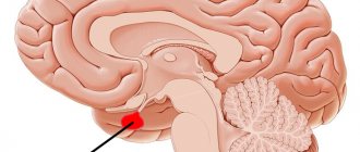
- tumors of the pituitary part of the brain;
- metabolism due to hormonal disorders;
- pathological changes in developmental functions.
A complete examination of the skull under the influence of electric sound radiation is necessary if there are clinical indicators in the form of:
- increasing headaches for no apparent reason;
- hormonal imbalances;
- nosebleeds;
- fainting, dizziness;
- pain in the jaws;
- pronounced asymmetry of the facial nerve.
The effectiveness of the method lies in determining the severity of cranial injuries and the possible presence of inflammatory processes in the pituitary gland.
Is it possible to do x-rays for pregnant women?
X-rays can be done for pregnant women, but only as prescribed by a doctor who will assess the duration of pregnancy, the organs that need to be examined, and take into account all possible risks.
The likelihood that x-rays taken during pregnancy will harm the baby is minimal. A diagnostic test is prescribed only when indicated, when other methods are not informative and do not make it possible to make an accurate diagnosis and begin treatment. Therefore, the benefits of x-rays are greater than the potential risk for the child. However, a large number of examinations of the abdominal organs performed shortly before pregnancy can negatively affect the development of the fetus.
X-rays of the head, limbs, or teeth do not expose the reproductive organs to radiation. The use of a protective lead apron and collar during the procedure reliably blocks scattered radiation. An exception is an abdominal x-ray, in which the abdomen and baby are exposed to direct x-rays.
The risk of harm to the fetus depends on the fetus' age and the intensity of exposure. Exposure to a high dose of radiation between the second and eighth weeks of pregnancy increases the risk of developmental disorders or birth defects, while exposure after the eighth week increases the risk that the baby will have learning and intellectual development problems.
However, the radiation dose received during an x-ray is much lower than what can cause these complications. Before taking an x-ray, you must tell your doctor that you are pregnant. Depending on the circumstances, he may delay the study or change it to reduce the amount of radiation.