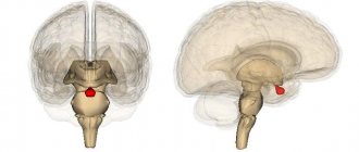
What is a pituitary microadenoma?
The anterior part of the pituitary gland, the adenohypophysis, is an important component for ensuring hormonal balance.
Hormones important for the female body are produced here:
- prolactin
, which ensures the mammary gland produces milk; - GTG
- it stimulates the process of functioning of the entire reproductive system; - TSH
– its task is to accelerate metabolic processes in the thyroid gland.
Neoplasms in the pituitary gland have a complex classification. This is primarily explained by the fact that from an anatomical point of view, the pituitary gland is part of the nervous system, and from a functional point of view, it is a component of the endocrine system.
Microadenoma is one of the types of pituitary adenomas, classified by tumor size
.
In total, depending on how large the formation is, there are 4 types of adenomas
:
- picoadenomas
- neither side of the tumor exceeds 3 mm in length; - microadenomas
– sizes in the range of 3-10 mm; - macroadenomas
- the sides of such adenomas are more than 10 mm; - giant adenomas
– their size exceeds 40 mm.
Therapy
Malignant neoplasms of the pituitary gland are one of the few cancer tumors that, even at stage 3, respond well to treatment with a properly developed treatment regimen and the patient’s compliance with all recommendations.
Surgery, which is performed under general anesthesia, allows you to completely eliminate the disease in the shortest possible time. If surgery is not possible for some reason, radiation therapy using a gamma knife is used.
Recovery with timely seeking medical help occurs in 80% of cases after the first operation. In some patients, it is impossible to immediately remove all pathological cells, and then a repeat operation is indicated. Since the disease affects the endocrine gland, all surgical actions must be coordinated with an endocrinologist.
If you suspect a malignant intracranial process, it is unacceptable to postpone visiting a specialist. With this pathology, time plays a decisive role in the healing process.
Oncological diseases must be treated exclusively in specialized institutions. But you can undergo diagnostics at the clinic on Komendantsky Prospekt in St. Petersburg. Our medical center is equipped with all the necessary equipment; we work with the best laboratories in the city. The results of studies and analyzes are delivered on the day of the study or within two days, depending on the complexity of the procedure. To make an appointment for diagnostics, call us and come to the center in the Primorsky district of St. Petersburg.
Causes of occurrence in women
One of the main causes of microadenoma of the pituitary gland of the brain is a change in hormonal levels. Most often this happens during pregnancy and breastfeeding. In addition, the following reasons can provoke deformation of gland tissue:
- disruptions in the activity of an internal organ associated with the hypothalamus;
- the hormonal function of the peripheral glands decreases, which provokes increased tissue growth and the appearance of compaction;
- hereditary factor;
- artificial termination of pregnancy and other surgical interventions in the pelvic organs;
- use of oral hormonal contraception;
- mechanical shocks, injuries that affect the nervous system;
- infectious processes in the brain;
- bad habits of a woman during pregnancy, as well as the ingestion of toxic substances into the body. This negatively affects the development of the embryo.
Rapidly growing pituitary tumors can destroy functioning cells, causing hormonal deficiency. Panhypopituitarism is accompanied by adrenal insufficiency, lack of sex and thyroid hormones, and retarded growth and development of children. Such patients are unprotected from the action of any stress factor; it provokes an acute drop in pressure and a state of shock.
When fighting benign, and especially malignant tumors, it is important to identify the disease in time - the outcome depends on how early or late treatment begins. Read more in the article: “Rea tumor marker: what is the norm for women.”
The role of diagnostics in the treatment of pituitary tumors
Malignant tumors of the pituitary gland are rare diseases, but they can have a very negative impact on human health, therefore, if an oncological process of an endocrine nature is suspected, a differentiated approach to examination is required. All procedures and tests performed in foreign diagnostic centers with maximum accuracy confirm or refute the pathology. High-quality diagnostics is one of the components when calculating the cost of cancer treatment abroad.
Today, examination for pituitary tumors usually includes molecular genetic testing, digital radiography, endoscopic examination, angiography, CT, MRI, PET-CT, and biopsy.
After the diagnosis is completed, a program with individual treatment tactics is drawn up.
Signs of microadenoma growth
Microadenomas of the pituitary gland of the brain do not manifest themselves as neurological and visual disorders, since they do not lead to compression of neighboring brain structures, but the situation changes with accelerated growth. Under the influence of stress, infections, pregnancy or childbirth, previously undiagnosed tumors can cause the following dangerous symptoms:
- decreased vision,
- headache,
- double vision,
- fainting states.
Rapidly growing pituitary tumors can destroy functioning cells, causing hormonal deficiency. Panhypopituitarism is accompanied by adrenal insufficiency, lack of sex and thyroid hormones, and retarded growth and development of children. Such patients are unprotected from the action of any stress factor; it provokes an acute drop in pressure and a state of shock.
Pituitary microadenoma can be hormonally active.
With a relatively small size, it can produce thyroid-stimulating, somatotropic, corticotropic and gonadotropic hormones, prolactin. As a result, the activity of the thyroid gland increases, body growth accelerates, and the formation of cortisol and sex steroids increases. Patients experience infertility and sexual dysfunction. Treatment uses drug therapy, radiation and surgery.
To correctly diagnose a gynecological disease, a specialist prescribes a smear test for microflora for women. Based on the microorganisms inhabiting the vagina, one can determine the state of women's health and the need for treatment. Read more in the article: “microflora in a smear: the norm in women.”
Symptoms of pressure on tissue
They form a clinical picture of ophthalmo-neurological syndrome. Features of focal symptoms depend on the location of the tumor and the direction of its growth. The main symptom, as a rule, is a dull headache arising from the pressure of the tumor on the walls of the sella turcica. A feature of pain with microadenomas is their constant nature, independent of the patient’s body position.
The pain is not accompanied by nausea, as with increased intracranial pressure, and is not relieved by taking analgesics.
The usual localization of headaches accompanying a pituitary adenoma is the temporal, frontal and posterior orbital regions. In the later stages of the disease, as well as when intratumoral hemorrhage occurs, the pain increases sharply, becoming unbearable. The occurrence of characteristic ophthalmological symptoms is associated with the proximity of the optic nerves, which are compressed by the tumor during its growth.
In this case, typical complaints from patients are limited fields of vision up to prolapse of the lateral halves, diplopia (double vision of visible objects), strabismus and other oculomotor disorders. Lateral tumor growth typically involves limited movement of one or both eyes up, down, or to the sides. Patients describe their visual sensations as looking through a pipe. Long-existing adenoma leads to atrophy of the optic nerves and a decrease in visual acuity that cannot be corrected.
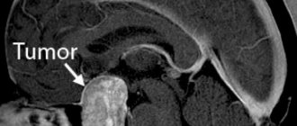
Pituitary tumor MRI
Brief description of the procedure
Duration: 30-60 minutes Necessity of using a contrast agent: as prescribed by a doctor Necessity of preparation for the study: no Presence of contraindications: yes Limitations: available Time to prepare a report: up to 1 hour Children: over 7 years old
Pituitary adenoma
One of the most common diseases of the pituitary gland, adenoma, grows from glandular cells. This is usually a benign tumor, in most cases developing in the anterior lobe of the pituitary gland, where hormones are produced and many important body functions are regulated.
Despite the slow and often imperceptible nature of development, an adenoma can disrupt the functioning of not only the pituitary gland, but also significantly affect the functioning of the brain.
However, due to its small size and asymptomatic development, pituitary adenoma is most often discovered by chance, for example, during the diagnosis of a completely different disease. Although in terms of frequency of occurrence, a pituitary tumor is in third place among brain tumors (the first two are glioma and meningioma).
Adenomas come in different varieties, so it’s nice to classify them by type. Taking as a basis some properties of the tumor, we can distinguish classifications according to:
- Size : microadenoma (tumor less than a centimeter in diameter) and macroadenoma (tumor more than a centimeter in diameter).
- Character - most adenomas are benign and slow in their development. But a rare type of tumor - atypical - grows very quickly, and often reappears even after a course of treatment. Malignant tumors have also been noted that have the potential to spread to other parts of the body. But this variety, due to its rare distribution, has not been fully studied.
- Hormone secretion - if pituitary adenomas produce hormones, they are classified as functional or hormonally active adenomas. In the absence of hormone production, they are classified as nonfunctional (hormonally inactive) adenomas.
There is also a classification of adenomas according to their location in the sella turcica (the back of the brain where the pituitary gland is located):
- within the boundaries of the sella turcica - endosellar;
- above the border of the sella turcica - endosuprasellar;
- below the border of the sella turcica - endoifrasellar;
- destroying the wall of the sella turcica - endolaterosellar.
A cyst stands apart - a neoplasm with liquid contents and a dense shell, the causes, symptoms and treatment of which are practically no different from other types of tumor.
Causes and symptoms of different types of adenomas
Pituitary adenoma is not classified as a hereditary disease and is not explained by genetic predisposition - this type of tumor develops spontaneously. At the moment, the exact reasons for the development of pituitary adenoma are not clear. However, there are predisposing factors for the appearance of a tumor:
- traumatic brain injuries;
- infectious diseases of the brain;
- uncontrolled use of hormones and hormonal drugs;
- exposure during fetal development.
But these same reasons can also influence the development of another type of tumor, for example, cysts of the intermediate velum of the brain. The symptoms of adenoma development are also very ramified. Depending on the type of tumor, symptoms are determined.
The presence of a hormonally active adenoma is indicated by:
For prolactinoma:
- cessation of menstruation;
- reduction in breast milk production during lactation;
- frigidity;
- decreased testosterone levels;
- weakening libido.
For a tumor that produces growth hormone:
- acromegaly (adult);
- gigantism (children);
- disproportionate development of hands and feet;
- diabetes;
- other visible changes in proportions.
For a tumor that produces adrenocorticotropic hormone:
- causeless weight gain;
- hematomas;
- hair growth on the face and body;
- diabetes;
- muscle weakness.
For thyrotropinoma:
- sudden weight loss;
- increased sweating;
- arrhythmia;
- hand tremors;
- insomnia.
If a microadenoma develops practically asymptomatically, then a macroadenoma, especially at the stage of active growth, is detected due to the following signs:
- sharp deterioration in vision (adenoma puts pressure on the optic nerve);
- deterioration of peripheral vision;
- decreased visual acuity;
- doubling in the eyes;
- disturbance of color perception (dimming of the brightness of shades).
Additionally observed:
- chronic headache (due to pressure on brain tissue);
- general signs of pituitary failure.
In addition, it is the development of macroadenoma that leads to complications such as:
- hydrocephalus;
- critical increase in intracranial pressure;
- blindness;
- hemorrhages into the cyst tissue (adenoma);
- cystic adenoma.
MRI for pituitary tumor
Depending on the type and size of the adenoma, it can be detected both during a preliminary examination and the results of laboratory tests, and during diagnosis on a magnetic resonance imaging scanner (primarily this applies to microadenoma). In any case, MRI is the preferred method for detecting tumors. This is explained not only by the fact that MRI sees a pituitary tumor more clearly and brightly than any other diagnostic method, but also by the ability to establish which body systems will be the first to suffer as a result of the development of a cyst (this is indicated by the location of the adenoma).
It is noteworthy that the cystic form of pituitary microadenoma can be seen with an MRI device even at a size of about 0.2 mm, which will significantly simplify further treatment and rehabilitation.
Regardless of the stage of development during which the tumor was discovered, with the help of modern drugs and devices it is possible to successfully influence its growth and development.
A pituitary cyst, and MRI visualizes it perfectly even without additional exposure to a contrast agent, is noticeable against the general background of the image in any section and in any projection (flat or volumetric). In addition to the fact that thanks to timely MRI diagnosis, the most competent treatment path can be prescribed, the result of this diagnosis can help to dismiss a number of diagnoses and tumors with symptoms similar to pituitary adenoma, for example:
- archanoid cyst;
- Rathke's pouch cyst;
- metal tumors.
MRI of the brain or pituitary gland should be performed only after consultation with neurosurgeons and endocrinologists, who, based on basic tests, will be able to determine the potential role of the pituitary gland in certain abnormalities of the body.
Alternative diagnostic options
Although MRI is the most comprehensive method for diagnosing any brain tumor, due to some contraindications, as well as the optionality of undergoing an uncomfortable procedure, other types of diagnostics are provided. For example, in addition to instrumental research methods (using a magnetic resonance imaging or computed tomograph), a biochemical blood test for hormones, as well as additional, basic neurological and physical studies, are mandatory. Sometimes, to suspect and note a potential adenoma, a general radiography of the skull is also used, in which changes or deformation on the side of the sella turcica can be seen.
If MRI and CT are necessary to visualize each layer of tissue, then a blood test performed several times and at the necessary time intervals is the main reason and prerequisite for prescribing instrumental diagnostics.
In addition to the standard biochemical blood test, additional tests are often prescribed:
- Adrenocorticotropic hormone stimulation test. ACTH is administered, which actively stimulates the adrenal glands, and after an hour the level of cortisol in the blood is checked. The adrenal glands are unable to release cortisol (in response to stimulation) if the pituitary adenoma interferes with ACTH production.
- Methyropane test. This consists of using 3 grams (12 tablets) of metyropane, taken by the patient the night before bedtime, to note the effect of the adenoma on ACTH production. As a final result, if the level of this hormone is elevated in the morning, this will indicate a direct effect of the adenoma on the production of the hormone.
- Insulin tolerance test. Involves the use of insulin to lower blood sugar levels. The healthy response should be the secretion of ACTH and growth hormone. If there is a pituitary adenoma, such a reaction will not be observed.
Such hormonal tests help to preliminarily confirm the presence of a functional adenoma, as well as try to classify it. This is especially important in the case when the adenoma is successfully treated without surgery (most cases of early detection of the cyst).
Treatment of adenoma
Due to the nature of the development of adenoma, the methods of its treatment are quite varied. The necessary treatment is determined only after a complete diagnosis. Among the most popular methods are surgery and radiation therapy. Thanks to the development of modern medicine, the traditional surgical method is increasingly being replaced by endoscopic access. The effectiveness of this method is up to 90% for microadenoma, and up to 30-70% for macroadenoma.
An innovative method, radiosurgery, is also gaining popularity. The essence of this type of radiation therapy is the versatile irradiation of the tumor with a low dose of radiation. Radiosurgery includes techniques such as cyberknife, gamma knife and Novalis.
Why are pituitary adenomas dangerous?
The danger from tumors in the anterior lobe of the brain is expressed in the following:
- Although most of the adenomas remain benign and do not degenerate into a malignant form, they cause many problems: as they grow, the tumors put pressure on other parts of the brain, which provokes endocrine and visual disturbances, as well as pathologies of the nervous system of varying complexity;
- A fairly common complication in such cases is the development of cystic tumors, as well as the risk of apoplexy.
Medical data show that every third woman over 20 years of age has various pathologies of the pituitary gland, and every fifth woman has adenomas themselves.
Depending on what stage the pituitary adenoma is at, symptoms in women, treatment, and prognosis differ significantly.
Classification and symptoms
When pituitary tumors appear, the symptoms largely depend on what type it is, and they may differ in women and men. According to functional activity, the division occurs depending on the production of which hormones they influence:
- Somatotropin-producing - cause gigantism if they develop in children; in adults - uneven growth of limbs and individual organs;
- prolactin-secreting - manifested by amenorrhea or galactorrhea in women, in men gynecomastia (enlargement of the mammary glands) is possible;
- ACTH-producing - provoke the development of Itsenko-Cushing syndrome, accompanied by characteristic weight gain, redness and dryness of the skin, etc.;
- Thyrotropin-producing - affect the functioning of the thyroid gland, often accompany hypothyroidism and thyrotoxicosis;
- gonadotropic - similar in symptoms to prolactin, can also lead to uterine bleeding and impotence;
- hormonally inactive.
Other common signs of a pituitary tumor include headaches, ptosis (drooping of the eyelid), nystagmus (“shifty eyes”) or restricted movement of the eyeballs, seizures, impaired consciousness, sleep disturbance, increased intracranial pressure, personality changes, and dementia.
These neoplasms are also divided into benign and malignant; another division option is based on size: intrasellar (within the sella turcica) and endosellar (extending its borders).
Reasons why the disease develops
Since the phenomenon of pituitary adenoma has not yet been fully studied, doctors only speculate on the reasons for its origin.
Factors that provoke the development of pathologies are considered to be:
- an infectious disease that affects the nervous system;
- head injury affecting some part of the brain or skull;
- there is a possibility that women who have been taking oral contraceptives for a long time have a tendency to form adenomas;
- genetic predisposition.
Symptoms of pituitary adenoma, diagnosis
It is important to understand that the symptoms of the pathology depend on what type of adenoma the woman is faced with.
In the first stages of development, pathologies do not manifest themselves, and are usually discovered by chance: most often during an ophthalmological examination, or taking tests for hormones.
Tangible differences have signs
hormonally active (which change hormonal levels, stimulating the production of a certain hormone) and inactive tumors:
- If hormonally active
pituitary microadenoma occurs, symptoms in women are expressed exclusively in changes in the concentration of a specific hormone; - Hormonally inactive
formations are expressed in a gradual but constant deterioration of vision and prolonged attacks of headaches.
If the slightest cause for concern is detected, a woman is recommended to consult a neurologist.
Diagnosis of the development of pathology usually consists of identifying the Hirsch Triad
– three groups of characteristic features:
- changes in the endocrine system;
- visual impairment;
- radiographic pathologies of the skull base.
Most often, diagnosis is made using MRI. The difficulty is that with this tomography, pituitary microadenomas can be visualized only in 60-75% of patients.
In the most difficult cases, doctors resort to computed tomography.
Pituitary microadenoma and pregnancy
There are certain types of microadenomas, the formation of which is accompanied by a significant excess of the hormone prolactin. In such cases, until the woman fully recovers, even the slightest chance of pregnancy is excluded.
In formations of other types, when the concentration of prolactin does not exceed normal limits, microadenoma
do not affect reproductive abilities: pregnancy can occur.
There are cases when pathology develops already during the period of gestation. Then a woman should definitely register with three main doctors: a gynecologist, a neurosurgeon, and an endocrinologist.
Pituitary microadenoma during pregnancy has the following features:
- During pregnancy and lactation, the pituitary gland area increases in size by approximately one and a half times. In this regard, the symptoms of pathology in a pregnant woman manifest themselves much more intensely;
- Approximately 5-25% of all tumors enlarge during this period. The most intensive growth is observed in the 2-3 trimesters. After the birth of a child, microadenomas usually become several times smaller.
How is pituitary microadenoma treated?
The choice of therapy method also depends on the hormonal activity of the microadenoma.
If a microadenoma in the brain does not secrete hormones, it is not so dangerous, sometimes it does not even require special treatment. It is imperative to undergo regular examinations and monitor its changes. To reduce symptoms, certain medications may be prescribed, or a folk remedy may be recommended.
to treat hormonally active or growing inactive tumors:
- medicinal;
- radiation therapy;
- removal of microadenoma using surgery (most often this is endoscopic removal through the nose; in cases of active tumor growth, various types of transcranial intervention are also used).
Surgery to remove tumors from the pituitary gland can have negative consequences and complications for a number of different reasons:
- there is a possibility of injury to healthy lobes of the pituitary gland of the brain;
- vision may deteriorate;
- disorders of blood circulation in the brain;
- infection entering the blood.
The endoscopic method of removal is the safest and is rarely accompanied by complications.
The prognosis for treatment of the pathology is very ambiguous:
- Minor vision problems caused by inactive growths usually disappear after surgery. More serious ophthalmological or endocrine disorders may remain unchanged;
- Depending on the type of pituitary tumor, the success rate of the operation ranges from 25 to 85%;
- It is believed that when adenomas larger than 2 cm form, their complete removal is impossible. Therefore, after 5 years, repeated treatment is recommended.
Stages of pituitary tumors and their symptoms
A low-quality tumor begins its formation with the so-called pre-invasive cancer. In these cases, the patient does not notice any symptoms; the tumor-like formation can exist for a long time without the active growth of cancer cells.
At this stage, a pituitary tumor in Israeli cancer centers is detected during preventive examinations.
Oncopathology at the first stage is mostly asymptomatic - this can continue for 2 or 3 years. Once diagnosed, it can be successfully treated.
In the second stage of the disease, rapid growth of a tumor-like formation in the pituitary gland is recorded. A sick person feels the first signs of illness: weakness and dizziness, headaches and nausea, sleep disturbances and increased fatigue.
The third stage of pituitary cancer is characterized by a significant increase in the tumor and damage to the occipital lymph nodes. At this stage, pronounced symptoms of the disease are noticed; usually the patient seeks medical help with the following symptoms:
- decreased visual function, temporary or permanent blindness due to compression of the visual center in the brain;
- headaches that are aching in nature;
- loss of consciousness and memory impairment;
- decreased sensitivity in the upper and lower extremities, as fluid circulation in the spinal cord is disrupted;
- hormonal imbalance;
- cachexia (severe exhaustion of the body).
Although the third stage is more difficult to treat, it is necessary to fight the tumor to prevent further complications.
At the fourth stage of a low-quality pituitary tumor, it reaches a large size with metastasis. Patients experience mental disorders, epilepsy and other serious symptoms.
Depending on the type of tumor, when treating pituitary tumors in Israel, doctors prolong the life of patients in 98% of cases, and in the most difficult situations provide palliative care.
Forecast and consequences
As with any disease, untimely intervention by medical specialists and refusal to treat hormonal imbalances can lead to irreversible consequences. Peripheral glands (thyroid and adrenal glands) produce hormones, which leads to pathological disorders. Timely detection of microadenoma and adequate therapy guarantee a favorable prognosis. Surgery and radiosurgery are more reliable than drug treatment.
Small tumors are cured faster and practically do not cause relapses.
The operation allows, in most cases, to restore lost vision, and the normalization of hormonal levels eliminates nervousness, improves sleep, regenerates the skin, and stimulates libido. The general condition of the body improves. Let's look at why pituitary microadenoma is dangerous in women.
Despite the fact that pituitary microadenoma is not a fatal disease, the disease cannot be left to chance. Overproduction of hormones, disrupting the normal background, leads to the following consequences:
- reproductive dysfunction - infertility.
- change in the cyclicity of menstruation.
- hormonal imbalance leading to accelerated aging.
- Excessive weight gain - obesity.
- sagging skin.
- compression of neighboring structures can provoke epilepsy.
- psycho-emotional disorders.
- dysfunction of the visual organs with subsequent blindness.
If you begin timely treatment of microadenoma, you can achieve complete recovery. In case of irreparable pathology of the visual apparatus and endocrine-metabolic disorders, the patient has the right to apply for disability.