Do you need preparation for MRI examinations?
Magnetic resonance imaging, as a reliable and accurate diagnostic method that is not affected by harmful X-ray radiation, has virtually no health contraindications. But there are a number of factors that cannot be ignored:
- the presence of a pacemaker located under the skin;
- metal clamps on vessels;
- the presence in the body of foreign metal objects of a fragmentary nature;
- metal crowns or pins in the mouth;
- presence of an implanted cochlear prosthesis;
- internal central nervous system stimulants;
- the presence of a compression-distraction device;
- built-in metal pins;
- the presence of a stationary prosthetic limb.
The above factors prohibit you from undergoing tomography. There are also conditional rules that you just shouldn’t forget about:
- When choosing comfortable clothes for the upcoming procedure, carefully inspect the items for the absence of metal buttons, zippers, hooks, buckles and decorations.
- When going for an MRI diagnosis, remove all jewelry from your body in advance.
- Check your pockets first so as not to miss even a small thing that could interfere with the diagnosis.
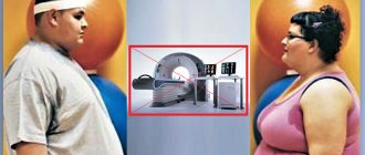
In addition to external reasons that prevent MR examination, there are also psychological factors that need to be taken into account.
It is important to warn the patient that he will be immobilized for a long time. The duration of the process can be 30-60 minutes.
What should you not do on the eve of an MRI?
It is forbidden to drink alcohol before the examination. This is especially important during the preparation phase for contrast-enhanced magnetic resonance imaging. Ethanol negatively affects vascular tone, distorts images and complicates the diagnosis of pathologies.
Two days before the examination, it is necessary to avoid taking energy drinks, reduce the amount of coffee consumed and other substances that have a tonic effect. It is not advisable to overeat in anticipation of a magnetic resonance imaging scan.
Self-administration of medications is unacceptable unless this has been previously agreed upon with the treating specialist. The patient is advised to get enough sleep before the examination and avoid overwork.
Preparing for an MRI with contrast
Contrast is a special dye solution administered intravenously. Before performing an MRI using a contrast agent, specialists need to make sure that the patient does not have problems with the kidneys or liver. A preliminary opinion from a urologist may be required.
It is important to exclude the presence of allergic reactions to the contrast agent. To do this, tests are carried out in advance for rejection of the composition.
Modern drugs are produced based on gadolinium salts. They do not cause allergies, but are excreted through the liver and kidneys. If you have chronic diseases of these organs, problems may arise. Therefore, preliminary consultation with a specialist is necessary.
Preparing for an abdominal MRI
To be prepared for diagnosing internal organs, you need to familiarize yourself with some rules:
- the procedure is carried out on an empty stomach, so it is advisable to refrain from eating 6-12 hours before the session;
- several days before the study you need to follow a diet that excludes raw fruits and vegetables, sweets, drinks containing gas, dairy products, legumes and mushrooms;
- two to three days before the tomography, take medications that reduce flatulence (Mezim or Espumisan);
- 30 minutes before the procedure you need to take an antispasmodic agent, for example No-Shpa.
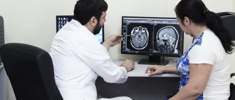
How to properly prepare for an MRI of the brain
The preparatory stage is carefully planned in advance. The radiologist has the final decision on the possibility, method, and tactics of MR imaging. No preparation is needed when performing an MR scan:
- Spinal column;
- Heads;
- Joints;
- Breasts.
To alleviate the patient's condition, doctors sometimes recommend drinking a glass of water 3 hours before the procedure. A light breakfast prevents discomfort due to an empty stomach. Planning contrast tomography excludes eating 4 hours in advance. Gadolinium rarely provokes hypersensitivity reactions (allergy, itching, dermatitis, rashes). Complications are minimized by following the doctor’s recommendations.
The patient must warn doctors about the presence of allergic diseases - asthmatic bronchitis, bronchial asthma. Even a small dose of a compound with increased sensitivity can provoke life-threatening complications, so a provocative test is performed before injecting the drug. A small dose of contrast is administered, and then the person's condition is monitored. Scanning continues in the absence of skin itching, rash, redness, or objective changes in health.
The occurrence of any complication requires immediate stop of the manipulation. There is a first aid kit in the treatment room for emergency medical care. Specialists immediately administer the necessary medications. For some time, the person is under medical supervision to provide emergency assistance in the event of life-threatening complications. Elimination of errors in MRI of the brain is achieved by proper preparation for the examination.
Paramagnetic substances are excreted through the kidneys. Renal failure is a contraindication to the contrast procedure. The possibility of MR scanning in patients with organ failure is determined after assessing the concentration of creatinine in the blood and urine. You can prepare properly only after assessing the test results.
Most of the substance is excreted through the urinary tract. An increase in the level of the drug in the blood indicates renal failure. Pathology excludes the possibility of contrast tomography. A biochemical blood test and a general urine test are mandatory diagnostic methods prescribed before a magnetic resonance scan of the brain and other organs.
At the preliminary stage, the results of a general analysis of urine, blood, and biochemical parameters are analyzed. Indicators are monitored for timely detection of renal or liver failure. Pathology slows down the removal of gadolinium and other contrast compounds. MR imaging should not be performed on patients with kidney or liver pathologies, as there is a high risk of poisoning.
Preparing for an MRI of the pelvis
This examination includes the study of organs such as the prostate gland, bladder and adjacent organs, the uterus and its appendages. To prepare for an MRI of the pelvic organs, perform the following procedures:
- cleaning the gastrointestinal tract with an enema;
- fasting for six hours;
- for the examination to be effective, it is necessary that the bladder is partially filled; for this, it is recommended to refrain from going to the toilet two to three hours before the examination and drink half a liter of clean water without gas an hour before the session;
- take the antispasmodic within 30-40 minutes.
What can you eat and drink before an MRI?
The main difficulty in preparation is scanning the bladder. The organ has a thin wall. Without filling, the walls stick together. Clear visualization is achievable by filling with water. It is recommended to drink 2-3 liters of liquid 4 hours before magnetic resonance imaging.
A cleansing enema is recommended a few hours before the intestinal scan. Removing feces and food debris improves the visibility of the wall of the gastrointestinal tract.
What can you eat before a brain MRI?
Four hours before a magnetic resonance imaging scan of the head with contrast, you can eat the following foods:
- Brown bread and other bakery products;
- Fresh vegetables;
- Caffeinated drinks.
Any products must be processed thermally. Steam fruits, vegetables, dill.
Before an MRI of the head, you can eat lean fish, meat, drink fermented milk drinks, and eat stewed vegetables. Regarding taking medications, you should first consult with your doctor.
Young children are allowed to eat 5 hours before the examination. A formula-fed infant should be optimally prepared before anesthesia. It is better to avoid introducing gas-forming mixtures.
Preparing for MRI in women
Additional rules exist for conducting research in women:
- If a woman is pregnant or suspects pregnancy, it is necessary to warn the doctor about this.
- If possible, avoid undergoing tomography during the menstrual cycle, when internal organs and arteries change in size and contours. Tomography results may be incorrect.
- If a session was carried out using a contrast agent, women who are breastfeeding must interrupt breastfeeding for two days until the dye is completely removed from the body.
Magnetic resonance imaging technique
Before an MRI with contrast, the patient is given a special drug that allows certain areas of the body to be illuminated. Immediately after this, the person enters the office where the examination equipment is located. During an MRI, the patient is in a horizontal position on the table. The diagnostic process is closely monitored by the operator. Sometimes he gives commands to better record the structure and condition of internal organs. Typically, he asks the patient to hold their breath for a few seconds or turn around on the table. Immediately after the procedure, the person returns to his normal life.
How to prepare children for MRI
Before going to the procedure with your child, relieve him of internal fears about an unfamiliar situation. The day before, you need to talk to the baby, tell him what awaits him. You need to convince him that the process is safe and painless.
MRI in children has some peculiarities. Since it is difficult for a child to remain motionless for a long time, the session is carried out with the help of mild sedatives or under general anesthesia. During the entire tomography process, the child’s condition is monitored by an experienced specialist – an anesthesiologist.
Before conducting diagnostics with a contrast agent, the presence or absence of allergic rejection to the drug is determined. A severe form of bronchial asthma in a child’s diagnosis may also be a contraindication.
Contraindications
Head tomography is not prescribed if the patient has absolute contraindications, but is allowed at the discretion of the doctor in case of relative contraindications. Diagnostics is prohibited if the following conditions exist:
- the patient has prosthetic heart valves, pacemakers or neurostimulators;
- presence of insulin pumps, inner and middle ear prostheses, cochlear implant;
- the patient has an Ilizarov apparatus;
- the presence of metal implants, ferromagnetic particles, fragments in the body.
Relative indications include:
- tremor, the patient’s inability to hold his breath for a long time during examination;
- presence of dental braces, dentures, stents, vena cava filters;
- heart failure, clip instead of gallbladder;
- pregnancy;
- claustrophobia;
- severe pain while standing still;
- coronary artery bypass grafting.
Preparing for an MRI of the brain
If a doctor sends a patient for an MRI of the brain, you also need to know that:
- if there are metal crowns, bridges, dentures, or braces in the oral cavity, visit the dentist in advance and temporarily remove these objects;
- all removable electronic and metal devices must be left outside the threshold of the room with the MR installation;
- emotionally prepare yourself for the upcoming procedure in order to avoid immersion in a stressful state, remember that the diagnosis is absolutely safe;
- If you cannot get rid of your anxiety, ask your doctor to give you a sedative.
By following the above rules, the patient will help specialists conduct an effective diagnosis. The MR procedure will be comfortable and high quality.