More detailed information about other diseases starting with the letter “R”: Radiation damage to the nervous system; Multiple sclerosis; Radiculitis; Spina bifida; Disseminated encephalomyelitis; Retrobulbar neuritis; Retinal migraine
What is retrobulbar neuritis?
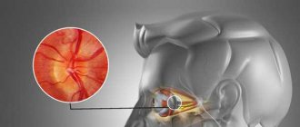
Retrobulbar neuritis is an inflammatory disease that affects the optic nerve, located between the orbit and the visual area. If there is an anomaly, the patient experiences a decrease in the level of visual acuity, the occurrence of prolapses, minimization of boundaries, and painful sensations during motor reflexes with the eyeballs. As research methods, doctors use complex neurological and ophthalmological techniques. Therapeutic therapy is performed using antibacterial/antiviral medications, glucocorticosteroids, diuretics and neuroprotective drugs. In some cases, detoxification procedures and physical therapy are prescribed.
Symptoms of retrobulbar neuritis
Retrobulbar neuritis refers to inflammation of the optic nerve, which occupies the area from the eyeball to the chiasm. There are three forms of this pathology: peripheral, transversal, axial. Each form of neuritis has its own symptoms. The most common is the axial form of retrobulbar neuritis, which is characterized by localization of inflammation in the aximal bundle. The patient experiences the appearance of central scotomas in the visual field and decreased vision.
The most severe form of the disease is considered transversal, since it is characterized by the spread of inflammation to the entire tissue of the optic nerve. The patient's vision gradually decreases and blindness may occur. Inflammation usually begins in the axial fascicle or at the periphery, after which it spreads through the septa to the rest of the tissue. The peripheral form of neuritis begins to develop with inflammation of the optic nerve sheaths, which then spreads to its tissue. The inflammatory process is characterized by the accumulation of exudative effusion in the subarachnoid and subdural space of the optic nerve. Patients note severe pain in the orbital area, which begins to intensify when the eyeball moves. Central vision remains normal. All types of neuritis are characterized by the absence of changes in the optic nerve head.
Retrobulbar neuritis can occur in acute and chronic forms. In the acute course of the disease, a decrease in visual acuity occurs a couple of days after the onset of inflammation. In the chronic course of the pathology, vision decreases gradually. Typically, a chronic course of the disease occurs in the case of diabetes mellitus. This form of the disease occurs mainly in men. Almost always the lesion is bilateral, and visual acuity decreases very slowly. At the beginning of the disease, the optic discs are normal, but their temporal pallor gradually appears.
Useful facts
Typically, retrobulbar neuritis acts as a secondary consequence after any abnormality occurring in the brain. In very rare situations, the disease is diagnosed as a primary true inflammatory process. In the neurological field, doctors are still studying the issue of this disorder in detail. Neuritis that is localized in the intraorbital region is called intrabulbar (it is examined by ophthalmologists). In both cases, therapy requires a joint solution of the problem between the neurologist and the ophthalmologist. The risk group includes people aged 20 to 40 years. According to average statistics, this type of disease occurs twice as often in females as in males.
Sign up for a consultation
After the examination, your doctor may order a magnetic resonance imaging scan of your head. This diagnostic method helps solve two important problems:
- Identify lesions in the brain that indicate the presence of multiple sclerosis. If they are detected, you will be prescribed medications that will help prevent multiple sclerosis and slow its progression.
- Find other possible causes of retrobulbar neuritis, such as intracranial tumors.
Before the test, you may be given a dye solution intravenously, which will make the optic nerve and other nerve structures more clearly visible in the pictures.
The causes of retrobulbar neuritis can also be inflammatory processes in the eye, general infections, and in rare cases, diseases of other internal organs. If your doctor suspects that retrobulbar neuritis is associated with neuromyelitis optica, you will have blood tests to look for specific antibodies.
Treatment of retrobulbar neuritis depends on the cause that caused it. If the cause cannot be determined, complex therapy is carried out. In most cases, vision can be restored.
Any sudden drop in vision should be a reason to urgently visit a doctor. To make an appointment with an ophthalmologist at the medical center in Presnya, call: +7 (495) 120-08-07.
Factors causing retrobulbar neuritis
Various problems provoke the development of abnormal vision impairment. Inflammation itself begins as a result of damage to the body by infectious diseases, chronic diseases caused by immunodeficiency. Etiological reasons include:
- Chronic stages of viral infections - the main causative agent is cytomegalovirus, which provokes the development of mononucleosis. The stable location of harmful agents causes deformations in immune properties. Due to the production of antibodies for one's own tissues, the process of autoimmune inflammation begins.
- Chronic infectious points - most often the anomaly is caused by unfavorable foci that are located nearby (for example, sinusitis, otitis media and others), much less often - by distant places (cystitis). The infection enters the nervous structure of the visual apparatus through blood transportation.
- Inflammatory phenomena in the orbit - provocateurs are inflammatory processes. Spread occurs in two ways: hematogenous and perineural, which causes damage to the retrobulbar perimeter of the nerve. Quite often, total optic neuritis is diagnosed.
Secondary progression of optic nerve disorder begins as a result of an anomaly that is localized in the cerebral region. The main etiofactors include the following diseases:
- Multiple sclerosis - selective damage to the myelin sheath of nerve fibers moves to the optical areas, which causes an inflammatory reaction. Neuritis of a retrobulbar nature is considered the initial stage in more than 65% of cases of disease.
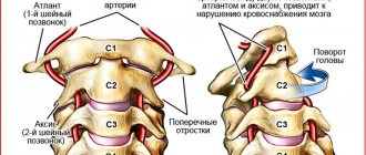
- Brain tumors - when voluminous tumors form in the area of the chiasm glioma, objects in another place begin to actively pressure the optic chiasm, disrupting the process of blood circulation in the body and metabolism in tissues. As a result, the patient develops symptoms of neuritis.
- Neuroinfections - Lyme disease, neuroAIDS, tuberculous meningitis can provoke abnormalities in the retrobulbar zone. Inflammatory phenomena are associated with the entry of infectious microorganisms into the body. In some cases, the disease may appear due to intoxication. Exogenous lesions are caused by the influence of toxic chemicals, methyl alcohol, iodine substances. Toxins appear as a result of dysmetabolic processes, renal failure or during pregnancy.
Etiology and pathogenesis of optic neuritis
The disease is provoked by inflammatory processes of the eyeball (retinitis, keratitis, iridocyclitis, panophthalmitis), orbit (periostitis, phlegmon) and brain (meningitis, arachnoiditis, encephalitis). The disease can also be provoked by infectious lesions of the nasopharynx: frontal sinusitis, sinusitis, tonsillitis, chronic tonsillitis, pharyngitis). Patients with malaria, tuberculosis, ARVI, gonorrhea, and diphtheria are at risk of developing neuritis of the facial nerve.
Common causes of the disease are blood diseases, traumatic brain injuries, alcoholism, diabetes mellitus, gout, and autoimmune disorders. Optic neuritis often occurs in multiple sclerosis. The basis of the disease in this case is the process of destruction of the myelon sheath of nerve fibers. This process is called demyelination. It is not inflammatory, but it is still classified as retrobulbar neuritis due to the similarity of symptoms.
The inflammatory process during neuritis covers both the optic nerve sheath and its trunk. Edema resulting from inflammation and infiltration cause compression of the optic fibers and their subsequent degeneration, which leads to deterioration of vision. After the inflammation is removed, some of the nerve fibers can restore their functions, so vision will improve. In severe cases of pathology, nerve fibers decay in patients. As a result, atrophy of the optic nerve occurs, which leads to irreversible consequences for the patient’s vision.
Mechanism of occurrence
When examining the visual department, one can understand that the nerve contains a large number of fibers (more than a million), which begin their journey from the retina and end in the subcortical ganglia of the eye. Various forms of pathogens can cause serious disorders, for example: ischemia, inflammation and tissue destruction. As a result, the patient experiences detrimental changes that affect the functions of transmitting information from the retinal receptors that receive the external image.
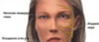
The clinical picture appears as a decrease in the level of vision, impaired color perception, and loss of boundaries. If you ignore the doctor’s recommendations or completely refuse treatment, the patient begins to experience irreversible abnormalities that fill the cavities with connective tissue growths. The last stage of the disease leads to complete loss of visual abilities.
Types of neuritis
There are different types of symptoms, which depend solely on the location of the lesion (central or peripheral lobe). In neurology, retrobulbar neuritis is divided into three classes:
- Axial - a common form of inflammatory reaction, localized only in the axial fascicle, characterized by a sharp decrease in central visual acuity and the appearance of scotomas.
- Peripheral - originates in peripheral structures, accompanied by the formation of intrathecal fluid, which provokes pain. A narrowing of the optical fields is characteristic.
- Transversal - a detrimental anomaly progresses in the periphery or axial region and then reaches the diameter of the nerve center. The patient is completely blind.
Another area of division of retrobulbar neuritis depends on the variability of the course:
- Acute form - manifests itself in the rapid loss of visual acuity. Most often it is diagnosed in the primary version of the lesion, which is typical for young people.
- Chronic form - symptoms have a gradual scale of increase, which are caused by a cerebral disorder. Patients experience some periods of remission or exacerbation. There is a possibility of the acute form becoming chronic.
Possible complications of optic neuritis
Acute inflammation of the optic nerve has no complications if treated correctly. If neuritis occurs against the background of multiple sclerosis, then it becomes chronic, accompanied by nerve atrophy and exacerbations.
Methods for preventing retrobulbar neuritis Regular preventive examinations with an ophthalmologist will help to timely determine the development of the disease. He will quickly assess the state of vision, its features, and also determine the thickness of the optic nerve.
Thus, there are no specific preventive measures. The main thing is to monitor your health and, at the first suspicion of developing a disease, contact a specialized specialist.
How does it manifest itself?
The main manifestations of the anomaly are various disturbances of optical perception. According to average statistics, in 60% of situations patients have vision problems. When diagnosing an acute form of neuritis, visual acuity decreases within a short time (from a couple of hours to two days), and in the chronic form it will take 7-14 days. Patients, talking about their sensations, report a “veil before their eyes” and “blurring of all objects”; some experience “light flashes”. The slow progression of the disease causes deterioration of visual adaptation at night.
Deformations of the optical boundaries provoke the appearance of dark dots before the eyes, narrowing the visible trajectory. They can be combined with a decrease in vision. Retrobulbar neuritis occurs with impaired color perception. Dychromatopsia is practically not noticed by patients, but is only detected during certain tests. Doctors say that more than 50% of patients complain of a feeling of pain in the area of the eye orbits, which increases with activity, in particular when turning their gaze upward.
Examination methods
The main goal of the diagnostic procedure is to make a correct conclusion with a clear identification of the etiology and the mandatory exclusion of the presence of multiple sclerosis in the patient. The study will require clarification of all symptoms, the rate of their development and previously experienced diseases, as well as full ophthalmological and neurological testing. Diagnostics:
- Visometric analysis helps determine vision loss; emerging problems can be corrected by wearing lenses.
- Color vision test - designed to accurately identify anomalies in the absence of changes in the optic disc.
- Perimetric scanning - scan the central, paracentral and peripheral sections.
- Ophthalmoscopy - looks for swelling, bleeding and the possibility of atrophy.
How to get rid of it?
Therapeutic measures for retrobulbar neuritis are based on an etiopathogenetic principle, which is selected taking into account the etiology of the disease. A secondary lesion requires elimination of the primary source, that is, removal of the tumor, therapy aimed at restoring immune abilities in multiple sclerosis, and getting rid of neuroinfections. The main treatment complexes include:
- Pharmacological antibiotics with a wide range of applications.
- Antiviral components.
- Introduction to a course of glucocorticosteroid therapy.
- To reduce swelling, diuretics and antihistamines are prescribed.
- A course of vitamin complexes (vitamins B and C, nicotinamide and others) is prescribed.
In case of toxic damage, detoxification will be required; for this, intravenous administration of saline solutions and dextran through a dropper is used. If the patient has no restrictions, it is recommended to carry out physiotherapeutic procedures.
The main course of treatment
If a disease of the optic neuritis category occurs and occurs in an acute form, it is necessary to hospitalize the victim, since the use of many drugs requires observation, and many of the prescribed drugs will have to be administered intravenously or using droppers.
Until the underlying causes of optic nerve dysfunction are determined in order for treatment to be more effective, medical workers are fighting the infection. To relieve symptoms, they try to stabilize the immune system, restore metabolic processes and metabolism of nervous system tissues.
Treatment with antibiotics is very effective. But there is a category of drugs that is contraindicated for use in various disorders of the optic nerve and related processes.
These are drugs such as:
- Neomycin;
- Streptomycin;
- Kanamycin;
- Gentamicin;
- and other medical products of the streptomycin group
Such drugs cure the resulting neuritis, but such treatment has side effects that lead to deterioration of vision and hearing impairment.
The streptomycin group of drugs is a good category of antibiotics. Their main disadvantage is the effects that affect not only the area of the optic nerve, but also the entire system of nerve endings, which is unacceptable for such a disease.
Medicines of a number of antibiotics are administered over several days under the supervision of specialists, since the patient may have the most unpredictable reactions of the body: from allergies to anaphylactic shock.
Treatment of an inflamed nerve by using steroid therapy, Piracetam or Neotropil, Solcoseryl intramuscularly leads to quick results. If vision is not restored, then a course of plasmapheresis is used instead of steroid drugs.
Treatment of retrobulbar neuritis
Retrobulbar neuritis has its own category of causes. Its symptoms are almost no different from other types of neuritis. But despite this, the treatment follows a certain scheme and has its own peculiarities. This type of neuritis is very often caused by the disease multiple sclerosis, so many specialists prescribe drugs prescribed by neurologists.
Neuritis in this category responds successfully to medications of the salicylic group. Together with it, complex therapy is used using the diaphoretic method using light-air baths. In this situation, a group of penicillin antibiotics is also used.
For problems associated with inflammation of the optic nerve, disorders of the nervous system and diseases with the prefix “neuritis” in different areas of the body, a complex of B vitamins and nicotinic acid are often prescribed, which are better absorbed thanks to injections and taking special pharmaceutical brewer’s yeast.