Poliomyelitis has a viral infectious cause. This disease has a long history and is known from characteristic descriptions from ancient written sources. Historical documents say that several thousand years BC, terrible epidemics among children periodically occurred in the world, leaving thousands of small disabled people everywhere. The disease brought real disaster to children at the beginning of the twentieth century, when there was no vaccine against the disease yet. Today, thanks to the global movement to eradicate and treat polio, the disease is very rare in developed countries, but in a number of African countries, as well as in Afghanistan, Pakistan, and Tajikistan, the disease still claims the lives of children or leaves them disabled.
General information about the disease
Poliomyelitis is caused by polio viruses. There are three types of poliovirus (type I, type II and type III). All three types of the virus are widespread and have been found in various countries and parts of the world. As a result of the disease, damage to the nervous system and gray matter of the spinal cord can occur, which leads to paralysis or even death, as well as inflammatory changes in the mucous membrane of the intestine and nasopharynx (these changes can be identified as an intestinal infection or acute respiratory infection), and most people with the virus polio cases do not experience any symptoms of the disease and will never become aware of the infection.
The polio virus is very stable in the external environment - it is not destroyed in the digestive system, is not killed by antibiotics, chemicals, is resistant to freezing and drying, but dies when boiled, exposed to ultraviolet irradiation and disinfectants.
The source of infection can be a sick person or a virus carrier. Infection occurs by airborne droplets (when coughing, sneezing, talking), oral-fecal (“dirty hand disease”), through food and water, and mechanically from flies carrying the virus.
Outbreaks of the disease often occur in the summer-autumn period, as with other classic viral intestinal diseases.
The vast majority of the disease affects children of preschool age, up to 5 years, and it does not depend on gender. But also in rare cases, adults become infected with polio, and then these are predominantly women; the reasons for this are not clear.
Symptoms of polio
Although polio is dangerous because it can cause partial or complete paralysis and even death, most people infected are asymptomatic. In addition, not all types of polio viruses lead to serious consequences. Strains of the virus that do not lead to paralysis are called non-paralytic; usually the disease in this case manifests itself as acute respiratory infections, acute respiratory viral infections or influenza.
The following symptoms of polio last up to 10 days:
- Temperature increased to 40°C, fever,
- Lethargy, loss of appetite,
- Sore throat, headache and joint pain,
- Pain, stiffness or difficulty moving in the back, neck, arms or legs,
- Nausea and vomiting
According to statistics, this is how the disease most often progresses, in a preparalytic form, within 1-2 weeks. But for every ten non-paralytic cases, there is one paralytic case, in which there will subsequently be problems with the musculoskeletal system: headaches and fever develop into muscle numbness. The paralytic form of the disease can be of several types: depending on the type of lesion:
- spinal cord (spinal form of polio),
- brain stem (bulbar form of polio),
- both types of lesions (bulbospinal form).
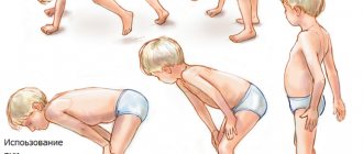
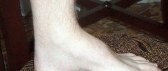
The development of the disease into a paralytic form is characterized by: severe muscle pain, weakness, decreased strength in the limbs, loss of reflexes. They are observed already in the first week of the disease. People who have had polio in childhood retain limited health and have a number of concomitant diseases associated with dysfunction of the musculoskeletal system - muscle weakness and atrophy, muscle pain, paralysis - temporary and permanent - of muscles, deformities of the hips, feet, ankles, breathing and swallowing disorders, sleep apnea. Children who survive polio often spend the rest of their lives with severe disabilities.
Polio
There are 4 types of reactions to the polio virus:
- development of immunity in the absence of symptoms of the disease (subclinical or silent infection);
- symptoms (in the viremia stage), which are in the nature of a general moderate infection without involving the nervous system in the process (abortive forms);
- the presence in many patients (up to 75% during the epidemic) of fever, headache, malaise, there may be meningeal phenomena, pleocytosis in the cerebrospinal fluid, but paralysis does not develop;
- development of paralysis (in rare cases).
In the subclinical form there are no symptoms. In the abortive form, the manifestations are indistinguishable from any general infection. Serological tests are positive.
The virus can be isolated. In other variants of the course of the disease, a preparalytic stage can be observed, which can sometimes develop into the stage of paralysis.
Pre-paralytic stage. During this stage, two phases are distinguished. In the first phase, fever, malaise, headache, drowsiness or insomnia, sweating, pharyngeal congestion, and gastrointestinal disorders (anorexia, vomiting, diarrhea) are observed. This minor illness phase lasts 1–2 days. Sometimes it is followed by a temporary improvement with a decrease in temperature for 48 hours, or the disease enters the “big illness” phase, in which the headache is more pronounced and is accompanied by pain in the back, limbs, and increased muscle fatigue. In the absence of paralysis, the patient recovers. In the cerebrospinal fluid, the pressure is increased, pleocytosis is noted (50–250 in 1 μl). Initially, there are both polymorphonuclear cells and lymphocytes, but after the 1st week there are only lymphocytes. The level of protein (globulins) increases moderately. Glucose levels are normal. During the 2nd week, protein levels increase.
Paralytic stage. In the spinal form, the development of paralysis is preceded by fasciculations. Pain in the limbs and increased muscle sensitivity to pressure are noted. Sometimes the preparalytic stage lasts up to 1–2 weeks. Paralysis may be widespread or localized. In severe cases, movements are impossible, except for very weak ones (in the neck, torso, limbs). In less severe cases, attention is drawn to asymmetry, “spotting” of paralysis; muscles can be severely affected on one side of the body and preserved on the other. Typically, paralysis is most pronounced during the first 24 hours, less often the disease gradually progresses. In “ascending” forms, paralysis spreads upward (from the legs), and a life-threatening situation may arise due to respiratory failure. “Descent” forms of paralysis are possible. It is necessary to monitor the function of the intercostal muscles and diaphragm. A test to identify respiratory paresis is a loud count in one breath. If the patient cannot count to 12–15, then there is severe respiratory failure; the forced respiratory volume should be measured to determine the need for assisted breathing.
Improvement usually begins by the end of the 1st week from the onset of paralysis. As with other neuronal lesions, there is loss or reduction of tendon and cutaneous reflexes. Sphincter disorders are rare, sensitivity is not impaired.
In the stem form (polioencephalitis), facial paralysis, paralysis of the tongue, pharynx, larynx and, less commonly, paralysis of the external eye muscles are observed. Dizziness and nystagmus are possible. There is a great danger of involving vital centers in the process. It is very important to distinguish respiratory disorders caused by the accumulation of saliva and mucus during paralysis of the pharyngeal muscles from true paralysis of the respiratory muscles.
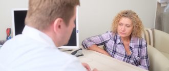
Diagnosis of polio
Most often, to make a diagnosis of “poliomyelitis,” a specialist only needs a cursory examination and history taking—symptoms such as muscle pain, stiffness of the neck and back, and difficulty swallowing and breathing are too alarming, eloquent and typical for a specialist. To confirm the diagnosis, a nasopharyngeal swab or stool examination for virological analysis is prescribed.
If alarming symptoms occur, the patient is hospitalized in an infectious diseases hospital, where he is examined by specialized doctors.
In a hospital setting, the following types of tests are prescribed:
- blood, urine, stool tests,
- analysis of samples from the nasopharynx,
- cerebrospinal fluid analysis
Tests help determine what exactly caused the disease.
Treatment options for polio
Unfortunately, there is no separate special way to cure polio, therefore, first of all, the patient is prescribed bed rest, symptomatic treatment aimed at reducing temperature, eliminating symptoms of fever, and pain relief. They strive to increase the level of comfort for the patient, speed up recovery, use mechanical ventilation if necessary, and then add feasible physical exercises (physiotherapeutic techniques, therapeutic exercises) to prevent deformation of the limbs and loss of muscle function.
With poliomyelitis, the death of nerve motor cells occurs, deviations in the function of the muscular system are observed, often accompanied by trophic and vascular disorders. The muscles of the lower extremities are most often affected.
Practice shows that the patient needs systematic restorative treatment aimed at eliminating pathological phenomena from the central nervous system and neuromuscular systems. In the treatment process, a combination of physical therapy is used - therapeutic exercises, physical exercises in water, walking, massage - with drug treatment and even surgery.
Exercise therapy for polio has an overall positive effect on the patient’s body and leads to the normalization of a number of vital functions, helps strengthen the affected muscles, and helps avoid the appearance of contractures and deformities of the limbs.
In addition, therapeutic physical education for existing disorders will help facilitate the patient’s development of mobility and self-care skills, and will contribute to the future physical development of the child.
An integrated approach to restoration consists of the following methods:
- Therapeutic exercise and massage aimed at improving tissue trophism and strengthening weakened muscles.
- Physiotherapeutic treatment: treatment with heat, paraffin baths, electrophoresis, magnetotherapy, electrical stimulation, darsonvalization, therapeutic swimming, hydromassage baths.
- Manual therapy
- Reflexology: acupuncture, laser therapy
A comprehensive examination and correct recovery methods after polio are carried out at the Center for Rehabilitation Medicine in Naberezhnye Chelny - the Center’s specialists will select the necessary ways to make life easier and prolong a comfortable life for a patient who has suffered polio.
Positive dynamics from physiotherapy, massages, and manual therapy are observed in all patients - these methods help build muscle mass, increase motor range, straighten gait, and avoid severe contractures. Methods of therapeutic physical culture and massage are applicable at various stages of the disease; they allow the patient to develop compensatory adaptability and give a chance to improve overall physical development and health. The Center for Rehabilitation Medicine employs highly qualified specialists who together will select a rehabilitation program for a patient who has suffered polio.
Poliomyelitis or Heine-Medina disease
Vomit
Diarrhea
2404 13 September
IMPORTANT!
The information in this section cannot be used for self-diagnosis and self-treatment.
In case of pain or other exacerbation of the disease, diagnostic tests should be prescribed only by the attending physician. To make a diagnosis and properly prescribe treatment, you should contact your doctor. Poliomyelitis: causes, symptoms, diagnosis and treatment methods.
Definition
Poliomyelitis (polios - gray, myelos - spinal cord) is an acute viral disease characterized by damage to the nervous system (mainly the gray matter of the spinal cord).
Thanks to vaccination, polio is now extremely rare. In 2021, about 180 cases of disease caused by wild virus were reported worldwide.
Causes of polio
The causative agents of poliomyelitis are polioviruses. People of any age who are not vaccinated in a timely manner are at risk of contracting polio caused by wild poliovirus. However, children under 1 year of age are predominantly affected.
Humans are the only carrier of wild polioviruses. Infection can occur by airborne droplets, neural and fecal-oral (by consuming contaminated food or water) routes.
The patient sheds viruses during the incubation period and the first week of the disease through the nasopharynx and for 1–6 months with feces. Flies usually serve as carriers of infection.
Polioviruses are stable in the external environment, persist for a long time in water, milk and feces, and do not lose viability at low temperatures. In the human body, it multiplies in the motor cells of the anterior horns of the spinal cord and the motor nuclei of the cranial nerves.
Wild poliovirus, entering the pharynx and/or gastrointestinal tract, attaches to receptors on epithelial cells, where it multiplies and accumulates. The virus then enters the lymphatic system, subsequently into the blood, spreads throughout the body and replicates (multiplies) in many organs and tissues: lymph nodes, spleen, liver, lungs, heart muscle and brown fat. Subsequently, through the endothelium of small vessels or along peripheral nerves, the virus penetrates into the central nervous system and the process involves motor neurons of the anterior and posterior horns of the spinal cord, motor cells of the cranial nerves (glossopharyngeal, vagus, facial, trigeminal), and proximal parts of the peripheral nerves. The reticular formation, vestibular nuclei and deep structures of the cerebellum in the brain are damaged. Nerve cells undergo dystrophic-necrotic changes, disintegrate and die. Cells of the brain stem, subcortical nuclei of the cerebellum and, to an even lesser extent, cells of the motor areas of the cerebral cortex and dorsal horns of the spinal cord undergo less pronounced changes. Paresis and/or paralysis of various muscle groups occur.
Restoration of the functions of paralyzed muscles initially proceeds at a rapid pace, and then slows down and can last from several months to 1-3 years.
After the end of acute events, the dead cells are replaced by scar tissue. The size of the spinal cord decreases.
The stage of residual effects is characterized by persistent flaccid paralysis, muscle atrophy, contractures and deformities of the limbs and trunk.
The transferred disease leaves a stable, type-specific immunity.
Classification of the disease
According to ICD-10, polio is divided as follows:
- acute poliomyelitis;
- acute paralytic poliomyelitis associated with vaccine virus;
- acute paralytic poliomyelitis caused by wild imported poliovirus (type I, II or III);
- acute paralytic poliomyelitis caused by wild local (endemic) poliovirus (type I, II or III);
- acute paralytic poliomyelitis of other and unspecified etiology;
- acute non-paralytic poliomyelitis.
Wild polio virus can be of three serotypes (1, 2, 3) - the presence of immunity from one serotype does not protect against infection by the other two.
Symptoms of polio
The incubation period of the disease lasts from 2 to 35 days. Poliomyelitis can occur with or without damage to the central nervous system (CNS). The prognosis depends on the clinical form and severity of the disease. The most unfavorable prognosis is for the paralytic form of poliomyelitis with respiratory failure.
Poliomyelitis without central nervous system involvement
may have two clinical forms: inapparent (virus carriage) and visceral (“minor disease”). The inapparent form is caused by virus carriage within the pharyngeal ring and intestines. This form has no clinical manifestations, so diagnosis is possible only on the basis of a virological examination.
The visceral form is manifested by a short-term increase in temperature, catarrhal (cough, runny nose, sore throat) and dyspeptic symptoms (nausea, vomiting, loose stools). The disease lasts about 3-5 days and is characterized by a favorable outcome. Diagnosis is possible only on the basis of epidemiological and virological data. From an epidemiological perspective, these two forms of polio are the most dangerous, since the patient, as a rule, is not aware of the disease and does not take precautions.
Poliomyelitis with central nervous system involvement
can occur in the form of a non-paralytic or paralytic form.
The non-paralytic form is manifested by serous meningitis or severe meningeal-radicular syndrome (with pain) without signs of inflammation of the soft meninges. The meningeal form is characterized by general cerebral symptoms (headache, repeated vomiting), fever and intoxication (weakness, weakness, malaise, decreased performance). The meningeal form can have a one- or two-wave course. In a single-wave course, against the background of high temperature and intoxication, meningeal and cerebral symptoms appear on the 1st-3rd day of illness. In a two-wave course, the first wave occurs as a visceral form without signs of damage to the meninges, and after 1-4 days of absence of elevated temperature, a second febrile wave is observed with the development of meningeal symptoms.
The course is favorable, recovery occurs in the third week of the disease.
The paralytic form occurs only in unvaccinated or incompletely vaccinated individuals. Depending on the level of damage, it is divided into several forms:
- spinal (cervical, thoracic, lumbar spinal cord);
- bulbar (nuclei of motor nerves located in the brain stem - III, IV, VI, VII, IX, X, XI, XII pairs);
- pontine (isolated lesion of the nucleus of the facial nerve - VII pair - in the area of the Varoliev bridge);
- mixed forms (bulbospinal, pontospinal).
The course of the paralytic form of polio has four stages:
1 - preparalytic (1-6 days), 2 - paralytic (1-3 days), - restorative (up to 2-3 years), - residual (over 3 years).
The preparalytic period lasts from the onset of the disease until the appearance of the first motor disorders. The disease begins acutely, and body temperature rises significantly. During the first 3 days, headache, malaise, runny nose, pharyngitis are observed, and gastrointestinal disorders (vomiting, loose stools or constipation) are possible. Then comes a period of temperature normalization, which lasts 2-4 days. In some patients this period may be absent. Then the temperature rises again to 39-40°C, the headache intensifies, pain in the back and limbs appears, severe hyperesthesia, confusion and meningeal phenomena, spontaneous pain in the spine, in the muscles of the neck, back, along the nerve trunks and roots, fascicular twitching of the muscles of the limbs, involuntary oscillatory movements of the eyes.
Then paralysis or paresis appears, which in most patients develops within a few hours.
The clinical picture of the paralytic period is determined by the localization of the central nervous system lesions. The legs are most often affected: the quadriceps, adductors, flexors and extensors are affected. On the arms there are the deltoid and triceps muscles, the instep supports of the forearm. Cyanosis, shortness of breath, limited mobility of the chest, retraction of the intercostal spaces and epigastric region during inspiration are characteristic of damage to the diaphragm and intercostal muscles.
With the development of paralysis, spontaneous muscle pain occurs. No sensory disturbances are observed.
When the bulbar form occurs against the background of fever, the functions of swallowing and speech are impaired, the pharyngeal reflex disappears, and asymmetry or immobility of the palatine arches, uvula, and soft palate is noted. There is increased secretion of mucus, which can accumulate in the upper respiratory tract and lead to breathing problems.
When the respiratory and cardiovascular centers of the brain are damaged, arrhythmic breathing with pauses, pathological breathing rhythms, cyanosis, fever, increased or decreased blood pressure, collapse, bradycardia or tachycardia appear. Possible death on the 1st-7th day of the disease.
From the 2nd week, the patients’ condition improves, bulbar symptoms decrease and subsequently may completely disappear.
The pontine form of polio is characterized by damage to the facial nerve. Asymmetry of the facial muscles appears, smoothness of the nasolabial fold on the affected side, drooping of the corner of the mouth, drooping of the eyelids. The course of the disease is favorable, with complete or partial restoration of the functions of facial muscles.
Of the mixed forms, the bulbospinal one is the most severe, since damage to the brain stem occurs in combination with paresis and paralysis of skeletal muscles. In 15% of patients, 10 years after the acute phase, after a period of stabilization, a progressive increase in muscle weakness and atrophy in previously unaffected muscles is detected.
Poliomyelitis in those vaccinated with oral live polio vaccine develops from 4 to 30 days after vaccination. Poliomyelitis can also develop in those in contact with vaccinated people up to the 60th day. Poliomyelitis progresses similarly to the disease caused by a wild virus. To eliminate vaccine-associated polio, WHO recommends switching to vaccination with inactivated polio vaccine.
Diagnostics
The diagnosis is established on the basis of clinical symptoms (meningeal symptoms, weakness of certain muscle groups, weakened tendon reflexes), epidemiological prerequisites (presence of polio in the patient’s environment), and laboratory test data.
All patients with acute flaccid paralysis under the age of 15 years, regardless of the suspected cause of the disease, are subject to examination for poliomyelitis.
Immediately after paralysis is detected, information about this is sent to the Territorial Department of Rospotrebnadzor. Two fecal samples collected (with an interval of 24–48 hours between samples) are sent to the laboratory for poliovirus diagnosis.
The final diagnosis is made by a special commission upon receipt of laboratory data. The final diagnosis is 60 days from the onset of the disease after repeated electroneuromyography.
Laboratory diagnosis of polio includes:
- virological study (study of nasopharyngeal swabs, feces, less often cerebrospinal fluid, blood);
- serological test (blood test for paired sera with an interval of 2-3 weeks; a 4-fold increase in antibody titer is considered diagnostic);
- antibodies to polio virus types 1 and 3;