Group of neurodegenerative diseases
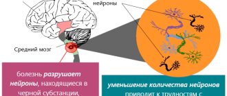
This group of diseases is characterized by damage to the central nervous system. A certain number of neurons die, and this leads to neurological pathologies. Neurodegenerative diseases can have different causes and different clinical manifestations. It is characteristic that all diseases in this category are incurable.
All therapy used is symptomatic. That is, it alleviates the manifestation of the disease, but does not in any way contribute to the regeneration of damaged areas. The most effective would be a transplantation of nerve tissue, but the method has both technical and moral obstacles. Similar diseases include Alzheimer's, Parkinson's, and Farah's diseases.
https://www.youtube.com/watch?v=https:accounts.google.comServiceLogin
A group of similar diseases is characterized by manifestations of central nervous system lesions. A certain number of neurons die, which leads to the development of neurological pathologies. Neurodegenerative diseases can occur for a variety of reasons and also have different clinical manifestations. A characteristic feature of all diseases from this group is their incurability. All therapeutic actions performed are symptomatic.
That is, therapy is aimed at alleviating the symptoms of the disease, but in no way can affect the regeneration of areas that are damaged. The most effective treatment would be tissue transplantation, but this method has several limitations, primarily technical and moral.
The most famous diseases of this group are Alzheimer's disease, Parkinson's disease, and Fahr's syndrome. Early diagnosis of these diseases is often difficult. In the diagnostic process, doctors mainly take into account patients' complaints and observe their behavior. The most modern and effective method for diagnosing diseases of this group is tomography, both computed tomography and magnetic resonance imaging.
How to deal with pathology?
Since the main source of Farah's disease has not yet been determined, therapeutic manipulations are reduced to reducing symptomatic manifestations. Drug therapy is prescribed, aimed at improving metabolism and metabolic processes. Drugs are prescribed individually by the treating specialist. If the main symptoms are parkinsonian changes, medications taken for Parkinson's disease are prescribed. Modern anticonvulsants are used to reduce the frequency of epileptic seizures. An additional supportive effect is achieved by resorting to systematic courses of exercise therapy, water therapy and mental activity training.
Inheritance of Fahr syndrome
Since this disease is a rather rare phenomenon, it is difficult for specialists to provide the correct clinical picture of the syndrome. Symptoms may appear if the patient is over 40 years of age. In younger people, Farah's disease symptoms may be vague and vague. Patients have impaired coordination of movement and parkinsonism.
Also, the development of the disease may be indicated by such signs as tremor, dystonia, rapid and erratic movements of the limbs (chorea), involuntary contractions of the hands and feet. Since the disease affects areas of the brain, performance and mental capabilities decrease. A person’s memory also suffers.
Speech is often impaired. Other neurological signs include pain and mental disorders. There is a juvenile form of the disease, which manifests itself in children and adolescents. The main symptoms are: dystonia, chorea, epileptic seizures. The senile form is typical for middle-aged and older people. In this case, parkinsonism, speech disorders, and other problems in the functioning of the nervous system are observed. Urinary incontinence may also occur.
The reasons why the disease may develop have not been precisely established. However, it is known that disorders of the thyroid and parathyroid glands have a huge impact on its occurrence. In this case, disruptions occur in the metabolic processes of calcium and phosphorus. Similar neurodegenerative diseases can also occur when the acid-base balance in the body is disturbed.
International Journal of Neurology 1 (79) 2021
References 1. Velichko M.A., Vasilyev V.V., Filippov Yu.L. Fahr syndrome in hypertension // Clinical medicine. - 1993. - No. 2. - P. 55-58.
2. Evtushenko S.K. Fahr's disease syndrome (somatoneurological manifestations) // Rare and difficult to diagnose diseases of the nervous system. - Svyatogorsk, 2003. - pp. 15-20.
3. Petelin L.S., Fokin M.A., Borzunovsa T.A., Shapovalova M.V. Fahr syndrome // Journal of Neurology and Psychiatry. - 1988. - No. 9. - P. 65-67.
4. Ponomarev V.V., Naumenko D.V. Fahr's disease: clinical picture and approaches to treatment // Journal of Neurology and Psychiatry. - 2004. - No. 3. - P. 42-45.
5. Fedulova M.V., Rusakova T.I., Ermolenko E.N. Farah's disease, identified during a forensic examination // Forensic medical examination. - 2006. - No. 5. - P. 35-37.
6. Trukhan D.I., Lebedov O.I. Changes in the organ of vision in diseases of internal organs. - M., 2014. - 208 p.
7. Yakhno N.N. Fahr's disease // Neurological questionnaire. - 2001. - No. 4. - P. 17-22.
8. Fahr T. // Zbl. Allg. Pathol. - 1930. - Bd. 30. - S. 129-133.
9. Gescywind DH, Loginov M., Stern JM et al. Identification of a locus on chromosome 14q for idiopathic basal ganglia calcification // Am. J.Hum. Genet. - 2009. - S. 764-772.
10. Guseo A., Boldizsar R., Gellert et al. Electron microscopic study of striatodental calcification // Acta neuropathology. - 1975. - No. 4. - S. 305-331.
11. Gescnwind DH, Loginov M. Identification of a locus on chromosome 14q for idiopathic basal ganglia calcification // Am. J.Hum. Genet. - 2005. - No. 65(3). - R. 764-772.
12. Goldscheider HG, Lischewski R. Clinical, endocrinologic, and computerized tomography scans for symmetrical calcification of the basal ganglia // Arch. Psychiat. Nervenktn. - 1980. - No. 228(1). - R. 53-65.
13. Cyseo A., Boldizsar F. Electron microscopic study of striatodental calcification // Acta Neuropathol. - 2006. - No. 31(4). - R. 305-313.
14. Maghraoul A., Birouk N. Fahr syndrome and dysparathytoidism // Presse Med. - 1995. - No. 24(28). - 1301-1304.
15. Proniska E., Kulczycki J., Rovinska E., Kuran W. Abolished phosphaturic response to parathormone in adult patients with Fahr disease and its restoration after propranolol administration // J. Neurol. - 1988. - No. 235(3). - R. 185-187.
16. Rossi N., Morena M. Calcification of the basal ganglia and Fahr disease. Report of two clinical cases and review of the literature // Recenti Prog. Med. - 2009. - No. 84(3). - R. 192-198.
17. Stellamor K., Stellamor V. Roentgen diagnosis og Fahr's disease // Rontgenblatter. - 1983. - No. 36(6). - R. 194-11.
18. Tardio E., Roldan ML, Pedrola D., Hierro FR Fahr disease and idiopathic pulmonary hemosiderosis in a 10 year old patient // An. Esp. Pediat. - 1980. - No. 13(7). - R. 599-604.
19. Taxer F., Haller R. Clinical early symptoms and CT findings in Fahr syndrome // Nervenarzt. - 2006. - No. 57(10). - R. 583-588.
Diagnostic methods
Before the advent of computed tomography, patients underwent X-ray examinations of the brain. Thanks to the advent of modern research methods, specialists received more informative pictures from the affected area. However, it is worth noting that computed tomography rather than magnetic resonance imaging has greater sensitivity for this clinical picture.
When analyzing most images, the affected areas are limited to the globus pallidus. They are small in size. Most often, the basal ganglia, thalamus, and cerebellum undergo changes. The level of calcification is approximately the same in young and elderly people. Also, no differences were found between groups in which Farah's disease is asymptomatic and those in which the syndrome is accompanied by pronounced symptoms.
When conducting a postmortem examination, the following picture is observed in the brain: the vessels have a whitish appearance (some branched areas). When touched by a knife, they make a sound similar to a crunch. For histological analysis, material is collected: sections of the cerebral cortex, basal ganglia, and cerebellum.
It is in the area of the latter that calcification occurs most often. The samples contain calcium salts. They are also most often detected on arteries (small, medium-sized) and capillaries. Less commonly (in isolated cases), Farah's disease affects the veins. Small calcium conglomerates are found along the entire length of the vessels, as well as in adjacent tissues. The presence of traces of arsenic, aluminum, cobalt, and mucopolysaccharides is also detected in the tissues.
Depending on whether Farah disease has symptoms, making a diagnosis can be somewhat more difficult. It is often diagnosed accidentally, when a tomography is performed to confirm a completely different disease. First of all, the specialist excludes calcium metabolism disorders and other developmental defects. Then a computed tomography scan (or x-ray examination) is prescribed.
Doctors note that one of the problems in making a correct diagnosis is hypoparathyroidism. This is a disease that occurs due to parathyroid hormone deficiency. As a result, the level of calcium in the blood decreases, and the level of phosphorus increases. If calcification of the striopallidodentate structures is observed on a CT scan, then additional tests are necessary to distinguish hypoparathyroidism from a condition such as Fahr's disease.
Forecast of the course and preventive recommendations
The disease is a chronic form of degenerative processes occurring in the brain, from which it is not yet possible to completely recover. If symptomatic treatment is started in time, the patient’s satisfactory condition can be maintained for many years. The asymptomatic course of the pathology has virtually no effect on the health and well-being of the patient. The anomaly in such cases is discovered by chance when diagnosing another disease using computed tomography of the brain.
There are no preventive measures to prevent Farah disease, since the etiology of the anomaly is not clear.
Consequences
What are the consequences of Farah disease?
All of the listed characteristics are realized only if there is complete dominance, and for the appearance of the trait, only one gene of a dominant nature will be sufficient.
https://www.youtube.com/watch?v=ytaboutru
Based on the listed features of the type of inheritance through which Farah syndrome is transmitted, we can say that the presence of a diseased gene in the future father can cause the disease to manifest itself in the child. In order to determine the likelihood with which your child may inherit Farah disease, you should contact a genetics specialist who will conduct all the necessary studies and calculations and give a fairly accurate result.
Causes
Why calcareous areas (calcifications) begin to appear in the subcortical parts of the brain is not completely clear. Mainly, the disease is caused by damage to either the parathyroid or thyroid gland, and a violation of hormonal calcium metabolism. It is not clear why, in this case, the structures of the brain are mainly affected with such high selectivity, and not, for example, calcareous stones are deposited in the kidneys.
There is an opinion about the genetic nature of Farah's disease. But it has not yet been possible to find the gene that is responsible for mineralization in the brain. In addition, calcifications located in the brain are not uncommon. In a large number of people, especially older people, calcifications can be found in the brain, for example, in the area of the sella turcica, without any clinical manifestations.
The difference between “healthy calcification” is that in Fahr’s disease the basal ganglia are predominantly affected, and the affected areas are symmetrical.
Clinical picture
Signs of the disease are dissociative with the morphological picture. This means that if the calcification is more severe, the observed symptoms may be less severe. Sometimes during a disease there are no symptoms at all, and only after opening and preparing appropriate preparations of the brain is a diagnosis made. Some researchers generally believe that the disease is considered rare because it manifests itself in only 1-2% of patients, the rest feel well, despite lifetime diagnosis using CT (computed tomography).
What symptoms do patients still experience? Most often detected :
- manifestations of parkinsonism: increased muscle rigidity;
- tremor of the limbs, which appears only at rest and disappears with voluntary movements, as well as during sleep (parkinsonian tremor);
- hyperkinesis appears, such as chorea, hemiballismus, athetosis, various tics
- in cases where, in addition to the basal ganglia, areas of the cortex are affected, episyndrome or convulsive seizures may occur.
We can say that the leading clinical syndrome will be parkinsonism syndrome. Secondary parkinsonism should not be confused with Parkinson's disease, since in this case the cause is known.
In second place in terms of frequency of occurrence are cognitive impairments. Impaired memory, social adaptation, and symptoms resembling dementia occur. Sometimes cerebellar symptoms are added (due to the appearance of calcifications in the dentate nuclei of the cerebellum, except in the basal ganglia). They are expressed in gait uncertainty, imbalance, and the appearance of intention tremor, usually symmetrically and in the limbs.
Since the disease is associated with a disorder of calcium metabolism, neurological manifestations are sometimes accompanied by muscle spasms.
In total, several variants of the course of this disease can be distinguished:
- young people aged about 30 years with signs of calcification;
- elderly patients with a “soft” CT scan and significant neurological impairment;
- patients with dysfunction of the parathyroid glands.
Fabry disease - symptoms and treatment
Main diagnostic criteria:
- burning pain in the distal extremities (feet and hands);
- characteristic skin manifestations;
- cataract;
- keratopathy;
- accumulation of trihexosylceramide in various tissues;
- decreased activity of the enzyme alpha-galactosidase A in blood plasma, leukocytes, tear fluid and fibroblast culture [1].
The first signs of the disease - constant neuropathic pain, sharp episodes of acroparesthesia (tingling and numbness in the feet and hands), intolerance to high and low temperatures and a feeling of lethargy - appear even in early school age [2].
Affected men are characterized by a specific appearance: prominent supraorbital arches and frontal tuberosities, prognathia (protrusion of the upper jaw), plump lips and a sunken bridge of the nose [2].
Detailed characteristics of clinical symptoms
Typical for this disease are painful attacks of acroparesthesia . The patient feels a burning sensation, tingling, discomfort in the feet and hands, which occurs with slight painful stimulation. The severity of such attacks increases significantly over the years, attacks occur more frequently and last longer. Sometimes attacks of such pain last for several days and are accompanied by low-grade fever (increased body temperature ranging from 37.1 to 38.0 °C) and signs of inflammation in a blood test [4]. Anderson-Fabry disease is characterized by the presence of pain crises - a periodic intense increase in pain symptoms and their spread to the proximal limbs and trunk. Stressful conditions, overheating, hypothermia, changes in atmospheric pressure, physical activity, and fatigue can provoke deterioration of the condition [2].
Angiokeratoma is a typical vascular lesion in Fabry disease. It appears as cherry-colored spots, slightly raised above the surface of the skin, painless. It consists of a conglomerate of several dilated vessels covered with superficial layers of skin. It is localized on the fingers of the upper and lower extremities, the area around the lips, in the genital area, anus, area around the navel, and on the knees. Does not change color under pressure. Over the years, their number and size increase. Angiokeratoma usually manifests at an early age and progresses over time [9].
It is possible to develop angiectasia (expansion of the lumen of a blood vessel) on the mucous membranes of the oral cavity and eyes [2].
Ophthalmological symptoms include an increase in the diameter and tortuous course of the vessels of the retina and conjunctiva, clouding of the cornea. When examining patients with a slit lamp, light vortex-like deposits of the substrate in the cornea are sometimes visualized, and infundibular keratopathy develops (pathology of the cornea, which provokes a decrease in its transparency and visual impairment). In the initial stages of the disease, visual acuity remains unchanged, but over time, significant clouding of the cornea develops, which can lead to blindness. Damage to the optic nerve is rare, but carries with it serious consequences.
Patients with Anderson-Fabry disease experience a sweating disorder such as hypohidrosis (decreased sweating) and sometimes anhidrosis (lack of sweating). This can lead to intolerance to high temperatures and the phenomenon of overheating of the body.
Many patients, especially men, complain of poor exercise tolerance and fatigue, which is associated with rapid overheating and increased intensity of paresthesias [1].
, the cardiovascular system is involved in the pathological process . The accumulation of sphingolipids in the muscle cells of the heart and the endothelium (inner layer) of blood vessels disrupts their functions and leads to the development of fibrosis (overgrowth of connective tissue). This is manifested by a feeling of shortness of breath, chest pain, arrhythmias, tachycardia, left ventricular hypertrophy, detected by ultrasound, MRI and ECG. Involvement of the cardiovascular system is the most unfavorable progression of the disease.
Kidney damage is detected in all hemizygotes (men with a single mutant X chromosome). Chronic renal failure progresses, which in the initial stages has a hidden course, there is no increase in blood pressure, a normal or slightly increased level of creatinine in the blood serum, and slight proteinuria (protein in the urine) are detected. Over time, renal failure reaches the terminal stage, severe uremia (severe self-poisoning of the body caused by renal failure), and hypertension occur.
One of the most obvious manifestations of the disease are neurological disorders. Patients complain of attacks of dizziness, headaches, and loss of consciousness. Due to the accumulation of sphingolipids in the lysosomes of nerve cells and vascular endothelium, ischemic conditions of the brain occur, as a result of which patients experience transient ischemic attacks, ischemic and hemorrhagic strokes. Often, ischemic stroke at a young age is the only manifestation of the disease, especially in heterozygous women (having one normal and one mutant chromosome) and patients with an atypical type of the disease [3]. As a result of ischemic changes in the brain, patients become forgetful, absent-minded, sloppy, and cognitive functions suffer.
Patients may complain of noise, ringing in the ears, a sharp decrease in hearing acuity, reaching deafness. Sensorineural hearing loss is observed[4].
Damage to the gastrointestinal tract is manifested by pain in the abdomen, dyspepsia (difficult and painful digestion), diarrhea, flatulence, and constipation. In most patients, episodes of diarrhea are replaced by constipation, and hemorrhoids develop. Gastrointestinal bleeding sometimes occurs [1].
Some patients have mild iron deficiency anemia that does not require correction [1]. In men, there is a lag in physical and sexual development.
Often, patients with Anderson-Fabry disease are diagnosed with rheumatic fever, which is associated with episodes of low-grade fever and damage to the skeletal system. Bone disorders are manifested by the involvement of the distal interphalangeal joints in the pathological process, a decrease in the mineral density of the vertebrae and aseptic necrosis of the heads of the femur and talus.
In severe cases of the disease, painful paresthesias, the psycho-emotional sphere of a person is disrupted, which leads to depressive and anxious states. In some cases, with intense painful manifestations of the disease, patients are prone to suicide.
Diagnostics
With the widespread use of neuroimaging techniques (CT), it has become possible to take three-dimensional “slices” of the brain and determine the exact location of calcifications. Previously, before the introduction of CT into clinical practice, although calcium was visible on radiographs of the skull, its precise localization was difficult.
MRI (magnetic resonance imaging) is of little help in diagnosing Fahr's disease . It is more indicated in the diagnosis of soft tissue formations, and calcifications are clearly visible using CT.
In addition to imaging techniques, the quantitative content of parathyroid hormone and thyrocalcitonin in the blood plasma is determined, and, if necessary, the cerebrospinal fluid is examined, but this is not a mandatory standard in diagnosis.
Causes of the disease
Given the rarity of the disease, the reasons why the syndrome develops have not been precisely elucidated to date.
It has been established that the main influence on the development of Farah syndrome is caused by pathological changes in the thyroid or other endocrine glands, which are located on the posterior surface of the thyroid gland. If their operation malfunctions, changes are triggered in the processes of calcium and phosphorus metabolism. Another cause of neurodegenerative disease may be a violation of the acid-base balance of the body, in which the content of acids decreases and, conversely, the amount of alkaline compounds greatly increases.
The hypothesis about the genetic nature of the disease is very controversial, but has a right to exist. Calcification of the basal ganglia can be caused by birth trauma. Occasionally, the disease is diagnosed in children with Down syndrome, in people who have undergone irradiation of the head, and also as a consequence of poisoning by poisons and lead.
The disease can appear at any age in all people, but men are more often affected. At risk are people with signs of cerebral calcification, patients with hypoparathyroidism, as well as elderly people who have slight vascular calcification.
About the basal ganglia professionally: