Activity of Purkinje cells in the cerebellar cortex
Purkinje cells provide output from the cerebellar cortex. Their dendrites contain many excitatory synapses from parallel fibers of granule cells (Fig. 40.10 and Fig. 40.11). In addition, each Purkinje cell receives extensive excitatory connections from a liana-shaped fiber, the processes of which intertwine their dendritic tree, forming numerous synaptic contacts. Signals from mossy fibers produce single action potentials (simple spikes) in Purkinje cells, while the responses of these cells to signals from an individual liana fiber are rhythmic discharges (complex spikes) (Fig. 40.12). Because the frequency of complex adhesions is low, they do not increase the mean firing rate of Purkinje cells, but do appear to alter their sensitivity to mossy fiber inputs. Such changes may be long-lasting and therefore must play a role in motor learning.
Purkinje cells receive inhibitory signals from basket cells and stellate cells (Fig. 40.10 and Fig. 40.11); as a result, inhibitory postsynaptic potentials are generated in them (Fig. 40.12). Purkinje cell axons descend through the granular layer into the white matter of the cerebellum (Fig. 40.11). Most of these axons terminate in one of the deep cerebellar nuclei. Some Purkinje cells in the vestibulocerebellum project to the lateral vestibular nucleus. Their discharge leads to inhibition of the activity of neurons in the deep cerebellar nuclei and lateral vestibular nucleus. GABA is released from Purkinje cells as an inhibitory neurotransmitter.
In Fig. Figure 40.13 shows some cerebellar neural networks formed by afferent fibers, interneurons and Purkinje cells. Mossy fibers cause activation of granule cells and Purkinje cells, as well as, through basket and stellate cells, direct (feedforward) inhibition of the latter (Fig. 40.13a). Liana-shaped fibers create powerful excitation of Purkinje cells (Fig. 40.13, c). Inputs from mossy fibers are regulated by inhibitory feedback to Golgi cells (Fig. 40.13c). Complex interactions occur between mossy and liana-like fibers (Fig. 40.13e). At first glance, the inhibitory nature of the output from the cerebellar cortex may seem paradoxical. However, it must be taken into account that all the pathways included in it give collaterals to the deep nuclei of the cerebellum. Therefore, the high activity of neurons in these nuclei is not surprising. The cerebellar cortex must modulate this activity. Thus, the real output from the cerebellum is through projections from the deep nuclei.
Story
For the first time, as you might guess, Purkinje cells were seen by a man named Purkinje. Or Purkina, as purists like to say. The Czech Jan Evangelista Purkinje was an amazing person, an encyclopedist, corresponded with Goethe, was a member of the Order of the Illuminati, discovered twilight vision, became one of the founders of fingerprinting and created a prototype of a movie camera, despite the fact that he was an older contemporary of Pushkin. He lived a long life and studied anatomy for just over two decades. In 1837, he described “ganglionic bodies” - Purkinje cells.
Important School No. 53 website
Jan Evangelista Purkinje
The most famous image of these branched cells was obtained by another great scientist - the 1906 Nobel laureate, Santiago Ramon y Cajal. His famous drawing, reproduced in all textbooks, depicts Purkinje cells and deeper granular cells in the cerebellum of a pigeon.
Purkinje cells (A) and granule cells (B) of the pigeon cerebellum. Drawing of Santiago Ramon y Cajal
Interneuronal connections of the cerebellum
Cerebellar neurons do not have direct output to spinal motor neurons, but act on them through cortical-stem motor centers.
Piriform neurons (Purkinje cells) form the middle layer of the cerebellar cortex and are the main functional unit with a pronounced integrative function. The structural basis of this function is the numerous branching dendrites of piriform cells.
Purkinje cells are the only efferent neurons of the cerebellar cortex and directly connect it with the intracerebellar and vestibular nuclei.
The main cerebellar afferents are liana and mossy fibers.
Liana-like fibers. The main source of liana-shaped fibers are neurons of the inferior olive of the medulla oblongata. Information comes to them from muscle and skin receptors, as well as from the motor cortex of the cerebral hemispheres. Each liana-shaped fiber establishes synaptic contacts with dendrites (transmitter aspartate) of one Purkinje cell. The number of synapses can reach three hundred, which explains the exceptionally strong excitatory effect of this afferent input (a large EPSP, at the top of which 3-5 action potentials are formed).
In addition, these fibers have a weak inhibitory effect on Purkinje cells through the basket and stellate cells of the superficial layer of the cerebellar cortex.
Mossy fibers send information to the cerebellar cortex from the cerebral cortex, proprioceptors of the musculoskeletal system, vestibular receptors and the reticular formation.
Mossy fibers form excitatory synapses on the dendrites of granule cells in the inner layer of the cerebellar cortex. Through the axons of granule cells, this afferent input has a direct excitatory effect (the mediator glutamate) and an indirect inhibitory effect on Purkinje cells. The activity of granule cells is regulated through the inhibitory Golgi cells of the inner layer of the cerebellar cortex according to the type of recurrent inhibition (GABA mediator).
In the cerebellum, the inhibitory nature of control dominates, which is explained by the fact that Purkinje cells, being inhibitory neurons, convert excitatory signals at the input into inhibitory signals at the output.
Thus, the efferent influence of the cerebellar cortex on the subsequent neural link is carried out through the mechanism of restraining neuronal activity.
When studying the connections of the cerebellar cortex with its nuclei, three symmetrical vertical zones were identified.
● Purkinje cells of the medial vermiform zone, as well as the flocculus and nodule, project to the tent nuclei.
The neurons of these nuclei are connected to the motor centers of the trunk, from which the vestibulo-, rubro- and reticulospinal tracts that regulate muscle tone go to the spinal motor centers.
● Purkinje cells of the intermediate zone of the cerebellum project to the globular and cortical nuclei.
The axons of the neurons of these nuclei go to the red nucleus of the midbrain, from which the rubrospinal tract begins, stimulating the tone of the flexor muscles through the spinal motor centers.
● Purkinje cells of the lateral zone of the cerebellar cortex project to the dentate nuclei of the cerebellum, from where pathways are directed to the ventrolateral nucleus of the thalamus, the neurons of which project to the motor areas of the cerebral cortex.
So, three paired cerebellar nuclei form the main efferent outputs of the cerebellum to the stem and cortical motor centers.
The neurons of these nuclei have high background activity. They are under the inhibitory influence of Purkinje cells and the excitatory influence of afferent inputs to the cerebellum, which enter these nuclei through collateral branches.
Fig.1. Scheme of the main interneuron connections of the cerebellum:
CP – Purkinje cells; kG – Golgi cells; kz – granule cells; kK – basket cells; zk – stellate cells. “+” - exciting and “-” inhibitory influences.
Functions of the cerebellum
The functions of the cerebellum form its three main influences on the body: on the motor apparatus, afferent systems and the autonomic system.
► The motor functions of the cerebellum include the regulation of muscle tone, posture and balance, coordination of posture and movement performed, as well as programming goal-directed movements.
●Regulation of muscle tone, posture and balance
carried out mainly by the ancient cerebellum and partly by the old cerebellum.
Receiving and processing impulses from vestibular receptors, proprioceptors of the movement apparatus, skin, visual and auditory receptors, the cerebellum is able to assess the state of the muscles, the position of the body in space and through the nuclei of the tent, using the vestibulo-, reticulo- and rubrospinal tracts, redistribute muscle tone, change posture body and maintain balance.
Impaired balance is the most characteristic symptom of damage to the ancient cerebellum.
●Coordination of posture and targeted movement performed
carried out by the old and new cerebellum. The cortex of this part of the cerebellum receives impulses from the receptors of the movement apparatus, as well as impulses from the motor cortex. By analyzing information about the movement program (from the motor cortex) and the execution of the movement (from proprioceptors), the cerebellum is able, through its intermediate nuclei, which have access to the red nucleus and the motor cortex, to coordinate the posture and the targeted movement being performed, as well as correct the direction of movement.
Impaired motor coordination (dynamic ataxia) is the most characteristic symptom of dysfunction of the intermediate zone of the cerebellum.
●The cerebellum is involved in programming goal-directed movements
. This function is carried out by that part of the cerebellar hemispheres that is included in the lateral zone.
Purkinje cells
The cortex of this part of the cerebellum receives impulses from the association zones of the cerebral cortex through the pontine nuclei. In the neocerebellar cortex, it is processed into a movement program, which, through the dentate nucleus of the cerebellum and the ventrolateral nucleus of the thalamus, enters the premotor and motor cortex of the cerebral hemispheres.
There, this program receives further processing and is implemented through the pyramidal and extrapyramidal systems as a complex, purposeful movement.
Fig.2. Scheme of cerebellar functions: a - regulation of tone, posture and balance; b – coordination of posture and targeted movements; c – programming of voluntary movements.
Control and correction of slower programmed movements is carried out by the cerebellum based on feedback afferentation from proprioceptors, as well as from vestibular, visual and tactile receptors.
Correction of fast movements occurs in a short time by changing the program in the lateral cerebellum, a network based on learning and previous experience.
► Afferent function of the cerebellum. The cerebellum has complex bidirectional connections with sensory systems. The mechanisms of the cerebellum’s influence on sensory functions are associated with its influence on the efferent control of the activity of the receptor apparatus and the switching center in sensory systems.
In the implementation of the influence of the cerebellum on the afferent systems of the body, projections of the cerebellar nuclei onto the specific and nonspecific nuclei of the thalamus as the main switching center in sensory systems play an important role.
► The role of the cerebellum in the regulation of autonomic functions. The cerebellum is the highest autonomic center. Autonomic functions are influenced by the old and ancient cerebellum (mainly the vermis), which receives part of the impulse from interoreceptors.
The cerebellum exerts efferent influences on the autonomic sphere through the nuclei of the tent. They are realized through the nuclei of the reticular formation of the trunk and can be excitatory, inhibitory and mixed. The specific mechanisms for processing interoceptive impulses by the cerebellum are unknown.
Cells of the third type
Histologists have identified several types of cells in the terminal sections of the conduction system of the heart. According to the classification considered here, cells of the third type will have a similar structure to those that make up the Purkinje fibers in the heart. They are more voluminous compared to other pacemakers, long and wide. The thickness of the myofibrils is not the same in all parts of the fiber, but the sum of all contractile elements is greater than in a normal cardiomyocyte.
Important Axonal polyneuropathy
Now we can compare cells of the third type with those that make up the Purkinje fibers. The histology (preparation obtained from tissue at the apex of the heart) of these elements differs significantly. The nucleus has an almost rectangular shape, and the contractile fibers are rather poorly developed, have many branches and are interconnected. In addition, they are not clearly oriented along the length of the cell and are located at large intervals. A meager number of organelles that are located around the myofibrils.
Differences in the frequency of generated impulses and the speed of their conduction require a phylogenetically developed mechanism for synchronizing the contraction process in all parts of the heart.
Cerebellum and motor coordination disorder
Human motor skills are characterized by the accuracy of purposeful movements, which is ensured by the proportional work of many muscle groups, controlled voluntarily and automatically. This complex multifunctional system is implemented by a multineural coordinating apparatus that controls the balance of the body, stabilizes the center of gravity, regulates the tone and coordinated various activities of the muscles.
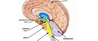
The center of coordination of movements is the cerebellum; functionally, it contains the body of the cerebellum, consisting of two hemispheres, the vermis and three pairs of peduncles.
In the implementation of voluntary movement, the main role of the cerebellum is to coordinate the fast and slow components of the motor act.
This becomes possible thanks to the bilateral connections of the cerebellum with the muscles and cerebral cortex. The cerebellum receives afferent impulses from all receptors irritated during movement (from proprioceptors, vestibular, visual, auditory, etc.).
Receiving information about the state of the motor system, the cerebellum influences the red nucleus of the brain, which sends impulses to the γ-motoneurons of the spinal cord, which regulate muscle tone. In addition, part of the afferent impulses through the cerebellum enters the cerebral cortex of the motor zone (precentral and frontal gyri).
The main function of the cerebellum is carried out at a subconscious level.
Efferent impulses from the cerebellar nuclei regulate proprioceptive stretch reflexes. During muscle contraction, the proprioceptor of the synergist muscles and antagonist muscles are excited. Normally, the transformation of voluntary movement into a complex reflex does not occur due to the inhibitory influence of cerebellar impulses. Therefore, when the cerebellum is damaged, disinhibition of segmental proprioceptive reflexes is manifested by movements of the limbs like ataxia.
The cerebellum has many afferent and efferent connections.
Posterior spinocerebellar tract (Flexig's tract). The first neuron is located in the spinal ganglion; the axon in the dorsal root, through the dorsal horn, approaches the cells of Clark's column. The fibers of these second neurons are directed to the outer layers of the posterior part of the lateral funiculus on their side, ascend along the entire spinal cord and, at the level of the medulla oblongata, as part of the inferior cerebellar peduncle, enter the cerebellar vermis.
This pathway is referred to as the posterior spinocerebellar tract. In the cortex of the cerebellar vermis there is a third neuron that contacts the piriform neurons of the cerebellar hemisphere cortex. The axons of the latter go to the dentate nucleus. The fibers of this fifth neuron are part of the superior cerebellar peduncle.
The right and left superior cerebellar peduncles cross - Wernicke's cross - and end at the red nucleus cells of the opposite side. The axons of the cells of the red nucleus are immediately directed to the opposite side of the midbrain and form a ventral decussation in the tegmentum of the midbrain - the decussation of Forel, pass as part of the lateral cord of the spinal cord, and reach the cells of the anterior horns.
The set of axons of the cells of the red nucleus is called the Monakov bundle.
Govers' anterior spinocerebellar tract. The first neuron is located in the spinal ganglion, the second neuron is a cell of the dorsal horn, but its axons move to the opposite side and go up the spinal cord, in the anterior part of the lateral cord, pass through the medulla oblongata, the pons of the brain, at the level of the superior medullary velum they pass to the opposite side side and as part of the superior cerebellar peduncle reach the cells of the cerebellar nuclei.
The cerebellum receives afferent proprioceptive impulses not only along the Flexig and Gowers pathways, they also arrive along the axons of the cells of the nuclei of the thin and cuneate fasciculi, some of which go through the lower cerebellar peduncles to its vermis.
In addition, the axons of the cells of the vestibular nuclei go to the cerebellum as part of the inferior peduncle - mainly from the vestibular lateral nucleus of Deiters, they end in the nucleus of the cerebellar clivus.
The fibers of the cells of this nucleus, as part of the upper and, possibly, lower cerebellar peduncles, approach the cells of the reticular formation of the brain stem and the vestibular lateral nucleus, from which the conductors form the descending tracts - vestibulospinal and reticular-spinal, ending at the cells of the anterior horns of the spinal cord.
This pathway regulates the balance of the body.
From the cerebellum, through the vestibular lateral nucleus, connections are established with the nuclei of the oculomotor nerves (as part of the medial longitudinal fasciculus).
The cerebellum has numerous connections with almost all lobes of the brain. There are 2 massive beams.
The fronto-pontine-cerebellar is a collection of axons of cells predominantly in the anterior sections of the superior and middle frontal gyri. In the depths of the lobe, they are collected in a compact bundle and form the anterior leg of the internal capsule.
Then the cerebral peduncles pass through the base and end with a synapse at the cells of the pons. The axons of the second neurons move to the opposite side of the pons and, as part of the middle cerebellar peduncle, enter its hemisphere, contacting the cells of the cerebellar cortex. The processes of these neurons approach the dentate nucleus. The fibers of the dentate nucleus cells in the superior cerebellar peduncle reach the red nucleus of the opposite side and carry impulses along the reticular-spinal tract that regulate human posture in an upright position, in particular standing and walking.
Occipitotemporal ponsocerebellar tract - its first neurons are located in the cortex of the occipital and temporal lobes; their axons gather in the subcortical white matter, then, as part of the posterior femur of the internal capsule, they go at the base of the midbrain to the pons nuclei of their side.
The axons of the pontine cells move to the opposite side and reach the cerebellar cortex along the middle peduncle. The fibers of these cells approach the dentate nucleus, which has connections with the brain stem. With the help of these tracts, coordination of the cerebellum with the organs of vision and hearing is ensured.
Ultimately, the existing decussations of the cerebellar afferent and efferent systems lead to homolateral communication of one hemisphere of the cerebellum and the limbs.
When the cerebellar hemisphere is damaged, disorders of its function occur on the same half of the body. Lesions in the lateral funiculus of the spinal cord also cause cerebellar disorders on its half of the body. The cerebral hemispheres are connected to the opposite hemispheres of the cerebellum.
Therefore, when the cerebral hemispheres or the red nucleus are damaged, cerebellar disorders are detected on the opposite side.
Definition
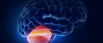
Purkinje cells are named after the Czech scientist who first discovered and described them, Jan Purkyně. The shape of the cell resembles a pear. Several dendrites extend from it, which in turn send out numerous branches with the formation of thousands of synapses. Purkinje cells are a cortical layer of the cerebellum, a section that is located below the occipital region of the brain.
: control of motor coordination, maintaining balance. Thanks to this small area (10% of the total volume) of the brain, a person performs all movements: plays football, holds cutlery and brings it to his mouth, writes written signs with his hand, types on the keyboard, moves his eyes, following objects in the outside world.
Important Soothing herbal teas: what to choose?
You can appreciate the significance of this brain structure by imagining how carefully movements must be synchronized in order for them to be precise, coordinated and smooth. The cerebellum adjusts the activity of skeletal muscles in order to achieve targeted precision of movement - up to a millimeter (in the spatial plane), up to a fraction of a second (in the time frame).
The cerebellum controls the movements of the head, limbs, body, eyes, while simultaneously receiving feedback sent by the nerve endings of the peripheral. By comparing the desired and actual results, he corrects motor activity. All processes occur instantly and unnoticed by humans.
THE CONCEPT OF ATAXIA, TYPES OF ATAXIA
Ataxia is a form of erratic movement, taxis - from the Greek. order, a- negation. It occurs when there is a violation of the coordination of the action of muscle groups - agonists (directly carrying out the movement), antagonists (in some phase counteracting the agonists), synergists (helping the work of either agonists or antagonists).
Movements lose coherence, accuracy, smoothness, proportionality and often do not achieve the goal. Such a patient's muscle strength remains sufficient, he has sufficient strength, he has no paresis.
The pathogenetic essence of ataxia consists of:
Violation of reciprocal innervation. The mechanism of reciprocal (coupled) inhibition of spinal motor centers is as follows: the axons of receptor cells (in the spinal ganglia) in the spinal cord are divided into branches, some of them excite motor neurons of the flexor muscles, and others contact with intercalary cells, which have an inhibitory effect on extensor muscle cells.
The complex integrative function of this mechanism also involves cerebellar impulses.
2. Termination of proprioceptive signaling (from muscle spindles, Golgi tendon bodies) along one or another ascending afferent pathway. Information about the degree of muscle tension at any given moment and about the results of the adaptive effects of functional systems ceases to be received.
The side of the motor function that is referred to as reverse afferentation is upset.
Types of ataxia.
1. Sensitive ataxia is associated with simultaneous suffering in coordination of movements and muscle-articular sensation. With severe ataxia in the upper limb, it is difficult to perform the simplest actions. At rest, involuntary movements are sometimes observed in the fingers of the hand, reminiscent of athetosis - pseudoathetosis. Coordination of movements is also impaired in the lower extremities, which is confirmed by missed hits and jerking movements when performing the heel-knee test.
Muscle tone in the affected limbs is reduced in both the flexor and extensor muscles. In a standing position, staggering is noted, especially when performing the Romberg test.
Movement becomes uncertain, the patient walks with his head down, controlling the act of walking with the help of vision. Thus, sensitive ataxia is always combined with a disorder of deep sensitivity and functional separation of individual segments of the limbs from the higher areas of the brain. Another characteristic feature of this type of ataxia is that it intensifies when the control of the visual analyzer is turned off.
Sensitive ataxia with damage to the posterior cords of the lower half of the spinal cord (for example, with syphilis, funicular myelosis - B12) may be accompanied by the disappearance of deep reflexes in the lower extremities, which is explained by degeneration of the collaterals of the thin bundle fibers, which are the afferent part of the deep reflex arc.
Cerebellar ataxia.
Associated with damage to the cerebellar systems. Considering that the cerebellar vermis takes part in the regulation of contraction of the muscles of the trunk, and the cerebral cortex of the distal limbs, two forms of cerebellar ataxia are distinguished.
- Static-locomtor - when the cerebellar vermis is damaged, mainly standing and walking are disrupted.
The patient stands with his legs wide apart and sways. When walking, the body deviates to the sides, the gait resembles the gait of a drunk. Turning is especially difficult. Deviations when walking are observed towards the cerebellar lesion. Stability is tested in the Romberg position.
If the cerebellar structures are damaged, the patient in this position sways in the appropriate direction; with staggering in the anteroposterior direction - characteristic of damage to the anterior parts of the cerebellar vermis.
The patient's walking in a straight line is examined, as well as the flank gait—stepping movements to the side. At the same time, pay attention to the clarity of the step and the ability to quickly stop in case of a sudden command. When the cerebellar structures are damaged, the combination of simple movements that make up a sequential chain of complex motor acts is disrupted. This is referred to as asynergy and is determined using the Babinski test.
- Dynamic ataxia - with it, the performance of various voluntary movements of the limbs is impaired.
This type of ataxia mainly depends on damage to the cerebellar hemispheres. This is detected when studying the movements of the upper extremities, for example, when performing the finger-to-toe, heel-knee, test for diadochokinesis, etc.
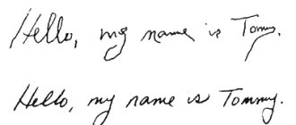
In addition to disruption of these tests with movements of the limbs, when the cerebellum is damaged, speech is disrupted - as a result of incoordination of the speech motor muscles, speech loses smoothness, becomes explosive, stress is placed on the wrong syllables - scanning speech; The handwriting changes - the handwriting becomes uneven, large - megalography.
Nystagmus is observed - a rhythmic twitching of the eyeballs when looking to the sides or up - a kind of intention tremor of the oculomotor muscles. When the cerebellar structures are damaged, the plane of nystagmus coincides with the direction of voluntary movements of the eyeballs - when looking to the side, the nystagmus is horizontal, when looking up and down - vertical.
Sometimes nystagmus is congenital. Such nystagmus is usually present not only when the eyeballs are abducted to the sides (with tension), but also when looking straight (“spontaneous nystagmus”).
When the cerebellar systems are damaged, muscle tone may change.
The most common symptom is muscle hypotonia: the muscles become flabby, flaccid, and joint hypermobility is possible. This may reduce deep reflexes.
Coordination of movements is impaired when the frontal and temporal lobes and their conductors are damaged. In this case, walking and standing are disrupted, the torso deviates posteriorly and in the direction opposite to the lesion. Missing in the arm and leg (hemiataxia) is detected. With this type of coordination disorder, other signs of damage to the corresponding lobes of the cerebral hemispheres are also found.
Cortical ataxia. Coordination of movements is impaired when the frontal and temporal lobes and their conductors are damaged. In this case, walking and standing are disrupted, the torso deviates posteriorly and in the direction opposite to the lesion. Missing in the arm and leg (hemiataxia) is detected. With this type of coordination disorder, other signs of damage to the corresponding lobes of the cerebral hemispheres are also found.
4. Vestibular ataxia - occurs when the function of the vestibular analyzer, in particular its proprioceptors in the labyrinth, is impaired.
With it, the balance of the body is upset; while walking, the patient deviates towards the affected labyrinth. Systemic dizziness, nausea, and horizontal rotatory nystagmus are characteristic. Hearing may be impaired on the side of the affected labyrinth.
Thus, a disorder of coordination of voluntary movements is observed with damage to both the cerebellum itself and the conductors through which impulses from the muscles, semicircular canals of the inner ear and cerebral cortex are brought to it and taken away from the cerebellum to the motor neurons of the brain stem and spinal cord.
Patients with damage to the cerebellar systems usually do not show any pathological manifestations at rest. Various types of incoordination appear in them only when muscles are tense.