Material and methods
In the period from 2014 to 2015, in the Department of Neurosurgery No. 2 of the National Medical Center named after.
N.I. Pirogov treated 65 patients (43 (66%) men and 22 (34%) women aged 32 to 68 years) with glial tumors of the brain. During the initial neurological examination, motor deficits of varying severity were detected in 46 (71%) patients (6 patients were diagnosed with hemiparesis with a decrease in strength to 1 point, in 15 - to 2-3 points, in 25 - to 4 points). 17 (26%) patients were diagnosed with aphasia (12 of them had a combination of speech and motor disorders, 5 had isolated speech disorders). In 40 (62%) patients, tumors were localized in the frontal and temporal lobes. Isolated damage to one lobe was present in only 9 patients: insular in 4, frontal in 2, and temporal in 3. In 37 (58%) patients, the tumor affected the left hemisphere, and in 24 (36%) - the right. In 4 (6%) patients, the tumor spread to both hemispheres of the brain (Table 1).
Table 1. Localization of glial tumors in operated patients
64 patients underwent primary surgery. One patient was operated on 2 times during the observation period: initially for a tumor of the frontal, temporal and insular lobes on the left (involving Broca's area and Wernicke's area) and again 16 months later due to continued growth of the tumor. Both operations were carried out using the asleep-wake-asleep protocol. Control of the radicality of tumor removal (MRI with contrast) was performed in all patients on the 1st day after surgery (Fig. 1).
Rice. 1. Images of preoperative planning at the navigation station. MRI brain, T1-weighted image with contrast. a - frontal; b - sagittal; c — axial projection. A space-occupying formation (1) of the left temporal and parietal lobes of the brain (in the projection of the superior temporal, angular and supramarginal gyri) with fuzzy, uneven contours, with pronounced perifocal edema, intensively accumulating contrast agent is determined. Arrows indicate the motor cortex (2), pyramidal tracts (3) and speech areas (4); d — 3D model of the brain with projection of the FZ and tumor.
Along with MRI in standard modes, 50 (77%) patients underwent fMRI at the stage of preoperative diagnosis due to the presence of speech disorders in 17 patients and involvement of the internal capsule in 4 patients (in addition, the group included 9 patients who had no manifestations of aphasia , but the risk of its development after surgery was high). The fMRI technique is based on recording regional hemodynamic changes that occur when the cerebral cortex is activated in response to specific stimulation (alternating phases of rest and motor, mental or other activity). Based on the results of the examination, the relationship of the tumor with the motor pathways, Wernicke’s and Broca’s areas was established in these patients (Fig. 2 and 3).
Rice. 2. Images from the navigation station MRI of the brain, T1-weighted image with contrast, sagittal (a) and axial (b) projections: a volumetric formation is determined (1) in the right temporal and parietal lobes of the brain, with fuzzy uneven contours, with pronounced perifocal swelling, intensively accumulating contrast agent. The pyramidal tracts (2) passing anterior to the tumor are indicated; c — 3D model of the spatial relationship of the tumor (1) and pyramidal tracts.
Rice. 3. Preoperative and control images MRI of the brain, T2-weighted image, sagittal and frontal projections. Preoperative T2 (a) and Flair (b) images: a picture of a space-occupying lesion in the left frontal lobe involving the premotor area and the anterior parts of the precentral gyrus; postoperative T1 (c) and Flair (d) images on the 1st day after surgery. The tumor was completely removed.
In patients with frontotemporal tumor localization in the dominant hemisphere and proximity to speech centers, in order to reduce the risk of the appearance or increase of postoperative motor, sensory and amnestic aphasia, tumor removal was carried out with intraoperative awakening (according to the asleep-awake-asleep protocol) (16 patients). For this purpose, a team of anesthesiologists and neurosurgeons had a conversation with the patient on the eve of the operation, during which all stages of the future surgical intervention were considered. Particular attention was paid to the moment of awakening in the operating room. The anesthesiologist sought to ensure that the patient had a conscious understanding of what he would see in the operating room. It was discussed what sensations he would experience when he woke up, what the peculiarities might be when waking up. In addition, testing with pictures was carried out, during which the patient named and described the objects depicted, and also practiced mental counting, memorizing a sequence of words, and reading. The criteria for selecting patients for the asleep-awake-asleep protocol were the absence of pronounced mnestic disorders, i.e., the ability to enter into full verbal contact, the absence of pronounced mental lability, and the absence of gross motor and sensory speech deficits. During testing, obvious mnestic disturbances or pronounced sensorimotor aphasia were revealed in 5 patients, as a result of which they decided to refrain from performing a conscious craniotomy.
During the operations, IONM was performed using the Natus Xltek protektor apparatus. The purpose of IONM is to dynamically monitor spontaneous and evoked bio-electrical activity of the brain (motor evoked potentials). During neurophysiological monitoring, motor evoked potentials were assessed using transcranial electrical stimulation based on the activation of pyramidal cells of the corticospinal tract (Brodmann area 4 of the precentral gyrus). Stimulating electrodes were located in the projection of points C3, C4, according to the international scheme 10-20 [3]. Depending on the surgical approach, different electrode placement options were used. To assess the conduction of electrical impulses along the pyramidal tracts, all patients underwent transcranial electrical stimulation, regardless of the severity of the neurological deficit. Motor responses were recorded using needle electrodes installed in muscles ( m .
Orbicularis Oculis , m . Orbicularis Oris , m . Deltoideus , m . Thenari / Hypothenari , m . Rectus Femori , m . Tibialis Anterior
). Transcranial stimulation (Fig. 4) was performed in single trains of 4 short high-frequency stimuli (500 Hz). Tension parameters were selected individually until representative M-responses were obtained from all tested muscles. Registration of M-responses was carried out under conditions of amplifier sensitivity of 50 μV/div with a notch filter of 50 Hz. During the operation, motor responses were recorded on both sides. The difference in amplitude and latency varied depending on the initial neurological deficit and options for installing stimulating electrodes.
Rice.
4. Monitoring of the pyramidal tracts (transcranial stimulation) during removal of glioma of the lower parts of the precentral gyrus of the frontal and lower parts of the postcentral gyrus of the parietal lobe of the left hemisphere. The test muscles were chosen. Deltoideus (a), m. Thenari (b, c), m. Tibialisanterior(d, d). The orange arrow indicates the M-response obtained from the muscle being tested. The red arrow indicates a series of stimuli. Motor cortical mapping (Fig. 5) was used in 50 patients with involvement of the precentral gyrus or premotor cortex. For this purpose, the technique of direct stimulation of the cortex with a bipolar electrode was used in packs of 4 stimuli with an internal frequency of 500 Hz and a stimulus strength of up to 25 mA with registration of motor responses from the tested muscles of the contralateral side.
Rice. 5. Motor mapping of the middle and lower parts of the precentral gyrus before encephalotomy in a patient undergoing removal of a glioma of the frontotemporal localization. The test muscles chosen were m. Longinalis superior (a), m. Orbicularis oris (b), m. Deltoideus (c), m. Thenari (g), m. Tibialis Anterior (e), m. Rectus.
Patients ( n
=16) with the tumor localized in the projection of the speech zones, tumor removal was carried out according to the asleep-awake-asleep protocol [4]. After depression of consciousness by intravenous administration of a propofol solution at a dose of 1.0-1.5 mg/kg, analgesia with a fentanyl solution at a dose of 2.5-3.0 mcg/kg and muscle relaxation with rocuronium at a dose of 0.7 mg/kg, tracheal intubation was performed and mechanical ventilation began in normal ventilation modes. Control of the patient's level of consciousness was ensured by inhalation of desflurane in doses of 0.3-0.6 MAC under conditions of monitoring the depth of depression of consciousness (target BIS values were 50-60). To assess the level of muscle relaxation, a neuromuscular conduction monitor using the TOF technique was used. The skin incision line was infiltrated with a solution of 0.2% ropivocaine in total doses not exceeding 300 mg, taking into account regional anesthesia. Anesthesia of the dura mater (DRM) was carried out by applying pads with lidocaine. After craniotomy was performed and the level of restoration of neuromuscular conduction, TOF 0.9 or more, was achieved, the supply of desflurane was stopped, the patient was awakened and extubated. Mapping of the FZZ of the brain was performed in 2 stages. Before awakening the patient, the motor areas bordering the formation were verified using direct electrical stimulation of the cortex. After awakening and extubation, verbal contact was established, and then speech zones were mapped (Fig. 6). For intraoperative verification of Wernicke's area, tasks were selected to repeat a series of frequency words in threes, which allows diagnosing disorders of phonemic hearing and the volume of auditory-verbal perception (as well as the pronunciation side of speech), testing with naming pictures, reading and counting. The intraoperative speech testing procedure was practiced with patients on the eve of surgery; only tasks that were available for stable, error-free execution were included in the test set. During stimulation of the cortex with a bipolar stimulator in Broca's and Wernicke's areas with rhythmic bursts of 4 stimuli with a strength of 15-20 mA, various types of speech impairment were recorded in some patients: speech-arest (5 patients), elements of motor aphasia, literal paraphasia with replacement of sounds by acoustic proximity and verbal paraphasia with substitution of words based on acoustic proximity, characteristic of phonemic hearing impairment (7).
Rice.
6. Intraoperative mapping of speech zones. Intraoperative photo (a). The trepanation window is formed in the left frontal-temporo-parietal region. The dura mater has been opened. After the patient woke up, mapping of speech zones was performed. Broca's area (22, 21, 25 - yellow outline), Wernicke's area (16-18, 20 - green outline) and the lower parts of the precentral gyrus (22, 26 - blue outline) were identified. On fMRI (b), the speech centers are indicated in green; the tumor is localized in the left insula. After verification of Broca's and Wernicke's areas, the tumor was removed with neurological and neurophysiological control of the preservation of speech functions and motor tracts (Fig. 7). In 2 patients, after mapping of speech zones, motor restlessness and emotional lability developed, which required resumption of endotracheal anesthesia (average time awake was 30±12 minutes).
Rice. 7. Intraoperative photo. Stage of tumor removal with intraoperative mapping of speech zones. At the awake stage, mapping of speech zones was carried out. The marks indicate Wernicke's area, discovered during direct stimulation of the cortex during the awakening stage. The green outline indicates the projection of the tumor.
In the remaining 14 patients, testing continued throughout the entire tumor removal (awake time was 120±20 minutes). After tumor removal, patients were again anesthetized, intubated with brief muscle relaxants, and surgery was completed under general anesthesia with continued monitoring via transcranial stimulation.
In 11 (17%) patients with tumor spread into the internal capsule, stimulation of the motor pathways was performed (Fig. 8). The stimulation threshold was decreased stepwise from 10 to 5 mA (according to the literature, a change of 1 mA corresponds to a change of distance of 1 mm). Changing the stimulus strength to a minimally representative one made it possible to remove the tumor in close proximity to the internal capsule. In 5 (8%) patients during tumor removal due to the appearance of a motor response to a stimulus strength of 5 mA, removal was stopped due to the extremely high risk of neurological deficit.
Rice. 8. Removal of a frontal lobe tumor with intraoperative mapping of motor areas. a — intraoperative photo: the stage of tumor removal of the left frontal lobe, the arrow indicates a bipolar stimulator used during mapping of motor pathways in the white matter. On fMRI, arrows indicate the motor representation of the arm (b) and leg (c), the tumor is located in close proximity to these zones and is in contact with the pyramidal tracts.
Regardless of the tumor location and the chosen anesthesia protocol (including awake craniotomy), all patients underwent background monitoring of transcranial motor evoked potentials. In 8 patients, the amplitude of the M response decreased by more than 50% during tumor removal. When assessing the postoperative neurological status, all of them were included in a group of 11 patients whose motor deficit increased. In 21 patients, a temporary decrease in amplitude was noted (by no more than 40%), which is apparently explained by the presence of a brain displacement effect after tumor removal or the effect of short muscle relaxants at the stage of re-intubation according to the asleep-awake-asleep protocol.
In 43 (66%) patients, the histological diagnosis corresponded to glioblastoma (WHO Grade IV), in 7 (11%) - fibrillary astrocytoma (WHO Grade II), in 6 (9%) - oligodendroglioma (WHO Grade II). Anaplastic astrocytoma (WHO Grade III) was verified in 9 (14%) patients, of which 2 patients were re-operated using mapping due to continued tumor growth.
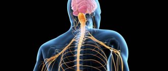
Practical work No. 2 Topic: Study of the structure of the brain.
Practical work No. 2
Topic: Study of the structure of the brain.
Target:
get acquainted with the departments and parts of the brain, structural features and functions performed.
Equipment:
table “Brain”, textbook, video and presentation “Human Brain”.
Progress:
1.
Using paragraph
50
, fill out the table “Brain”
| № p/p | Divisions and parts of brain regions | Structural features | Functions performed |
| 1 | Trunk: medulla oblongata; bridge; | ||
| 2 | midbrain; | ||
| 3 | diencephalon | ||
| 4 | Cerebellum | ||
| 5 | Forebrain (left and right hemisphere) |
2.
(Write down the conclusion) Conclusion: Brain - ………………………………………………………………
Practical work No. 2. EXAMINATION.
Topic: Study of the structure of the brain.
Target:
get acquainted with the departments and parts of the brain, structural features and functions performed.
Equipment:
table “Brain”, textbook, video and presentation “Human Brain”
Progress:
1. Using paragraph 50, fill out the “Brain” table.
| № p/p | Divisions and parts of brain regions | Structural features | Functions performed |
| 1 | Trunk: medulla oblongata; bridge; | – gray matter is located in separate clusters of nuclei; – the place where the nerve fibers are located; | reflex arcs pass through the nuclei: cough reflex; sneezing reflex; tear reflex, etc.; in the nuclei there are centers responsible for the act of swallowing, the functioning of the digestive glands, the regulation of respiration, the activity of the heart and blood vessels; conduct impulses to the cerebral cortex, cerebellum, medulla oblongata and spinal cord; |
| 2 | midbrain; | provides a reflex change in the size of the pupil, the curvature of the lens depending on the brightness of the light; | |
| 3 | diencephalon | conducts impulses to the cerebral cortex from skin receptors and sensory organs; responsible for the feeling of thirst and hunger; for maintaining a constant internal environment, for the work of the endocrine glands and the autonomic nervous system | |
| 4 | Cerebellum | consists of hemispheres and a worm connecting them; the surface has numerous transverse depressions - grooves and narrow elevations between them - convolutions. This is the cerebellar cortex | takes part in the coordination of movements, making them accurate and purposeful; provides balance to the body |
| 5 | Left hemisphere | Oral and written communication; information analysis; generalization, decision making | |
| Right hemisphere | Imaginative thinking; musical and artistic creativity; perception of music |
2. Conclusion: Brain -
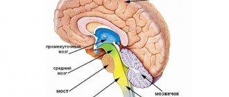
is the main regulator of all body functions and ensures higher nervous activity in humans. The brain consists of five sections: the medulla oblongata, the midbrain (sometimes another section is distinguished in the midbrain - the pons, or pons), the cerebellum, the diencephalon, and the cerebral hemispheres.
results
At the stage of neurophysiological mapping, the results obtained were compared with the MRI picture of the representation of functional zones, synchronized at the navigation station (Table 2).
Table 2. Results of direct electrical stimulation of the cortex in the FZZ area and white matter near the pyramidal tract
When comparing the mapping results with fMRI and tractography data, in 12 (18%) patients a wider representation of speech zones was revealed, which, in our opinion, is explained by the error in performing test tasks during fMRI and the existing preoperative neurological deficit (elements of sensory aphasia). All patients in On the 1st day after surgery, contrast-enhanced MRI control was performed (Figs. 9 and 10). Total tumor removal (100% of the tumor) was achieved in 39 (60%) patients (see Fig. 1, 9, 10), subtotal (80-95% of the tumor) - in 16 (24%), 10 (16%) patients partial removal of the tumor was performed (the tumor had invaded the internal capsule, speech centers or primary motor centers). At the time of discharge, the patients' condition on the Karnofsky scale was 80±10 points. The patient's hospital stay after surgery was 7±1 days. In 47 (72%) patients, there was no deterioration in the neurological status. In 18 (28%) patients in the early postoperative period, an increase in neurological deficit was noted in the form of an increase in paresis by 1-2 points or aggravation of speech disorders: in 11 patients the motor deficit increased by 1 point, in 7 patients the severity of aphasia increased (the group included 5 patients who did not undergo awake, and 2 patients who underwent surgery with mapping of speech zones). In 8 patients, partial regression of paresis was noted within 7 days after surgery (by an average of 1 point). When tracking the follow-up after 4 months in 18 patients in the group with increased neurological deficit, 10 of them showed regression of the neurological deficit. After 4 months, neurological deficit at the postoperative level remained in 5 patients: 3 had hemiparesis of 2-3 points, more pronounced in the arm, 3 had elements of motor aphasia, 2 had hemiparesis of 3-4 points, more pronounced in the leg. Thus, persistent neurological deficit after 4 months persisted in 8 (12%) patients, which corresponds to world literature data.
Rice. 9. Preoperative and control images: MRI of the brain, T1-weighted image with contrast, axial projection. a — preoperative image: a picture of a space-occupying formation of the left insular lobe, intensively accumulating contrast agent; b — postoperative image on the 1st day after surgery: the tumor was completely removed, there was no accumulation of contrast agent.
Rice. 10. a — preoperative images, T2-weighted image in axial projection before surgery: a volumetric formation of the left frontal lobe is determined (in the projection of Broca’s area) with pronounced perifocal edema; b — postoperative images, T-weighted image on the 1st day after radical removal of the tumor of the left frontal, insular and temporal lobes of the brain: there are no signs of residual tumor tissue.
Structure and functions of the brain (medulla oblongata and hindbrain) - NERVOUS SYSTEM - 9th grade
Lesson objectives:
educational: study the structure and functions of the medulla oblongata, pons and cerebellum; develop skills in conducting simple experiments to identify the functions of the medulla oblongata and cerebellum;
developmental: develop the ability to use various sources to obtain knowledge; compare and summarize knowledge;
educational: create conditions for the formation of a negative attitude towards alcohol consumption; healthy lifestyle skills.
Lesson type: combined.
Teaching methods: practical, partially exploratory.
Basic terms and concepts: medulla oblongata, hindbrain, pons, cerebellum.
Materials and equipment: tables on anatomy, physiology and hygiene, model and dummies of the brain, electronic learning tool “Man and his health”.
List of knowledge, skills and abilities that are expected to be developed in this lesson:
- characterize the role of the brain in the life of the body, the structure and functions of the human medulla oblongata and hindbrain;
- recognize the pons and cerebellum of the hindbrain.
- Checking homework
Ø Teacher's activities
Explains task 1 (for everyone).
The structural components of the spinal cord are given: 1 - white matter; 2 - gray matter; 3 - posterior roots of nerves; 4 - anterior nerve roots. Consider the structures and functions listed below among them.
A - sensory neurons, B - pathways; B - motor neurons, D - neuron bodies; D - contact of sensory and motor neurons; E - impulse transmission to muscles; F - impulse transmission from the receptor to the brain; 3 — impulse transmission “up” and “down”; I is the conductor function; K - reflex function.
Explains task 2 (for those who completed the previous task first).
Read the list of parts of the nervous system. A - motor neurons; B - sensitive neurons; B - anterior root of the spinal nerve; G - posterior root of the spinal nerve; D - spinal cord.
Determine and write down in the form of a code, when which neurons and parts of the human nervous system are damaged, the following movement disorders occur: 1) the leg moves, but does not feel pain; 2) the leg does not move, but feels irritation and pain; 3) the leg has lost sensation and is paralyzed; 4) loss of sensitivity and complete paralysis of the body below the waist.
Ø Student activities
Complete tasks.
Suggested correct answers to task 1: 1) B, 3, I; 2) G, D, K; 3) A, F, I; 4) B, E, I.
Suggested correct answers to task 2: 1) B, D; 2) A, B; 3) A, B, C, D; 4) D.
- Organizing time
Ø Teacher's activities
Communicates the goals and objectives of the lesson. Introduces the main issues that will be addressed in the lesson.
III. Learning new material
Ø Teacher's activities
Considers the following questions with students: comparative analysis of brain models of chordates, finding similarities and differences in the structure of the brain of different classes of animals. Clarifies the role of the brain in the life of the body.
Gives tasks:
— read the material “Brain” § 10 of the textbook;
- determine where the brain is located, what its parts are;
- find the parts of the brain on the model and tables, in the drawing of the electronic device.
Describes the structural features of the medulla oblongata (location, size, distribution of gray and white matter).
Ø Student activities
They remember everything they remember about the structural features of the brain in animals of different classes. Write down the names of the parts of the brain in workbooks.
Ø Teacher's activities
Gives the task: to identify during experimental work some functions of the medulla oblongata.
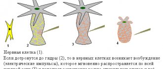
Experiment 1. Make several swallowing movements in a row. What are you observing? (Make sure that in the absence of a stimulus it is impossible to make a swallowing movement.)
Experience 2. Take two or three quick and deep breaths and exhale, observe your condition. (Make sure that the next breath is involuntarily delayed.) Concludes: “During the experiment, we found out what vital functions the medulla oblongata performs. However, these are just a few of them.”
Ø Student activities
Carry out the suggested experiments.
Ø Teacher's activities
Suggests reading the material “Medulla oblongata” (§ 10 of the textbook), the text of the electronic tool, to find out the functions of the medulla oblongata.
Ø Student activities
Write down the formulations of new concepts in workbooks: nuclei, centers.
Ø Teacher's activities
He suggests working with Figure 12 (§ 10 of the textbook). Asks to define what is included in the concept of “hindbrain”, and to find the pons and cerebellum in the drawing of an electronic device and a model of the brain.
Ø Student activities
Write down in notebooks: “The hindbrain consists of the pons and cerebellum.”
Ø Teacher's activities
Invites students to work in pairs and study the structure and functions of the pons and cerebellum.
Ø Student activities
They work in pairs. One student studies the structure and functions of the pons, the second - the cerebellum, then there is mutual learning in pairs.
Ø Teacher's activities
Gives the task: to identify during experimental work some functions of the cerebellum.
Experiment 1. Close your eyes, extend your right hand forward with your index finger extended, and clench the remaining fingers into a fist. After this, touch the tip of your nose with the tip of your index finger. Did you succeed?
Experiment 2. Work in pairs. One (the subject) bends his arm at the elbow. Another (experimenter) grabs his forearm near the hand and invites the subject to pull his hand towards himself, overcoming resistance. Then, unexpectedly for the subject, the experimenter releases his hand. What's happening to the hand?
Ø Student activities
Under the guidance of the teacher, they perform an experiment.
Ø Teacher's activities
Asking questions. When performing the finger-nose test, about 30 muscles are involved, sequentially bending and unbending, but the movement is smooth, why? You know from physics lessons that an unexpectedly released hand, due to inertia, should have hit the shoulder, why didn’t this happen?
Ø Student activities
Answer questions.
Suggested Correct Answer: The cerebellum controls precise, coordinated movements and balance.
Ø Teacher's activities
Describes the effects of alcohol on the cerebellum.
Ø Student activities
Listen to the teacher's story.
- Reinforcing the material learned
Ø Teacher's activities
Invites students to fill out the table and compare departments by structure and function.
| Divisions of the brain | Structure | Functions |
| Oblong | ||
| Bridge | ||
| Cerebellum |
Ø Student activities
Fill out the table.
Ø Teacher's activities
Asks students to answer the questions:
- What similarities did you see in the structure of all departments?
- How does the structure of the cerebellum differ from the structure of the medulla oblongata and the pons?
- What function is missing in the cerebellum, but is present in the medulla oblongata and pons?
- What general function do the spinal cord and medulla oblongata perform?
- Why shouldn’t small children be given small objects to play with?
- A patient suffering from hypertension has impaired coordination of movements. However, his mental abilities were not affected. Dysfunction of which part of the brain led to this result?
- Why do drunk drivers often cause accidents?
- The child has a high fever and cough accompanied by vomiting. The parents consulted a doctor with a suspected food problem. And after examining the patient, the doctor diagnosed a respiratory disease. Why is a child’s severe cough often accompanied by vomiting?
Ø Student activities
Answer questions.
Suggested correct answers:
- There is gray and white matter.
- The cerebellum has hemispheres and a vermis, the hemispheres are covered with cortex.
- Unlike the cerebellum, the medulla oblongata and the pons have a conductive function.
- Reflex.
- Children can swallow small objects, since swallowing is an unconditioned reflex and its centers are located in the medulla oblongata.
- Cerebellum.
- Alcohol primarily affects the cerebellum, causing loss of smoothness and coordination of movements.
- The centers of cough and vomiting are located close to each other in the medulla oblongata.
- Reflection
Ø Teacher's activities
Invites students to think and answer the following questions:
— What did I learn about my body in class today?
— How can this knowledge help me protect my own health?
— Drinking alcohol, even in small doses, is dangerous because...
— How do you evaluate your activities in class?
Ø Student activities
Answer questions.
- Homework
Ø Teacher's activities
Offers you to complete the following task. Study the material in § 10 of the textbook, p. 31-32.
Creative tasks (optional):
— Make up a dialogue between the cerebellum and the medulla oblongata.
— Develop a “Lie Detector” game for the cerebellum or medulla oblongata.
Ø Student activities
Write down homework.
Discussion
The main goal of surgical treatment of brain gliomas is maximum resection of tumor tissue. Total or near-total removal of tumors, as well as obtaining a histological diagnosis, are key factors to improve the quality of life and increase the patient's life expectancy [5]. According to studies [6, 7], the average life expectancy of a patient after total resection of astrocytomas of I-II malignancy is on average 30 months longer than after subtotal resection (life expectancy varied from 61.1 to 90.5 months). In addition, adjuvant therapy (radiation, chemotherapy, immunotherapy) is more effective when the tumor size is small or the amount of residual tissue is small [8]. However, when the tumor is localized in the FZ of the brain, such as the motor cortex, speech areas, subcortical nuclei and internal capsule, the risk of developing neurological deficit with total removal reaches 30% [9]. Given the high risk of patient disability, it is extremely important to use all available medical technologies to perform the maximum possible tumor resection, with minimal risk of developing persistent neurological complications in the postoperative period. A detailed understanding of the relationship between the FZ and tumor tissue undoubtedly makes it possible to reduce the percentage of neurological deficits after surgery. The use of fMRI and tractography data during intraoperative navigation allows one to track the distance to the FZ in real time, but does not provide an idea of the state of functions and does not always allow one to prevent functional disorders. IONM has established itself as a method that defines the boundaries of the maximum possible functional tumor resection. Currently, neurophysiological control is actively developing and is widely used in neurosurgical practice.
The technique of intraoperative neurophysiological mapping of the cerebral cortex originates from epilepsy surgery in the first half of the twentieth century, when neurosurgeon W. Penfield and electrophysiologist H. Jasper [10] used stimulation of the cerebral cortex to localize the focus of epileptic activity. Subsequently, W. Penfield generalized the results of cortical stimulation data, obtaining an accurate representation of the motor function of different parts of the body. P. Merton and N. Morton were the first to use a non-invasive method for studying motor responses in 1980, introducing the concept of transcranial electrical stimulation. For a long time, locoregional anesthesia (pain relief based on a combination of local anesthesia along the skin incision line with the injection of a local anesthetic solution into the exit points of the sensory nerves innervating the scalp) made it possible to effectively solve the problem of pain relief for neurosurgical patients. Later, when endotracheal anesthesia was introduced into practice, this technique was forgotten and was used only in epilepsy surgery. However, in the 80s of the 20th century, thanks to the work of M. Berger and G. Ojemann [11], who introduced the concept of “cortical mapping” into surgical neuro-oncology, operations again began to include the stage of awakening the patient at the time of tumor removal. This method made it possible to prevent persistent neurological deficits in patients undergoing surgery for epilepsy. Over time, neurophysiological control techniques have improved, and different anesthesia protocols have been developed for conscious brain surgery.
According to T. Reithmeier et al. [12], the use of neurophysiological mapping of the cerebral cortex makes it possible to reduce the incidence of speech disorders after removal of intracerebral tumors by 2 times compared with the same operations without neurophysiological control - from 29 to 14%. N. Duffau et al. [13] when analyzing their material, found that the incidence of postoperative neurological disorders decreased from 17 to 6%. We managed to reduce the incidence of neurological deficits to 12%. The combination of neurophysiological monitoring with the use of the asleep-awake-asleep anesthesiological protocol makes it possible to more clearly monitor and prevent motor and speech disorders. Preoperative planning data (synchronization of fMRI with navigation MRI) does not always coincide with the data of neurophysiological mapping of the FZ. Mapping the cortex and marking functional and silent zones allows us to adjust and select the optimal area of encephalotomy with minimal risk of damage to the FZ. To directly monitor the state of the pyramidal tracts, the technique of direct subcortical stimulation is used. In this case, the strength of the stimulus is directly proportional to the depth to which it penetrates, and therefore to the distance to the motor pathways. Thus, 1 mA stimulus corresponds to approximately 1 mm [14]. Reducing the stimulus strength in the range from 10 to 3 mA makes it possible to remove the tumor as close as possible to the pathways with constant monitoring of their reactivity. According to the authors [15], this permissible distance is reduced from 8-10 to 2 mm. We used a range of 5-10 mm (5-10 mA). Subcortical stimulation was supplemented by transcranial and transcortical stimulation, which made it possible to monitor not only the reactivity of the conductors at the subcortical level, but also the continuity of the tract from the cortical level. At the same time, a decrease in the amplitude of M-responses by 50% or lower, contrary to the opinion of some authors [16], in our work did not always entail the appearance or increase of a neurological deficit. A decrease in reactivity during transcranial stimulation is explained in some cases by the effect of displacement after decompression and tumor removal, which creates an additional air gap between the cerebral cortex and the dura mater. Transcortical stimulation does not have these disadvantages. In turn, maintaining the reactivity of the pyramidal tracts at depth does not guarantee the prevention of vascular disorders near the internal capsule, which can also cause postoperative neurological deficit.
When evaluating each of the methods of preoperative planning and IONM, their inherent shortcomings are revealed, but the combination of these methods makes it possible to complement the accuracy of each of them and ultimately improve the functional outcome without sacrificing the radicality of tumor removal.
Today, IONM allows not only to prevent and detect intraoperative functional disorders, but also provides neurophysiological navigation. In combination with navigation planning in our work, this made it possible to optimize the access trajectory and perform total or subtotal removal of the FZD brain tumor with a satisfactory functional outcome in the majority of patients. Minimal or insignificant postoperative neurological deficit after subtotal tumor removal has made it possible to increase the number of patients referred for chemotherapy and radiation therapy. The main result of the work is that we were able to confirm: intraoperative mapping of the cortex and pathways with a dynamic change in stimulus strength makes it possible to obtain an idea of the location and proximity to the trajectory of the FZ intervention and stop tumor resection in time.
A comment
The work of the authors under the guidance of Dr. A.A. Zueva is devoted to the surgery of intracerebral tumors of functionally significant areas of the brain (both cortical areas and pathways using the example of the pyramidal tract). The authors have accumulated sufficient clinical material (65 patients), of which 14 operations with intraoperative awakening were performed. Neurophysiology was used (a combination of bipolar stimulation and transcranial evoked potentials). All patients underwent MRI control in the early postoperative period with an assessment of the extent of tumor resection; both early and 4-month follow-up were followed in all patients.
The dependence of current strength and pulse penetration depth is analyzed in detail, and modern Russian and foreign works on this topic are presented.
A small remark that does not reduce the value of the study is a small list of references, and the second thing to mention in the work, in addition to the pyramidal tract, is the need to map the associative conductors of the white matter.
The work is of scientific and practical interest; it should, of course, be supported and the authors wish further success in their work.
S.A. Goryainov, A.A. Potapov (Moscow)