The main task of the human body is to maintain a constant internal environment (i.e. homeostasis), as well as adaptation (i.e. adaptation) to the environment. These tasks are performed by 3 body systems: 1. The immune system, which is responsible for protecting against foreign genetic information. 2. The humoral (endocrine) system is responsible for the slow regulation of the activity of individual organs and tissues of the body. 3. The nervous system, which appeared later than the previous two; is responsible for the rapid and precise regulation of the activity of individual organs and tissues.
Content
- Functions of the nervous system
- NS departments
- Reflex as the basic principle of the nervous system
Neurophysiology views the nervous system as part of a living system that specializes in the transmission, analysis and synthesis of information, and neuropsychology as the material substrate of complex forms of mental activity, formed on the basis of the integration of various parts of the brain into functional systems.
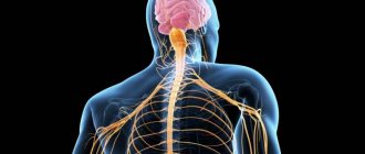
The nervous system (NS) is a set of anatomically and functionally interconnected nervous structures that provide regulation and coordination of the activities of the human body and its interaction with the environment.
The structural unit of the NS is a cell with a process ( neuron , or neurocyte). The nervous system is a collection of neurons that communicate with each other through synapses.
Functions of the nervous system
The nervous system occupies a special position among other body systems. It ensures the relationship of the organism with the environment . Receptors respond to any signals from the external and internal environment, converting them into streams of nerve impulses that enter the central nervous system. Based on the analysis of the flow of nerve impulses encoding information, the brain forms an adequate response.
Together with the endocrine glands, the nervous system regulates the functioning of all organs . This regulation is carried out due to the fact that the spinal cord and brain are connected by nerves to all organs through bilateral connections. The central nervous system receives signals from the organs about their functional state, and the nervous system, in turn, sends signals to the organs, correcting their functions and ensuring all vital processes - movement, nutrition, excretion, etc. The nervous system ensures coordination of the activities of cells, tissues, organs , organ systems. In this case, the body functions as a single whole.
The nervous system is the material basis of mental processes : attention, memory, speech, thinking, etc., with the help of which a person not only learns about the environment, but can also actively change it.
Thus, several functions of the nervous system can be distinguished: 1. Connects the body with the environment (perception and transmission). 2. Provides interaction between tissues of organs and body systems and their regulation.
Functions of the central nervous system
Among the main functions of the central nervous system are the following:
- Coordination . This is coordinated work between various organs and systems, which is ensured by the central nervous system. This includes all forms of various body movements, movement of the body in space, maintaining a certain position and posture, labor activity, as well as a certain number of adaptive general biological reactions.
- Integration _ This is the unification of all body functions. This function is divided into three types. Nervous - unification occurs due to the peripheral and central nervous systems. Humoral - functions in the body are combined mainly with the help of humoral factors. Mechanical - responsible for the performance of functions in the body in the presence of organ integrity (if damage or fractures are observed in any organ, the function is considered impaired).
- Correlation . This function provides interconnection between various individual functions, organs and systems.
- Regulation . This includes self-regulation, various types of reflexes, the formation of functional systems, which, in turn, provide a positive adaptive result associated with changes in the conditions of the internal and external environment in the body. The regulatory influence in the central nervous system can manifest itself in the form of triggering, corrective and trophic metabolic processes.
- Establishing and maintaining the relationship between the environment and the body.
- Labor and cognitive processes of the body. Such functions are responsible for the adequacy of the body in environmental conditions.
NS departments
Central nervous system and peripheral nervous system
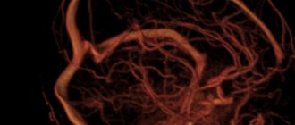
According to topographical principles, the nervous system is divided into central and peripheral. The central nervous system (CNS) includes those parts that are contained in the cranial cavity and the spinal canal, i.e. brain and spinal cord. The spinal cord is a tube with a small canal in the middle, surrounded by neurons and their processes. The brain is an extension of the spinal cord. The topographic boundary with the spinal cord is a plane passing through the lower edge of the foramen magnum. The average brain weight is 1400 g with individual variations from 1100 to 2000 g.
The peripheral nervous system (PNS) includes all the nerve structures located outside of it. These are nodes and bundles of fibers that connect the central nervous system with sensory organs and various effectors (muscles, glands, etc.), i.e. ganglia and nerves. The peripheral nervous system connects the spinal cord and brain with receptors and effectors. It consists of 12 pairs of cranial and 31-33 pairs of spinal (spinal) nerves.
Somatic nervous system and autonomic nervous system
According to the functional classification, the nervous system is divided into somatic nervous system and autonomic nervous system. Both somatic and autonomic nervous systems include central and peripheral parts.
The somatic nervous system includes the parts of the nervous system that regulate the functioning of skeletal muscles. Responsible for connecting the body with the external environment, providing sensitivity and movement, causing contraction of skeletal muscles. It regulates primarily the functions of voluntary movement. Its neurons are located in the anterior horns of the spinal cord, and their axons through the anterior roots of the spinal cord are directed to the skeletal (striated) muscles.
The autonomic (autonomic) nervous system is a collection of nerves and ganglia through which the heart, blood vessels, internal organs, glands, etc. are regulated. Internal organs receive double innervation (supply of organs and tissues with nerves, which ensures their connection with the central nervous system) - from the sympathetic and parasympathetic divisions of the autonomic nervous system. These two departments have excitatory and inhibitory influences, determining the level of activity of the organs.
The vegetative nervous system provides metabolism, respiration, and excretion. Influencing the metabolic activity in various organs and tissues in accordance with the changing conditions of their functioning and the external environment, it carries out an adaptive-professional function.
The autonomic nervous system innervates smooth muscles; it is also called the visceral nervous system. The division of the peripheral nervous system into somatic and autonomic is quite arbitrary, since in the central nervous system there is a significant overlap of projections of both, and somatic and autonomic reactions are equal components of any behavioral reaction.
In the autonomic nervous system, there are 2 divisions that are functional antagonists : sympathetic and parasympathetic . They differ in the localization of centers in the brain and peripheral nodes, as well as the nature of the effect on internal organs.
Differences between sympathetic and parasympathetic nervous system
1. The fibers of the sympathetic nervous system emerge from the thoracic and lumbar spinal cord, where the first sympathetic neuron lies. They then converge on the sympathetic ganglia located along the spine, where the second sympathetic neuron is located. The fibers of the parasympathetic nervous system begin in the spinal cord above or below the place where the sympathetic nerves exit the cranial and sacral regions, and then converge in ganglia located not along the spinal column, but close to the innervated organ.
2. The peculiarities of the location of the ganglia of these two systems suggest a difference in the effect they have. The action of the sympathetic nervous system is more diffuse, while the parasympathetic nervous system is more specific, since it is associated only with changes in the organ next to which the ganglion is located.
3. These systems also differ in the mediators involved in synaptic transmission. The main transmitter for the sympathetic nervous system is adrenaline, and for the parasympathetic nervous system it is acetylcholine.
4. The results of the activity of these two systems are largely opposite. While the main function of the sympathetic nervous system is to mobilize the body for fight or flight, the parasympathetic nervous system primarily ensures the maintenance of homeostasis.
5. Activation of the sympathetic nervous system underlies the behavior of a person eager to fight. The stimulation of the parasympathetic nervous system ensures digestion in a person lying on the couch after a hearty lunch.
The sympathetic nervous system excites, and the parasympathetic nervous system inhibits the activity of the heart, the first weakens the motor activity of the intestines, the second strengthens it. At the same time, they can act at the same time: together they increase the motor activity of the salivary and gastric glands, although the composition of the secreted juice varies depending on the share of participation of each system.
Anatomy of the Human Central Nervous System - information:
The central nervous system (CNS) is the main part of the nervous system of animals and humans, consisting of a collection of nerve cells (neurons) and their processes.
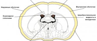
The central nervous system consists of the brain and spinal cord and their protective membranes. The outermost is the dura mater, under it is the arachnoid (arachnoid), and then the pia mater, fused with the surface of the brain. Between the pia mater and the arachnoid membrane is the subarachnoid space, which contains cerebrospinal fluid, in which both the brain and spinal cord literally float. The action of the buoyant force of the fluid leads to the fact that, for example, the adult brain, which has an average mass of 1500 g, actually weighs 50–100 g inside the skull. The meninges and cerebrospinal fluid also play the role of shock absorbers, softening all kinds of shocks and shocks that tests the body and which could lead to damage to the nervous system.
The central nervous system is made up of gray and white matter. Gray matter is composed of cell bodies, dendrites, and unmyelinated axons, organized into complexes that include countless synapses and serve as information processing centers for many functions of the nervous system. White matter consists of myelinated and unmyelinated axons that act as conductors transmitting impulses from one center to another. The gray and white matter also contains glial cells. CNS neurons form many circuits that perform two main functions: they provide reflex activity, as well as complex information processing in higher brain centers. These higher centers, such as the visual cortex (visual cortex), receive incoming information, process it, and transmit a response signal along the axons.
The result of the activity of the nervous system is one or another activity, which is based on the contraction or relaxation of muscles or the secretion or cessation of secretion of glands. It is with the work of muscles and glands that any way of our self-expression is connected. Incoming sensory information is processed through a sequence of centers connected by long axons that form specific pathways, for example pain, visual, auditory. Sensory (ascending) pathways go in an ascending direction to the centers of the brain. Motor (descending) tracts connect the brain with motor neurons of the cranial and spinal nerves. The pathways are usually organized in such a way that information (for example, pain or tactile) from the right side of the body enters the left side of the brain and vice versa. This rule also applies to the descending motor pathways: the right half of the brain controls the movements of the left half of the body, and the left half controls the right. There are, however, a few exceptions to this general rule.
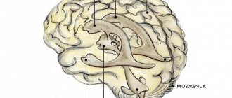
The brain consists of three main structures: the cerebral hemispheres, the cerebellum and the brainstem.
The cerebral hemispheres - the largest part of the brain - contain higher nerve centers that form the basis of consciousness, intelligence, personality, speech, and understanding. In each of the cerebral hemispheres, the following formations are distinguished: underlying isolated accumulations (nuclei) of gray matter, which contain many important centers; a large mass of white matter located above them; covering the outside of the hemispheres is a thick layer of gray matter with numerous convolutions that makes up the cerebral cortex.
The cerebellum also consists of an underlying gray matter, an intermediate mass of white matter, and an outer thick layer of gray matter that forms many convolutions. The cerebellum primarily provides coordination of movements.
The brainstem is formed by a mass of gray and white matter that is not divided into layers. The trunk is closely connected with the cerebral hemispheres, the cerebellum and the spinal cord and contains numerous centers of sensory and motor pathways. The first two pairs of cranial nerves arise from the cerebral hemispheres, while the remaining ten pairs arise from the trunk. The trunk regulates vital functions such as breathing and blood circulation.
Spinal cord . Located inside the spinal column and protected by its bone tissue, the spinal cord has a cylindrical shape and is covered with three membranes. In a cross section, the gray matter is shaped like the letter H or a butterfly. Gray matter is surrounded by white matter. Sensitive fibers of the spinal nerves end in the dorsal (posterior) parts of the gray matter - the dorsal horns (at the ends of the H, facing the back). The bodies of motor neurons of the spinal nerves are located in the ventral (anterior) parts of the gray matter - the anterior horns (at the ends of the H, distant from the back). In the white matter there are ascending sensory pathways ending in the gray matter of the spinal cord, and descending motor pathways coming from the gray matter. In addition, many fibers in the white matter connect different parts of the gray matter of the spinal cord.
The main and specific function of the central nervous system is the implementation of simple and complex highly differentiated reflective reactions, called reflexes. In higher animals and humans, the lower and middle sections of the central nervous system - the spinal cord, medulla oblongata, midbrain, diencephalon and cerebellum - regulate the activity of individual organs and systems of a highly developed organism, carry out communication and interaction between them, ensure the unity of the organism and the integrity of its activities. The highest department of the central nervous system - the cerebral cortex and the nearest subcortical formations - mainly regulates the connection and relationship of the body as a whole with the environment.
Basic features of the structure and function of the central nervous system is connected with all organs and tissues through the peripheral nervous system, which in vertebrates includes cranial nerves extending from the brain and spinal nerves from the spinal cord, intervertebral nerve ganglia, as well as the peripheral part of the autonomic nervous system - nerve ganglia, with nerve fibers approaching them (preganglionic) and extending from them (postganglionic).
Sensitive, or afferent, nerve adductor fibers carry excitation to the central nervous system from peripheral receptors; along the efferent efferent (motor and autonomic) nerve fibers, excitation from the central nervous system is directed to the cells of the executive working apparatus (muscles, glands, blood vessels, etc.). In all parts of the central nervous system there are afferent neurons that perceive stimuli coming from the periphery, and efferent neurons that send nerve impulses to the periphery to various executive effector organs.
Afferent and efferent cells with their processes can contact each other and form a two-neuron reflex arc that carries out elementary reflexes (for example, tendon reflexes of the spinal cord). But, as a rule, intercalary nerve cells, or interneurons, are located in the reflex arc between the afferent and efferent neurons. Communication between different parts of the central nervous system is also carried out using many processes of afferent, efferent and intercalary neurons of these parts, forming intracentral short and long pathways. The CNS also includes neuroglial cells, which perform a supporting function in it and also participate in the metabolism of nerve cells.
Reflex as the basic principle of the nervous system
I. M. Sechenov in 1863 in his work “Reflexes of the Brain” he developed the idea that the reflex is the basic principle of the functioning of not only the spinal cord, but also the brain.
A reflex is the body’s response to irritation with the participation of the central nervous system. Reflexes are divided into: 1) unconditioned reflexes: innate (hereditary) reactions of the body to stimuli carried out with the participation of the spinal cord or brain stem; 2) conditioned reflexes: temporary reactions of the body acquired on the basis of unconditioned reflexes, carried out with the obligatory participation of the cerebral cortex, which form the basis of higher nervous activity.
Each reflex has its own reflex arc - this is the path along which excitation passes from the receptor to the effector (executive organ).
The reflex arc is represented by a chain of neurons that provide the perception of irritation, the transformation of the energy of irritation into a nerve impulse, the conduction of a nerve impulse to the nerve centers, the processing of incoming information and the implementation of a response.
Any reflex arc consists of 5 components
1. A receptor is a specialized cell designed to perceive a stimulus (sound, light, chemical, etc.). 2. Afferent pathway, which is represented by afferent neurons. 3. Part of the central nervous system, represented by the spinal cord or brain; 4. The efferent pathway consists of the axons of efferent neurons extending beyond the CNS. 5. An effector is a working organ (muscle, gland, etc.).
The simplest reflex arc includes 2 neurons and is called monosynaptic (based on the number of synapses). A more complex one is represented by 3 neurons and is called three-neuron or disynaptic. However, most reflex arcs include a large number of interneurons and are called polysynaptic.
Reflex arcs can pass through the spinal cord only (for example, withdrawing a hand when touching a hot object) or through the brain only (for example, closing the eyelids when a stream of air is directed at the face), or through both the spinal cord and the brain.
Reflex arcs are closed into reflex rings using feedback connections. The concept of feedback and its functional role was indicated by Bell in 1826. He wrote that two-way connections are established between the muscle and the central nervous system. With the help of feedback, signals about the functional state of the effector are sent to the central nervous system.
The morphological basis of feedback is the receptors located in the effector and the afferent neurons associated with them. Thanks to feedback afferent connections, fine regulation of the effector’s work and an adequate response of the body to environmental changes are carried out.