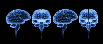
What parts does the brain consist of?
The human brain consists of several sections. Each of them performs its function, ensuring the vital functions of the body.
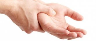
Table 1. Main departments.
| Name | Description |
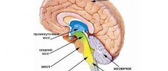
| Oblong | This part is a continuation of the spinal cord. It consists of gray matter nuclei and white matter tracts. It is this part that determines the connection between the brain and the body. |
| Average | It consists of 4 tubercles, two of which are responsible for vision and two for hearing. |
| Rear | The hindbrain includes the pons and cerebellum. This is a small section in the back of the head, which weighs around 140 grams. Consists of two hemispheres connected to each other. |
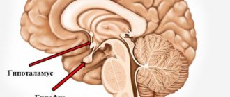
| Intermediate | Consists of the thalamus, hypothalamus. |
| Finite | This section forms both hemispheres of the brain, connected by the corpus callosum. The surface is full of convolutions and grooves covered with bark. The hemispheres are divided into lobes: frontal, parietal, temporal and occipital. |
The last section occupies more than 80% of the total mass of the organ. The organ can also be divided into 3 parts: the cerebellum, the brainstem and the cerebral hemispheres.
In this case, the entire brain is covered in the form of a shell, divided into three components:
- Arachnoid (cerebrospinal fluid circulates through it)
- Soft (adjacent to the brain and full of blood vessels)
- Hard (in contact with the skull and protects the brain from damage)
All components are important in the regulation of life and have a specific function. But the activity regulation centers are located in the cortex.
The human brain consists of many sections, each of which has a complex structure and performs a specific role. The largest of them is the final one, which consists of hemispheres. All this is covered with three shells that provide protective and nourishing functions.
Learn about the structure and functions of the brain from the video provided.
Structure
The morphological structure of the cerebral hemispheres within the brain suggests the presence of a cortical layer consisting of gray matter and subcortical sections formed by white matter. Within the mass of white matter there are local areas of gray - the nuclei. White matter serves as the basis for nerve fibers:
- Associative. They unite different functional areas within one hemisphere.
- Commissural. They connect areas of different hemispheres, often symmetrically located.
- Conductive. They connect the parts of the brain - the brain and the spinal cord - thereby forming a single central nervous system network.
The structure of the cortex covering the cerebral hemispheres suggests the presence of grooves (depressions relative to the surface) and convolutions (elevations relative to the surface). In anatomy, the area of the cortex covering the cerebral hemispheres increases due to the presence of grooves. The duration and depth of the furrows are individual characteristics of a person.
The cortical layer, formed from gray matter, contains control centers that are responsible for higher mental functions, which are interconnected with processes such as cognitive and mental activity. The cortex, covering the cerebral hemispheres within the brain, coordinates the functions of the body, forming adaptive responses to external influences. The cortex is formed by 6 types of cells.
The cytoarchitectonics (location of cells in the tissue) of the cortex suggests the presence of molecular, granular (external, internal), pyramidal (external, internal), multiform plates. Within the brain, the surfaces of the hemispheres are distinguished: superolateral (covers the upper lateral areas), medial (located in the middle region), basal (adjacent to the base of the skull).
The superolateral surface has a convex shape and is adjacent to the bone structures that form the cranial vault. The flat medial surface is located opposite the similar surface of the second hemisphere. Each hemisphere consists of 5 lobes. The frontal lobe of the cortex, covering the cerebral hemispheres, is the largest part of the telencephalon. The central sulcus, anatomically transversely crossing the brain, is the border between the frontal and parietal lobes within the cerebral hemispheres.
The Sylvian fissure, also known as the lateral fissure, runs within the cerebral hemispheres perpendicular to the central one, separating the temporal lobe of the telencephalon from two segments - the frontal and parietal. The parieto-occipital sulcus is the border of the parietal segment on the posterior side. The insular lobe within the hemispheres of the brain is located deep in the Sylvian fissure. The insular lobe is visible if you lift the areas of the frontal and temporal lobes that cover it.
Important What diseases cause dysphagia (difficulty swallowing)?
The Sylvian fissure, also known as the lateral fissure, which separates the temporal lobe from the brain structures of the frontal and parietal, is one of the largest. It is formed at the 14th week of intrauterine development. Small grooves dividing the lobes of the cerebral cortex into convolutions are formed at 24-38 weeks, forming a peculiar relief of the surface of the hemispheres, individual for each person.
The sulci and gyri of the 3rd order continue to develop after birth, most intensively in the 1st year of life. In the frontal lobe of the cortex within the cerebral hemispheres, a precentral groove runs parallel to the central one, separating and limiting the precentral gyrus - the center where the initiation and regulation of conscious motor activity, directed by volitional effort, occurs.
The precentral gyrus is perpendicularly crossed by the frontal sulci (superior, inferior). They divide the frontal gyri into segments. In the posterior part of the frontal gyrus, located below, there is Broca's center, which is responsible for speech function. In the parietal lobe, the postcentral sulcus runs parallel to the central one.
The postcentral gyrus, located between these grooves, is a center of sensitivity, which is responsible for the body’s reactions to pain, tactile, and temperature influences. The temporal grooves (superior, inferior) lie in the temporal lobe and are located parallel to the Sylvian fissure.
The temporal sulci are divided into segments by the temporal gyri, where in the upper part there is a center responsible for hearing function and Wernicke's center, responsible for speech function. The calcarine groove, which provides visual function, lies in the occipital lobe, where it borders on the parieto-occipital groove.
Brain
It is difficult to list all the functions, because this is an extremely complex organ. This includes all aspects of the human body. However, it is possible to identify the main functions performed by the brain.
The functions of the main organ include all human senses. These are vision, hearing, taste, smell and touch. All of them are performed in the cerebral cortex. It is also responsible for many other aspects of life, including motor function.
Human speech is performed in the cerebral hemispheres, namely in Broca's and Wernicke's centers. The hemispheres also perform many other functions.
The back of the brain, which includes the cerebellum, regulates balance and coordination. However, all centers important for life are located in the medulla oblongata. Here breathing, the work of the heart, blood vessels, all food and protective reflexes, as well as the regulation of muscle fibers are regulated.
Vision and hearing are not processed only in the cortex. The midbrain is also responsible for this task, regulating processes at the lower level. The same applies to motor function.
Sensitivity is regulated by the diencephalon, namely the thalamus.
The hypothalamus is the main element of the endocrine system, which regulates nerve signals and transforms them into endocrine ones. It also regulates the autonomic nervous system.
There are a lot of functions of the human brain, all of them are performed in its departments. However, most of the active activity is located in the cerebral cortex. These include hearing, smell, touch, sight and taste.
Functions of neocortex
The new cortex is responsible for performing higher nervous functions (thinking, speech, processing information from the senses, creativity, etc.). Clinical trials have shown that each area of the cerebral cortex is responsible for strictly defined functions. For example, human speech is controlled by the left frontal gyrus. However, if any of the areas is damaged, the neighboring one can take over its function, although this requires a long period of time. Conventionally, there are three main groups of functions performed by the neocortex - sensory, motor and associative.
Sensory
This group includes a set of functions with the help of which a person is able to perceive information from the senses.
Each sense is analyzed by a separate area, but signals from others are also taken into account.
Signals from the skin are processed by the posterior central gyrus. Moreover, information from the lower extremities goes to the upper part of the gyrus, from the body to the middle part, from the head and hands to the lower part. In this case, the posterior central gyrus processes only pain and temperature sensations. The sense of touch is controlled by the superior parietal region.
Vision is controlled by the occipital region. Information is received in field 17, and in fields 18 and 19 it is processed, that is, color, size, shape and other parameters are analyzed.
Hearing is processed in the temporal region.
Charm and taste sensations are controlled by the hippocampal gyrus, which, unlike the general structure of the neocortex, has only 3 horizontal layers.
It is worth noting that in addition to the zones of direct reception of information from the senses, next to them there are secondary ones, in which the relationship between the received images and those stored in memory occurs. When these areas of the brain are damaged, a person completely loses the ability to recognize incoming data.
Motor
This group includes the functions of the neocortex, with the help of which any movement of the human limbs is carried out. Motor skills are controlled and controlled by the precentral region. The lower limbs depend on the upper parts of the central gyrus, and the upper limbs depend on the lower ones. In addition to the precentral, the frontal, occipital and superior parietal regions are involved in movement. An important feature of the performance of motor functions is that they cannot be performed without constant connections with sensory areas.
Associative
This group of neocortical functions is responsible for such complex elements of consciousness as thinking, planning, emotional control, memory, empathy and many others.
Associative functions are performed by the frontal, temporal and parietal regions.
In these areas of the brain, a reaction to data coming from the senses is formed and command signals are sent to the motor and sensory areas.
To receive and control, all sensory and motor areas of the cerebral cortex are surrounded by associative fields, in which the received information is analyzed. But at the same time, it is worth considering that the data coming into these fields is already primarily processed in the sensory and motor areas. For example, if there is a disruption in the functioning of such an area in the visual area, a person sees and understands that there is an object, but cannot name it and, accordingly, make a decision about his further behavior.
In addition, the frontal lobe of the cortex is very tightly connected to the limbic system, which allows it to control and manage emotional messages and reflexes. This makes it possible for a person to develop as a person.
The performance of associative functions in the neocortex is possible due to the fact that the neurons of this part of the central nervous system are able to retain traces of excitation based on the feedback principle and can persist for a long time (from several years to a lifetime). This ability is memory, with the help of which associative connections of the information received are built.
Cortex
The human brain has a small top layer, about 3-4 mm thick. This is its bark - the main difference between humans and animals. It performs many functions, being used in all aspects of life. It is the action of the cortex that most affects a person’s behavior and consciousness.
Functions of the cerebral cortex include:
- Human interaction with the outside world through reflexes
- Thinking and consciousness
- Regulation of internal processes of the body, including organ function and metabolism
- Definition of human behavior
In fact, the cerebral cortex determines a person’s consciousness, controls all his thought processes, ensures interaction with the environment and the functioning of the body. It creates a relationship with the world based on reflexes, which allows a person to develop and adapt.
Each part of the cerebral cortex is determined by its functions. The limbic system is the most ancient among them. It is responsible for the regulation of behavioral reactions, the formation of sleep, emotions, memory and the control of autonomic processes.
The functions of the cortex include regulating and processing human feelings. These are sight, hearing, smell, taste and touch. Although these functions are partially divided between the cortex and midbrain.
The cerebral cortex has many functions. It is she who determines a person’s consciousness, regulates his behavior and allows him to think. It also allows you to interact with the outside world at the level of reflexes. The cortex controls organ function and metabolism. However, its functions are much broader and affect many aspects of human activity.
Physiology of the cerebral cortex
The finite or large brain, which has reached its highest development in humans, is rightly considered the most complex and most amazing creation of nature.
The functions of this section of the central nervous system are so different from the functions of the trunk and spinal cord that they are classified as a special chapter of physiology - the physiology of higher nervous activity. This term was introduced by I.P. Pavlov. The activity of the nervous system, aimed at uniting and regulating all organs and systems of the body, was called lower nervous activity by I. P. Pavlov. By higher nervous activity (mental), he understood behavior, activity aimed at adapting the body to changing environmental conditions, at balancing with the environment. In the behavior of an animal, its relationship with the environment, the leading role is played by the telencephalon, the organ of consciousness, memory, and in humans - the organ of mental activity and thinking.
The great achievements of I.P. Pavlov in the field of studying the functions of the cerebral hemispheres are explained by the fact that he proved the reflex nature of the activity of the cortex and discovered a new, qualitatively higher type of reflexes inherent only to it, namely conditioned reflexes. Having discovered the basic mechanism of activity of the cerebral cortex, he thereby created a fruitful, progressive method for studying its functions - the method of conditioned reflexes. As it turned out later, conditioned reflexes are those elementary acts, those “bricks” from which the behavior of humans and animals is built.
The significance of the hemispheres in various animals before I.P. Pavlov was studied by surgically removing them. The results of removing the cerebral hemispheres of birds and dogs showed that autonomic functions: blood circulation, breathing, digestion, etc., are not significantly impaired. With careful care, the animal lives a long time. Its connection with the external environment is disrupted. To directly acting stimuli: a pin prick, irritation of the oral mucosa with food, a completely adequate reaction occurs: the animal withdraws its paw, swallows the food, i.e. the animal retains its innate unconditioned reflexes. All acquired behavioral reactions and conditioned reflexes developed in the process of individual life are irretrievably lost.
Localization of functions in the cerebral cortex. To study the localization of functions in the cerebral cortex or, in other words, the significance of individual zones of the cortex, various methods are used: partial removal of the cortex, electrical and chemical stimulation, recording of brain biocurrents and the method of conditioned reflexes.
The stimulation method made it possible to establish the following zones in the cortex: motor (motor), sensitive (sensory) and silent, which were later called associative.
Motor cortex areas. Movements occur when the cortex is stimulated in the area of the precentral gyrus. Electrical stimulation of the upper part of the gyrus causes movement of the muscles of the legs and torso, the middle part of the arms, and the lower part of the facial muscles. The size of the cortical motor area is proportional not to muscle mass, but to the accuracy of movements. The area that controls the movements of the hand, tongue, and facial muscles is especially large (Fig. 119).
Rice. 119. Location and size of motor areas in the cerebral cortex (according to Penfield and Rasmussen). The sizes of body parts correspond to the sizes of motor representation. 1 - foot; 2 - lower leg; 3 - knee; 4 - thigh; 5 - torso; 6 — brush; 7 — thumb; 8 - neck; 9 - face; 10 - lips; 11 - tongue; 12 - larynx
Sensory areas of the cortex. Extirpation of various areas of the cortex in animals made it possible to establish in general terms the localization of sensory (sensitive) functions. The area of the cortex where this type of sensitivity is projected is called the primary projection zone.
Human skin sensitivity, feelings of touch, pressure, cold and heat are projected into the postcentral gyrus. In its upper part there is a projection of the skin sensitivity of the legs and torso, lower - the arms and even lower - the head.
The absolute size of the projection zones of individual areas of the skin is not the same. Thus, the projection of the skin of the hands occupies a larger area in the cortex than the projection of the surface of the body (Fig. 120).
Rice. 120. Location and size of sensitive zones in the cerebral cortex (according to Penfield and Rasmussen). The sizes of body parts correspond to the sizes of sensory representation. 1 - genitals; 2 - foot; 3 - thigh; 4 - torso; 5 - shoulder; 6 — brush; 7 - index and thumb; 8 - face; 9 - lips; 10 - teeth; 11 - tongue; 12 - pharynx and internal organs
The magnitude of the cortical projection is proportional to the significance of a given receptive surface in behavior. Interestingly, the pig has a particularly large projection into the cortex of the snout.
Articular-muscular, proprioceptive sensitivity is projected into the postcentral and precentral gyri.
The visual cortex is located in the occipital lobe. When it is irritated, visual sensations arise - flashes of light; removing it leads to blindness. Removal of the visual zone on one half of the brain causes blindness in one half of each eye, since each optic nerve is divided at the base of the brain into two halves (forming an incomplete decussation): one of them goes to its half of the brain, and the other to the opposite.
The ability to see is an innate ability, but the ability to recognize objects is developed throughout life.
The hearing function is provided by the temporal lobes of the cerebral hemispheres. Their irritation is caused by simple auditory sensations. Removal of both auditory zones leads to deafness, and unilateral removal reduces hearing acuity.
The olfactory cortex is located at the base of the brain, in the region of the parahippocampal gyrus.
The projection of the taste analyzer is apparently localized in the lower part of the postcentral gyrus, where the sensitivity of the oral cavity and tongue is projected.
Limbic system. The telencephalon contains the formations that make up the limbic system: the cingulate gyrus, hippocampus, amygdala, fornix, and septum pellucidum. They are involved in maintaining the constancy of the internal environment of the body, regulating autonomic function and forming emotions and motivations. This system is otherwise called the “visceral brain.” Information from internal organs comes here. When the stomach and bladder are irritated, evoked potentials arise in the limbic cortex.
Electrical stimulation of various areas of the limbic system changes autonomic functions: blood pressure, respiration, movements of the digestive tract, tone of the uterus and bladder.
The destruction of individual parts of the limbic system leads to behavioral disturbances: animals can become calmer or, on the contrary, aggressive, easily reacting with rage, and sexual behavior changes. The limbic system has extensive connections with all areas of the brain, the reticular formation and the hypothalamus. It provides cortical control of all autonomic functions: cardiovascular, respiratory, digestive, metabolism, energy.
Association areas of the cortex. The projection zones of the cortex occupy a small area of the surface of the human cerebral cortex. The rest is occupied by so-called associative zones. The neurons of these areas are not connected either to the sense organs or to the muscles; they communicate between different areas of the cortex, integrating, combining all impulses entering the cortex into integral acts of learning (reading, speech, writing), logical thinking, memory and providing the possibility of expedient behavior reactions.
When associative zones are violated, agnosia appears - the inability to recognize and apraxia - the inability to perform learned movements. For example, stereoagnosia is expressed in the fact that a person cannot find either a key or a box of matches in his pocket by touch, although visually he immediately recognizes them. If the outer surface of the occipital lobe, the associative visual zone, is damaged, vision is preserved, but recognition disorder occurs (visual agnosia). The patient, being literate, cannot read what is written, recognizes a familiar person after he speaks. When areas of the auditory cortex are damaged, auditory agnosia can occur: a person hears, but ceases to understand the meaning of words.
In case of disruption of the associative speech zones of the cortex, aphasia—loss of speech—is possible. Aphasia can be motor or sensory. Motor aphasia occurs when the posterior third of the inferior frontal gyrus on the left, the so-called Broca's center, is damaged (this center is located in right-handed people only in the left hemisphere). The patient understands speech, but cannot speak himself.
With agraphia, a person forgets how to write, and with apraxia, he forgets how to perform learned movements: light a match, fasten a button, sing a melody, etc.
The role of each hemisphere in their joint activity is not the same. If in right-handed people the left hemisphere predominates in the motor functions of the body, then the right hemisphere predominates in the sensitive ones. (Recall that the left hemisphere innervates the muscles of the right half of the body and receives information from the receptors on the right side; the left half of the body is innervated by the right hemisphere.)
The motor asymmetry of the brain is most clearly revealed: the right arm and leg (in right-handed people) are stronger, more dexterous and more accurate in movements than the left. Most people are right-handed. Forcibly retraining left-handed children is not recommended.
The left hemisphere in right-handed people provides understanding and formation of oral and written speech, mathematical abilities and abstract, verbal, logical thinking (its defeat is accompanied by aphasia and agraphia). The right “illiterate” hemisphere is characterized by figurative, concrete thinking. Musical and artistic abilities are mainly determined by the function of the right “emotional” hemisphere.
The division of people into “thinkers” and “artists”, noted by I.P. Pavlov, i.e. persons with a predominance of a logical or figurative type of thinking, is associated with the predominance of the function of the corresponding hemisphere.
Bioelectrical activity of the cerebral cortex. Fluctuations in the electrical potentials of the cortex were first recorded by V.V. Pravdich-Neminsky in 1913. Currently, their study has become one of the leading methods for studying the brain in physiology and clinical practice. Fluctuations in cortical potentials are recorded using a multichannel cathode oscilloscope. In humans, potentials are usually recorded from the scalp using electrodes secured in various ways, such as adhesive tape. In this case, the electrode displays the total activity of tens of thousands of cortical neurons located underneath it. Usually, biocurrents are diverted from many symmetrical areas of the brain, for example, frontal, parietal, and occipital. The resulting recording is called an electroencephalogram (EEG).
The EEG distinguishes between waves of different frequencies and sizes (amplitudes). Based on the frequency of oscillations per second, they are distinguished: delta waves - the slowest, 0.5 - 3.5; theta waves - from 4 to 7; alpha waves - from 8 to 13; beta waves - more
13. The higher the frequency of the waves, the smaller their amplitude. Consequently, the smallest fluctuations are beta waves and the largest are delta waves (Fig. 121).
Rice. 121. Basic rhythms of the electroencephalogram. I - beta rhythm; II - alpha rhythm; III - theta rhythm; IV - delta rhythm; V - convulsive discharges
On the EEG, depending on the state of the brain and the recording area, one or another rhythm predominates. When a person is at rest and his eyes are closed, an alpha rhythm is recorded from the occipital lobes with an oscillation frequency of 8 - 13 per 1 s. When the eyes are open, the alpha rhythm disappears and a more frequent beta rhythm appears. It occurs when solving difficult problems, emotional arousal, intense attention. Such an episode is described. Einstein's EEG was recorded. The alpha rhythm was recorded. At the same time, Einstein solved problems that were not difficult for him. Suddenly a beta rhythm appeared on the EEG. When asked what happened, Einstein replied that he remembered a gross error in yesterday's calculations.
During sleep, the waves become slower and higher, and the delta rhythm predominates. However, during sleep, periods occur when brain activity increases and frequent waves of alpha and beta rhythms are recorded (paradoxical sleep). It has been established that these changes in the EEG are combined with dreams.
The cessation of blood supply to the brain leads to the disappearance of electrical activity within 15 s. EEG recording is used in a surgical clinic to monitor the state of the brain during major operations, especially on a “dry heart”. During deep anesthesia, alpha and beta rhythms are completely absent.
In some brain diseases, the EEG pattern changes. For example, patients with epilepsy have a characteristic type of EEG. Using electroencephalography, you can determine the location of the tumor in the brain.
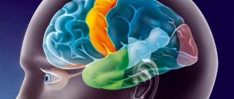
Lobes of the cerebral cortex
Among them:
- Frontal lobe. This is the main part of the cortex in which the motor centers, mental functions and speech center are located. Analytical activities and the area responsible for speech motor skills are also located here.
- Temporal lobes. These areas are located on the sides of the cortex. They contain the main centers of feelings, the center for understanding speech, as well as emotional centers responsible for joy, fear, pleasure and other emotions.
- Occipital lobe. It processes visual data.
- Parietal lobe. Contains sensory activity centers as well as a center for musical understanding.
Cell layers
There are six layers of bark, starting with the top:
- Molecular - mostly made up of fibers
- Grainy
- Pyramidal - consists of pyramidal type neurons
- Second grainy
- Second pyramidal - consists of pyramidal type neurons reaching the molecular layer
- Multimorphic - consists of small polymorphic cells that pass into the white matter
Each layer of the cortex has its own function, being unique levels of action. The entire work of the cerebral cortex is based on them.
Cortical zones
Another type of classification of areas of the cerebral cortex is also noted. It marks three zones of the cortex, differing in purpose and structure.
Among them:
- Primary zone. Consists of highly differentiated cells and receives data from receptors.
- Secondary zone. Responsible for processing the received information and consists of departments of analyzer cores.
- Associative. Forms conditioned reflexes and helps to understand the world around us.
This determines not only the individual structure of the zones, but also the individual functions for each of them.
The human cerebral cortex has a complex structure, distributed into lobes and layers. Each section is responsible for its own functions, regulating various life processes. In total there are 5 lobes and 6 layers, which together make up the cortex.
Diseases associated with disorders of its activity
There are many diseases that affect the human brain. Some of them affect its cortex, disrupting its processes and reducing performance. However, not much is known about them.
A common disease of the cortex is atrophy or Pick's disease. This disease develops in older people and is characterized by the death of neurons. The external condition of the brain is similar to Alzheimer's disease and resembles a dried walnut. The disease is not curable; individual symptoms are eliminated.
There are also diseases that indirectly affect the cortex. With hypertension, foci of excitation appear in the cortex, creating powerful vasoconstrictor impulses. This leads to increased blood pressure.
In addition, diseases can occur against the background of external infections. The same meningitis that occurs due to infections of pneumococcus, meningococcus and the like. The development of the disease is characterized by pain in the head, fever, pain in the eyes and many other symptoms such as weakness, nausea and drowsiness.
Many diseases that develop in the brain and its cortex have not yet been studied. Therefore, their treatment is complicated by a lack of information. So it is recommended to consult a doctor at the first non-standard symptoms, which will prevent the disease by diagnosing it at an early stage.
There are many diseases of the cerebral cortex. Among them are infectious diseases, illnesses associated with other diseases of the body, as well as diseases with an unclear cause. But most of them can be cured with medicine. Therefore, it is recommended not to delay if you feel unwell and undergo a bark examination, which is carried out in many clinics.
Tick-borne encephalitis
This disease has a viral etiology and is preceded by an increase in temperature, the development of headaches and malaise, weakening of muscle tissue, which causes rigidity. With an advanced form of tick-borne encephalitis, the symptoms worsen: the patient often exhibits hallucinations and obsessions, he becomes aggressive and agitated, and suffers from seizures.
Measles
With the development of measles encephalitis, damage to the cerebral hemispheres is manifested by a sharp increase in temperature, confusion, as a result of which the patient cannot navigate in time and space, visual hallucinations and nervous excitability. Characteristic symptoms of measles encephalitis are also convulsions that develop in all parts of the body, paresis and weakness of the muscles of the limbs: there is a risk of coma.
Lethargic
If damage to the cerebral cortex is caused by the development of lethargic encephalitis, the patient’s temperature rises, speech deteriorates, and catatonia occurs - a pathological condition in which the body freezes in one position for several hours. Lethargic encephalitis is also characterized by symptoms such as apathy, malaise, and drowsiness caused by disruption of the daily routine.
Important Serenate tablets: instructions for use, analogues, reviews