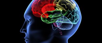
Brain structure
It is customary to divide human intelligence into five main parts: the cerebral hemispheres, the cerebellum, the medulla oblongata, the median and the pons. In some textbooks you can find a different classification. It states that the brain consists of the forebrain, midbrain, hindbrain, and brainstem. Its composition is quite simple. It’s even funny that such an important organ consists of nothing but water, minerals, lipids and proteins. Today we will talk in more detail about the structure and functions of the forebrain.
to contents ^
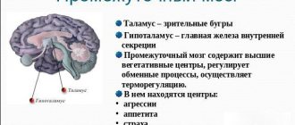
Diencephalon
This department is part of the front part of the organ and controls and switches all incoming information. The functions of the forebrain are the adaptive capabilities of the human body (external negative factors) and the regulation of the autonomic nervous system.
The diencephalon includes:
- Thalamic region;
- Hypothalamic-pituitary system (hypothalamus and posterior pituitary gland);
- Epithalamus.
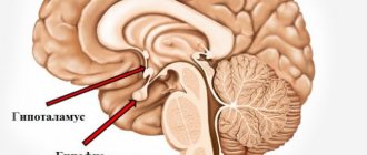
The hypothalamus regulates the functioning of internal organs and systems and is the center of pleasure. This part is presented in the form of a small cluster of neurons that transmit signals to the pituitary gland.
The thalamus processes all signals coming from sensory receptors, redistributing them to the appropriate parts of the central nervous system.
The epithalamus synthesizes the hormone melatonin, which is involved in the regulation of human biorhythms and emotional background.
The hypothalamus is part of an important system of the central nervous system - the limbic system. This system performs a motivational-emotional function (adapts when habitual conditions change). The system is closely connected with memory and smell, evoking clear memories of a vivid event or reproducing a favorite smell (food, perfume).
Forebrain and its structure
The forebrain is quite complex. Everyone knows it very well, and when we mention this organ, a picture of two hemispheres immediately comes to mind. It's right. Gray matter is divided into sections: the cerebral hemispheres and the diencephalon. If we talk about a more detailed division and go deeper into this topic, we can single out: the basal ganglia, the cerebrum, the hippocampus and the limbic system - a complex that consists of structures responsible for visceral, motivational and emotional sensations. Such a fairly extensive structure of the human forebrain will be of little interest to a person far from medical science, so in this article we will refer to the first classification and talk about the structure of which in more detail.
Useful to know: Midbrain: structure, functions, development
to contents ^
Table “Structure and functions of the brain”
Structure
The main organ of the nervous system consists of three parts:
- two hemispheres;
- trunk;
- cerebellum.
It also has five departments:
- final, constituting 80% of the mass;
- intermediate;
- rear;
- average;
- oblong.
Each section consists of a specific set of cells (white and gray matter).
White matter is presented in the form of nerve fibers, which can be of three types:
- association – connect cortical areas in one hemisphere;
- commissural - connects the two hemispheres;
- projection – connect the cortex with the underlying formations.
Gray matter consists of neuron nuclei, their functions include the transmission of information.
Rice. 2. Lobes of the cerebral cortex.
The following table will help you understand in more detail the structure and functions of the brain:
Table “Structure and functions of the brain”
| Department | Structure | Functions |
| Finite | Located from the occipital to the frontal bone. It consists of two hemispheres, which have many grooves and convolutions. On top they are covered with a bark consisting of lobes. | The right hemisphere is responsible for the left side of the body, and the left hemisphere is responsible for the right side. The temporal lobe of the cerebral cortex regulates hearing and smell, the occipital lobe regulates vision, the parietal lobe regulates taste and touch; frontal – speech, thinking, movement. |
| Intermediate | Consists of the hypothalamus and thalamus. | The thalamus is a mediator in the transmission of stimuli to the hemispheres and helps to adequately adapt to changes in the environment. The hypothalamus regulates the functioning of metabolic processes and endocrine glands. Manages the work of the cardiovascular and digestive systems. Regulates sleep and wakefulness, manages food and drink needs. |
| Rear | It consists of the cerebellum and the pons, which is presented in the form of a white thick cushion located above the oblongata. The cerebellum is located behind the pons and has two hemispheres, the inferior and superior surfaces and the vermis. | This section provides a conductive function during the transmission of impulses. The cerebellum controls the coordination of movements. |
| Average | Located from the anterior edge of the bridge to the optic tracts. | Responsible for hidden vision, as well as the work of the orienting reflex, which ensures the body turns in the direction of the heard sharp noise. |
| Oblong | Presented as a continuation of the spinal cord. | Controls coordination of movements, balance, regulates metabolic processes, breathing, blood circulation. Controls the process of coughing and sneezing. |
Rice. 3. Functions of parts of the brain.
The brainstem consists of the medulla oblongata, midbrain, diencephalon and pons. The trunk is the connecting link between the spinal and head sections of the central nervous system. Its functions include controlling articulate speech, heartbeat and breathing.
Components and its functions
Hemispheres of the brain. Some of the important components that are separated by the posteroanterior cavity. The parts are connected by the corpus callosum - this is a white wall. The upper ball itself is covered with a shell of neurons and gray matter, arranged in columns in several layers. The surface of the hemispheres has the form of folds, convolutions and depressions, which are called grooves. It is these depressions that divide the brain into the temporal, frontal, parietal and occipital parts. They are named after the bones they are adjacent to. In neurons, the analysis of nerve connections that come from outside is carried out, these are visual, auditory, and neurons responsible for muscle activity. Gustatory and olfactory neurons are located in the temporal lobe, while behavioral neurons are located in the anterior gray matter. The central zone is responsible for human activity.
The main feature of the hemispheres is that they differ significantly from each other. For right-handed people, the neurons responsible for speech are located in the left hemisphere, and the right hemisphere is responsible for actions, logical chains, recognition of faces, songs, paintings and other things. Under the influence of external stimuli, experience is created and accumulated. In the hemispheres, to summarize and say briefly, the main centers are formed that interact with the most complex patterns of behavior, instincts and memory.
The diencephalon consists of three parts: lower, upper and central. Everyone has heard the word thalamus at least once - this is precisely the upper part of the diencephalon. It, in turn, is made up of the ventricle and paired formations. This is where all information from outside comes in, the initial assessment takes place and then passes further into the cortex of the human intellect. The hypothalamus is the lower part, which performs the function of metabolism and regulates brain energy. In the centers of the hypothalamus there are nuclei that are responsible for various sensations. In combination with the components of gray matter in impulses supplied for motor activity.
Useful to know: How the diencephalon functions and why it is needed
to contents ^
BRAIN
BRAIN, anterior center. nervous system of vertebrates and humans, occupying the cranial cavity. Regulates the most important functions of the body, controls behavioral reactions, is the basis of memory and higher nervous activity. In invertebrate animals that have a center. nervous system, these functions are performed by the cephalic ganglion, which in the most highly organized (insects and mollusks) is often called the G. m. Among chordates, the primitive G. m. in the form of accumulations of nerve cells at the head end of the body appears for the first time in the lancelet. Cyclostomes already have all the basics. sections of the brain: anterior, middle and posterior. In fish and terrestrial vertebrates, the same structural plan of the brain is retained. At the same time, in amphibians and reptiles, the middle and anterior sections of the brain undergo significant changes. In the latter, the diencephalon and two symmetrical hemispheres intensively develop. It means in birds. The cerebellum, basal ganglia and specif. develop. the structure of the forebrain cloak is the hyperstriatum (in mammals it corresponds to the neocortex). In mammals, the forebrain undergoes dramatic differentiation; Means. development reaches the cerebral cortex.
In mammalian genetics there are 5 main types. departments: medulla oblongata (borders the spinal cord); hindbrain (includes pons and cerebellum); midbrain (represented by the quadrigeminal and cerebral peduncles); the diencephalon (includes the epithalamus, thalamus and hypothalamus, with which the pituitary gland is connected) and the telencephalon (cerebral hemispheres). The first three sections together (with the exception of the cerebellum) form the brain stem.
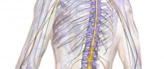
Phylogenetically, G. m. develops from the anterior end of the neural tube. It is a derivative of the three head bubbles. The forebrain is divided into two, of which one forms the telencephalon, including the cerebral hemispheres and basal ganglia, and the second forms the diencephalon. The midbrain vesicle gives rise to the midbrain. The cerebellum, pons and medulla oblongata are formed from the posterior one. Brain tissue consists of gray matter (the bodies of neurons and neuroglia), which forms the cortex and nucleus, and white matter, which consists primarily of brain tissue. of nerve fibers that communicate between nerve cells. The brain is covered with three membranes: soft, arachnoid, and hard (external). The soft and arachnoid membranes contain many vessels; between them is the so-called. subarachnoid space communicating with the ventricles of the brain, in which cerebrospinal fluid is formed and circulates. The ventricles occupy the center. part of the G. m. and are connected to each other and to the spinal canal. With the participation of cerebrospinal fluid, an exchange occurs. substances that travel with the blood from the vessels to the brain and back. The blood vessels of the brain, which provide its nutrition, receive arterial blood from the carotid arteries and vertebral arteries; venous blood enters the jugular veins.
The trunk of the brain contains sensory and motor nuclei of the cranial nerves, projection (for example, visual and auditory systems) and switching (from sensory pathways to neurons of the primary sensory zones of the cortex) nuclei, nuclei of the reticular formation, as well as nerve fibers and their closely located groups (tracts). The cranial nerves depart from the trunk of the brain. In the structures of the brainstem there are nerve centers that control the functions of respiration, blood circulation, swallowing, vomiting, etc. (medulla oblongata), vision and hearing (tubercles of the quadrigeminal midbrain), regulation of biological. rhythms (upper diencephalon). In the brain stem there are tracts connecting various. parts of the brain and spinal cord.
The diencephalon consists of many interconnected nuclei. Functionally closely connected with the forebrain. The nuclei of the thalamus are involved in processing information coming from the internal. organs and hypothalamus, sensory information, in the higher integrative functions of the brain (together with the cortex of the brain), regulation of its excitability, as well as in the organization of motor activity. The hypothalamus includes nuclei that control metabolism, ensuring the constancy of internal. body environment (homeostasis), thermoregulation, vascular tone, etc. Functionally, it is connected with the pituitary gland.
The hemispheres of the brain consist of the cortex, white matter, and basal nuclei. They are responsible for the development of acquired behaviors. acts and higher nervous activity. Each hemisphere is divided into lobes. Their cortex forms grooves and convolutions, significantly increasing its surface, and reaches its greatest development in humans, monkeys and cetaceans (the volume of the basal ganglia also increases). The cortex is divided into sensory, motor (motor), and associative zones. The hemispheres of the brain are connected to each other by a large bundle of nerve fibers—the corpus callosum. A powerful system of ascending and descending fibers provides connections between the hemispheres and the underlying parts of the brain. Information from the olfactory receptors comes directly to the hemispheres. The cortex forms a single functional whole with the basal ganglia and the nuclei of other parts of the brain and is involved in the processing of sensory information, ensures intersensory integration, purposeful and coordinating. motor reactions are the main the substrate of learning, memory, intellectual functions, such as consciousness, thinking, speech, etc. The basal ganglia, located in the thickness of the white matter of the hemisphere, together with the cortex are involved in the organization of coordination. movements and sensorimotor integration. Some formations of the brain are combined into the extrapyramidal system, which is responsible for ensuring movement and regulating muscle tone and posture. The structures united in the limbic system are responsible for a number of innate behaviors. acts, some types of memory, the formation of emotions and motivations. All departments of the brain muscle are united into a single system, in which the cerebral cortex of the brain muscle plays a leading and organizing role.
Human brain (right half, left view): 1 – cerebral hemisphere; 2 – thalamus; 3 – hypothalamus; 4 – corpus callosum; 5 – pituitary gland; 6 – quadrigeminal; 7 &nd...
Human brain. The average weight of the hemorrhoids of an adult male is 1400 g, that of a female is 1250 g, which corresponds to 2–2.5% of body weight. Newborn baby's weight is approx. 350 g. An increase in its mass is achieved due to the formation of soft myelin sheaths around nerve fibers, the formation of new synapses (contacts between neurons) and the proliferation of cells of auxiliary nervous tissue (neuroglia). Max. G. m. reaches its mass between 18 and 30 years of age, then it gradually decreases. A child begins to talk when the mass of his brain is approximately 750 g. Based on this, it is assumed that the most ancient people (for example, Pithecanthropus), whose brain mass corresponded to this value, could have had the rudiments of speech. However, according to available data, specific. The characteristics of a person’s nervous activity and his abilities are associated not only with the mass of the brain, but also with the number of connections between neurons. G. m. is the most intensively working structure of the human body, while it uses up to 20% of the oxygen entering the body.
Brain cavities and cerebrospinal fluid
Brain cavities
. Inside the brain and spinal cord there are cavities of various diameters, which are remnants of the initially hollow neural tube of the embryo (see Fig. 13). Inside the spinal cord is the spinal canal, which, passing into the brain, expands in the medulla oblongata and forms the fourth ventricle. At the level of the midbrain, the ventricle passes into a narrow canal - the aqueduct of Sylvius. In the diencephalon, the aqueduct of Sylvius expands, forming the cavity of the third ventricle, which smoothly passes at the level of the cerebral hemispheres into the lateral ventricles through the foramina of Monro (I and II). All of these cavities are filled with liquor.
Liquor
(cerebrospinal or cerebral fluid) - viscous, transparent, colorless liquid: consists of 89% water, 2.2% organic substances, 8.8% inorganic substances; has a slightly alkaline reaction. Liquor contains very little protein, and its salt composition resembles blood plasma. CSF pressure – 100-150 mm.water column. In an adult, approximately 100-150 ml circulates. cerebrospinal fluid (20-40 ml are in the ventricles of the brain).
The functions of cerebrospinal fluid are that it creates a hydraulic protective membrane around the brain; maintains intracranial pressure; participates in the removal of metabolic products.
Basically, cerebrospinal fluid is formed in the choroid plexuses of the lateral ventricles of the brain due to the activity of ependymal cells, but it can also be formed in other ventricles of the brain. At the same time, there is an outflow of cerebrospinal fluid into the venous sinuses of the subarachnoid space. This ensures continuous circulation of cerebrospinal fluid and its renewal. In some diseases, this circulation is disrupted (for example, with hydrocephalus, when cerebrospinal fluid is still formed, but its outflow is difficult), which, in turn, leads to disturbances in the functioning of the central nervous system.
In general, the circulation of cerebrospinal fluid
occurs as follows: from the lateral ventricles - into the third ventricle, from the 3rd through the cerebral aqueduct into the 4th ventricle.
From the holes in the posterior wall of the fourth ventricle - into the cistern between the cerebellum and the medulla oblongata, that is, into the subarachnoid space. In children, there is also a flow of cerebrospinal fluid through the central spinal canal. In adults it partially heals. Next, the cerebrospinal fluid enters the bloodstream (through protrusions of the arachnoid membrane, which penetrate the lumen of the venous vessels of the dura mater and further into the internal jugular veins), as well as into the blood capillaries at the site of the exit of the cranial and spinal nerves from the cranial cavity and from the spinal canal .
The movement of cerebrospinal fluid is facilitated by changes in the position of the torso, head, limbs, as well as the pulse wave and respiratory movements. It is possible that the flow of fluid is facilitated by the fact that the largest quantities are still formed in the lateral ventricles.
Thus, the cerebrospinal fluid creates a kind of “water cushion”, so that the brain actually floats inside the cavities, being additionally fixed by the meninges.
If desired, see illustrations for the textbook “Anatomy of the Central Nervous System” by Dykhan L.B. on topics of lectures 2-3
[1] In this case we are talking about organisms with a central nervous system. There are exceptions: local reflex reactions of the skin, in which the central nervous system is not involved.
[2] in more primitive organisms, the arachnoid and vascular membranes formed a single soft membrane, then they separated, and now the soft membrane is called the vascular membrane (the exact name is secondary soft).