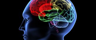
The cerebellum is a part of the brain responsible for coordinating movements and maintaining posture, tone and balance of the body.
Cerebellar stroke is less common than other forms of cerebrovascular pathology, but represents a significant problem due to insufficient knowledge and diagnostic difficulties. The proximity of the brain stem and vital nerve centers makes this localization of strokes very dangerous and requires quick, qualified help.
Causes of ischemic stroke of the cerebellum
Disruption of blood flow through the vessels of the cerebellum leads to either their blockage, which happens much more often, or rupture, in which case the result is a hematoma.
Ischemic stroke of the cerebellum, or infarction, occurs due to thrombosis or embolism of the vessels supplying the organ. Embolism is most common in patients suffering from cardiac pathology.
Thus, there is a high risk of blockage of the cerebellar arteries by thromboembolism during atrial fibrillation, recent or acute myocardial infarction. Intracardiac thrombi with arterial blood flow enter the brain vessels and cause their blockage.
Thrombosis of the cerebellar arteries is most often associated with atherosclerosis, when there is a proliferation of fatty deposits with a high probability of plaque rupture. With arterial hypertension during a crisis, so-called fibrinoid necrosis of the arterial walls is possible, which is also fraught with thrombosis.
Hemorrhage into the cerebellum, although less common than a heart attack, causes more problems due to tissue displacement and compression of surrounding structures by excess blood. Typically, hematomas occur due to arterial hypertension, when, against the background of high pressure numbers, the vessel “bursts” and blood rushes into the cerebellar parenchyma.
Other causes include arteriovenous malformations and aneurysms that form during fetal development and remain undetected for a long time because they are asymptomatic. There have been cases of cerebellar stroke in young patients associated with dissection of a section of the vertebral artery.
Expert opinion
Author: Andrey Igorevich Volkov
Neurologist, Candidate of Medical Sciences
Cerebellar stroke accounts for the smallest proportion of all strokes - no more than 1.5%. It is also the least studied type of acute cerebral circulatory disorder. The disease occurs 3 times more often in men than in women and rarely affects young people.
The clinical picture of isolated cerebellar strokes boils down to uncoordination of movements, unsteadiness of gait, and loss of balance. However, these symptoms do not always appear in small areas of cerebellar stroke. The only reliable method for diagnosing and identifying the area of brain damage is MRI. Based on the examination results, it is necessary to immediately prescribe adequate drug therapy to avoid complications.
The Yusupov Hospital has a neurology clinic, where patients suspected of having a stroke undergo diagnosis, treatment and a set of rehabilitation measures necessary for the complete restoration of damaged functions. According to research, with qualified rehabilitation, it is possible to completely eliminate coordination problems and voluntary movements due to cerebellar stroke. Thanks to timely effective therapy, the risk of recurrent attacks is reduced.
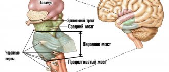
Structure of the cerebellum
The structure of the cerebellum is complex. The organ consists of many parts - lobes of the hemispheres, vermis, cortex, nuclei and legs, which perform certain functions and connect the cerebellum with parts of the brain. The anatomy of the cerebellum is represented by two hemispheres, lying on either side of the vermis. The cerebellar cortex is formed from three areas:
- Archicerebellum. Otherwise called the flocculonodular lobe.
- Paleocerebellum. Bilaterally interacts with the receptors of the spinal cord and the sensorimotor area of the cortex covering the cerebral hemispheres.
- Neocerebellum. Bilaterally interacts with cortical neurons that make up the cerebral hemispheres, receptors of the visual and auditory apparatus.
The cortex consists of an inner (granular), outer (molecular) and middle layer formed from Purkinje cells. The layered structure of the organ indicates integration processes that reflect the active interaction of different parts of the brain. The layers of the cerebellum provide its connection with different parts of the central nervous system. The thickness of the cerebellum contains 3 pairs of nuclei - lateral dentate, median tent nuclei, rounded and cork-like in the space between the first two pairs.
The cerebellum is connected to the parts of the brain through the peduncles. The fiber composition of the peduncles located in the cerebellum includes the nuclei of the vestibular nerve, as well as nerve bundles - the gentle and sphenoid. The lower legs connect the organ with a portion of the medulla oblongata. The middle legs in the form of a spacious, protruding part of the cerebellum are located laterally and pass into the pons. The cortical structures of the hemispheres located in the cerebrum control the functioning of the cerebellum using the middle peduncles. The flattened tufts of the upper legs are directed in both directions.
Worm
The structure and functions of the cerebellum cannot be imagined without the vermis, which connects both hemispheres of the organ together. The vermiform structure is composed of white matter nerve fibers. Pathological changes in the tissues of the worm cause ataxia.
Slices
The lobes of the hemisphere located in the cerebellum are delimited by grooves. The worm and hemispheres contain 8 lobules. The anterior, as well as the flocculonodular lobes, located in the cerebellum, are connected by working functions with the brain of the spinal column and the vestibular apparatus. The cerebellar hemispheres receive impulses from muscle and joint tissue receptors. On the front side, a small formation is adjacent to the hemisphere - a shred.
Cores
The nuclei located in the cerebellum control the axial and proximal muscles of the limbs. The idea of movement appears in the associative cortical structures of the brain, then is transmitted through afferent pathways towards the elements of the musculoskeletal system. The program of planned movement is formed in the dentate nucleus and simultaneously in the cerebellar hemispheres, then redirected to the motor centers of the cortex using the thalamic nuclei. The reproduced movement cannot be controlled using the somatosensory feedback system. Damage to the nuclei causes convulsive contraction of the muscles that control the limbs.
Symptoms of ischemic stroke of the cerebellum
Damage to the cerebellum is primarily manifested by impaired coordination and balance. There are two types of cerebellar stroke: isolated in a certain area and extensive.
Symptoms of isolated cerebellar stroke:
- Disorders of the vestibular apparatus, gait, fine motor skills of the hands;
- Impaired speech and hearing, the victim is unable to pronounce words or make sounds;
- Severe dizziness and nausea;
- Acute pain in the occipital region;
- Coordination of movements and sense of balance are impaired.
Symptoms of extensive cerebellar stroke:
- Acute headache, vomiting and nausea (general cerebral symptoms);
- Coordination of movements and hand motor skills are impaired;
- The patient's speech suffers;
- The patient is unable to maintain balance;
- Respiratory and cardiac functions are impaired;
- The patient is unable to control the functioning of the tongue and make swallowing movements.
Treatment and prognosis
For any type of lesion, therapy is divided into symptomatic and etiological. The chances of restoring dead cells are very small. Therefore, doctors only treat the symptoms of the disease. Medicines and surgical techniques are used. The prognosis for the disease is unfavorable.
How to develop the cerebellum?
Children with cerebellar insufficiency may lag behind their peers in development. And in older people, frequent falls are a source of dangerous complications. How to develop a “small” brain?
- The most ancient method is rocking a baby in a cradle. It’s not for nothing that our ancestors made cradles for the birth of their first child. This is not only caring for the mother, but also the development of the child’s vestibular apparatus;
- Next, a swing is suitable for training a two-year-old. They must be modern and safe. Just play with your baby and develop not only vestibular skills, but also conversational speech;
- After 5-7 years of age, children are taught on Dr. Bilgow’s balancing board. You can make it yourself or buy it in a store. It’s better to attend courses with an experienced exercise therapy instructor;
- There's more to the board than just balancing. Exercises are used with throwing and catching bags of sand, a ball and other devices.
Important 8 recipes for folk remedies made from garlic for cleaning blood vessels from cholesterol
It is important to exercise regularly (3-4 times a week). Lesson duration: at least 30 minutes
If you feel well, such training is also available for older people.
Diagnostics
A cerebellar stroke is often accompanied by headache, dizziness and nausea. Since the cerebellum in the human body is responsible for coordination and balance, if the cerebellum is damaged, the patient cannot perform simple actions, for example: raise two arms at the same time and hold them, move the arm to the side.
The main instrumental method for diagnosing cerebellar strokes is neuroimaging. However, in the most acute period of the disease (during the first 8 hours), ischemic zones are not yet determined by CT. MRI is a more sensitive method for visualizing cerebellar infarction, since the images do not show artifacts from bone structures.
Make an appointment
Functions of the cerebellum:
Movement coordination
. Most body movements require coordination of several muscle groups. The cerebellum allows the body to move smoothly.
Maintaining balance
. The cerebellum detects changes in movement balance. It sends signals to the body to adjust to movement.
Eye coordination
.
The cerebellum helps the body learn movements that require practice and fine tuning. For example, the cerebellum plays a role in learning the movements needed to ride a bicycle.
Researchers believe the cerebellum influences thinking and is linked to language and mood, but these functions are not yet well understood.
First aid
First aid for a cerebellar stroke at the prehospital stage is to immediately call an ambulance.
Before the medical team arrives, you need to do the following:
- lay him down so that his head is higher than his body;
- provide access to fresh air, remove all tight clothing;
- measure blood pressure, most likely it will be elevated, so the person needs to be given a drug that he takes “for blood pressure” constantly;
- if the heart rhythm is disturbed and breathing becomes difficult, it is imperative to begin resuscitation (even if the victim is conscious).
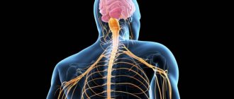
Physiology of the cerebellum, its influence on motor and autonomic functions of the body
The cerebellum is a suprasegmental formation that does not have a direct connection with the executive apparatus. The cerebellum is part of the extrapyramidal system. It consists of two hemispheres and a worm located between them. The outer surfaces of the hemispheres are covered with gray matter - the cerebellar cortex, and accumulations of gray matter in the white matter form the cerebellar nuclei.
The cerebellum receives impulses from receptors in the skin, muscles and tendons through the spinocerebellar tract and through the nuclei of the medulla oblongata (from the spinobulbar tract). Vestibular influences also come from the medulla oblongata to the cerebellum, and visual and auditory influences from the midbrain. The corticopontine-cerebellar tract connects the cerebellum with the cerebral cortex. In the cerebellar cortex, the representation of various peripheral receptors has a somatotopic organization. In addition, there is an orderliness in the connections of these zones with the corresponding perceptive areas of the cortex. Thus, the visual zone of the cerebellum is connected with the visual zone of the cortex, the representation of each muscle group in the cerebellum is connected with the representation of the muscles of the same name in the cortex, etc. This correspondence facilitates the joint activity of the cerebellum and the cortex in controlling various functions of the body.
Efferent impulses from the cerebellum travel to the red nuclei of the reticular formation, medulla oblongata, thalamus, cortex and subcortical nuclei.
The cerebellum is involved in the regulation of motor activity. Electrical stimulation of the surface of the cerebellum causes movements of the eyes, head and limbs, which differ from cortical motor effects in their tonic nature and long duration. The cerebellum regulates the change and redistribution of skeletal muscle tone, which is necessary for the organization of normal posture and motor acts.
The functions of the cerebellum were studied in the clinic with its lesions in humans, as well as in animals by removal (extirpation of the cerebellum) (L. Luciani, L. A. Orbeli). As a result of loss of cerebellar functions, movement disorders occur:
• atony (sharp drop and improper distribution of muscle tone);
• astasia (impossibility of maintaining a stationary position, continuous rocking movements, trembling of the head, torso and limbs);
• asthenia (increased muscle fatigue);
• ataxia (impaired coordinated movements, gait, etc.).
The cerebellum also influences a number of autonomic functions, such as the gastrointestinal tract, blood pressure levels, and blood composition.
Thus, the integration of a wide variety of sensory influences, primarily proprioceptive and vestibular, occurs in the cerebellum. The cerebellum was even previously considered the center of balance and regulation of muscle tone. However, its functions, as it turned out, are much broader—they also cover the regulation of the activity of vegetative organs. The activity of the cerebellum occurs in direct connection with the cerebral cortex, under its control.
Reticular formation of the brainstem. Descending and ascending influences of the reticular formation of the brainstem. Participation of the reticular formation in the formation of the integral activity of the body.
Reticular formation of the brainstem
– accumulation of polymorphic neurons along the brain stem.
Physiological feature of neurons of the reticular formation:
1) spontaneous bioelectrical activity. Its causes are humoral irritation (increased levels of carbon dioxide and biologically active substances);
2) fairly high excitability of neurons;
3) high sensitivity to biologically active substances.
The reticular formation has extensive bilateral connections with all parts of the nervous system; according to its functional significance and morphology, it is divided into two sections:
1) rastral (ascending) section – reticular formation of the diencephalon;
2) caudal (descending) – reticular formation of the hindbrain, midbrain, and pons.
The physiological role of the reticular formation is the activation and inhibition of brain structures.
The influences of the Russian Federation can be generally divided into downward and upward. In turn, each of these influences has an inhibitory and excitatory effect.
The ascending influences of the RF on the cerebral cortex increase its tone and regulate the excitability of its neurons without changing the specificity of responses to adequate stimulation. RF affects the functional state of all sensory areas of the brain, therefore, it is important in the integration of sensory information from different analyzers.
RF is directly related to the regulation of the wakefulness-sleep cycle. Stimulation of some structures of the Russian Federation leads to the development of sleep, stimulation of others causes awakening. G. Magun and D. Moruzzi put forward the concept according to which all types of signals coming from peripheral receptors reach the RF collaterals of the medulla oblongata and the pons, where they switch to neurons that give ascending pathways to the thalamus and then to the cerebral cortex.
Excitation of the RF of the medulla oblongata or pons causes synchronization of the activity of the cerebral cortex, the appearance of slow rhythms in its electrical parameters, and sleep inhibition.
Excitation of the midbrain RF causes the opposite effect of awakening: desynchronization of the electrical activity of the cortex, the appearance of fast low-amplitude β-like rhythms in the electroencephalogram.
G. Bremer (1935) showed that if the brain is cut between the anterior and posterior colliculi, the animal stops responding to all types of signals; if a transection is made between the medulla oblongata and the midbrain (while the RF retains its connection with the forebrain), then the animal reacts to light, sound and other signals. Consequently, maintaining an active analyzing state of the brain is possible while maintaining connections with the forebrain.
The activation reaction of the cerebral cortex is observed upon stimulation of the RF of the medulla oblongata, midbrain, and diencephalon. At the same time, irritation of some nuclei of the thalamus leads to the emergence of limited local areas of excitation, and not to its general excitation, as happens with irritation of other parts of the Russian Federation.
RF of the brain stem can have not only an excitatory, but also an inhibitory effect on the activity of the cerebral cortex.
The descending influences of the brainstem RF on the regulatory activity of the spinal cord were established by I.M. Sechenov (1862). They showed that when the midbrain is irritated by salt crystals, the frog's paw withdrawal reflexes arise slowly, require stronger stimulation, or do not appear at all, i.e., they are inhibited.
G. Magun (1945-1950), applying local irritations to the RF of the medulla oblongata, found that when certain points are irritated, the flexion reflexes of the forepaw, knee, and cornea are inhibited and become sluggish. When the RF was stimulated at other points of the medulla oblongata, these same reflexes were evoked more easily and were stronger, i.e. their implementation was facilitated. According to Magun, inhibitory influences on spinal cord reflexes can only be exerted by the RF of the medulla oblongata, and facilitatory influences are regulated by the entire RF of the brainstem and SC
.27. Thalamus. Functional characteristics and features of nuclear groups of the thalamus.
The thalamus is a paired formation, the largest accumulation of gray matter in the diencephalon.
Topographically, anterior, middle, posterior, medial and lateral groups of nuclei are distinguished.
By function they distinguish:
1) specific:
• switching, relay. They receive primary information from various receptors. The nerve impulse travels along the thalamocortical tract to a strictly limited area of the cerebral cortex (primary projection zones), due to which specific sensations arise. The nuclei of the ventrabasal complex receive impulses from skin receptors, proprioceptors of tendons, and ligaments. The impulse is sent to the sensorimotor zone, and the body’s orientation in space is regulated. The lateral nuclei switch impulses from visual receptors to the occipital visual area. The medial nuclei react to a strictly defined sound wavelength and conduct an impulse to the temporal zone;
• associative (internal) nuclei. The primary impulse comes from the relay nuclei, is processed (an integrative function is carried out), transmitted to the associative zones of the cerebral cortex, the activity of the associative nuclei increases under the action of a painful stimulus;
2) nonspecific nuclei. This is a nonspecific pathway for transmitting impulses to the cerebral cortex; the frequency of the biopotential changes (modeling function);
3) motor nuclei involved in the regulation of motor activity. Impulses from the cerebellum and basal ganglia go to the motor zone, effecting interconnection, coordination, sequence of movements, and spatial orientation of the body.
The thalamus is the collector of all afferent information, except for olfactory receptors, and is the most important integrative center.
7) behavioral reactions. Irritation of the starting emotional zone (anterior nuclei) causes a feeling of joy, satisfaction, erotic feelings, the stopping zone (posterior nuclei) causes fear, feelings of anger, rage.
Drug and surgical treatment
Treatment of patients with cerebellar stroke includes effects on cerebral vessels and neurons, basic therapy. In particular, the effect on cerebral vessels implies recanalization (thrombolysis, mechanical thrombectomy, acetylsalicylic acid) and prevention of thrombus formation, while the effect on neurons implies neuroprotection and stimulation of neuronal plasticity. Basic therapy consists of correcting breathing disorders, regulating the functions of the cardiovascular system, controlling glucose metabolism and body temperature, normalizing water-electrolyte balance, preventing and treating complications.
Unsuccessful conservative measures require the use of more radical methods of treatment, for example, to remove a blood clot when it is detected or to remove blood clots, reducing intracranial pressure.
Symptoms of tissue damage in the frontal lobe of the brain
Among the most severe lesions of the tissues of the frontal lobes are ischemic, tissue structure, stroke with a focus of ischemia or hemorrhage in this area. Impaired blood supply to this part of the brain negatively affects cognitive abilities. The main reasons for the deterioration and cessation of blood flow in this area:
- Exacerbation of arterial hypertension.
- Atherosclerotic damage to vascular walls.
- Developmental anomalies and acquired defects of elements of the circulatory system (vascular malformations, aneurysms).
- Disorder in the hemostasis system (increased thrombosis, blood clotting disorder).
Frontal lobe syndrome occurs due to damage to the tissues of the prefrontal region, which is responsible for multi-stage thought processes, behavior and cognitive abilities. General cerebral symptoms of pathology:
- Acute pain in the frontal plane of the head.
- Nausea, often leading to bouts of vomiting.
- Dizziness, confusion.
- Sometimes increased body temperature.
Frontal lobe syndrome is classified as an organic personality disorder in the international medical classification of diseases. It often develops against the background of other pathologies - Pick's disease and Alzheimer's disease, brain tumors, trauma to the head, damage to the vascular system in this area. Specific signs of the disease:
- Activation of rudimentary reflexes (sucking, grasping, searching). Occurs when a large area of tissue is affected.
- Loss of self-control, inability to plan and manage actions.
- Loss of awareness of one's own personality.
- .
- Impossibility of abstract thinking and planning.
- Perseveration. Unconscious repeated repetition of one word, phrase, or action.
- Impaired concentration and memory functions.
Important Classification of cerebral sinuses
Similar disorders are often observed in patients with Alzheimer's disease, who experience pathological structural changes in tissues - the formation of protein structures around neurons that interfere with and disrupt the relationships between nerve cells. Alzheimer's disease often causes dementia.
Rehabilitation and restoration of the cerebellum after stroke
Stroke rehabilitation should be provided by a physician trained in rehabilitation and physical therapy, as well as by other physicians with experience or primary training in stroke rehabilitation—occupational therapists, physical therapists, nurses, social workers, and nutritionists. The patient and his family are also included in the core team.
Immediately after a patient has had a stroke, doctors usually allow local massage procedures. It is advisable to start massage after a stroke as early as a person’s health allows. If the patient is in normal condition, then the procedure begins on the second day. They start with segmental and point massage. The flexion groups are massaged on the lower extremities, and the extensor groups are massaged on the upper extremities.
The first massage sessions last for 5-10 minutes, with a gradual increase in time up to half an hour. When prescribing a massage, it is very important to remember the contraindications: fever, high blood pressure, headaches, heart problems, problems with the respiratory system.
Symptoms of cerebellar damage
Failure in the functioning of this organ can be determined by minor changes in motor activity or the inability to hold a posture in one position. Thus, the patient may not have a reflex to point his leg in the direction of falling, but a small push is enough for him to fall.
In medicine, this phenomenon is called static ataxia, and its cause lies in the damage of the worm. In this state, the patient tries to spread his legs as wide as possible to maintain balance. To test this reflex, the doctor asks the sick person to stand up and bring their legs together, then close their eyes and stretch their arms forward.
If the cerebellar vermis is truly damaged, then the body usually tilts backward; if the hemispheres are damaged, then the sick person leans towards the affected lobe. If the condition is severe, the patient will not be able to stand up, and difficulties will arise in maintaining a sitting posture.
With extensive damage to the hemispheres, the appearance of dynamic or kinetic ataxia is noted. In this case, the patient loses the ability to accurately carry out movements. Diagnosis of such disorders consists of performing certain exercises or tests under the supervision of a doctor.
With eyes closed, the patient is asked to stand up straight, then stretch his arms straight out in front of him and touch the tip of his nose. If one of the lobes is damaged, the index finger deviates towards it.
It is proposed to rotate your hands simultaneously and in one direction with your eyes closed; if one of the hemispheres is disrupted, the hand on its side will lag behind.
In the supine position, you need to lift one of your legs and then lower the heel of that leg onto the knee of the other. If everything went well, the doctor suggests lowering the heel further down the bone. If the leg begins to slip, this indicates the development of pathology.
Another simple way to test the functions of this organ is to be able to hold a full vessel of water without spilling a drop.
The patient has a deterioration in speech: rhythm appears, sentences lose meaning, and words are stressed incorrectly. Tremors in the limbs and changes in handwriting are also observed.
If the disturbances affect the cerebellar nuclei, then the patient experiences convulsive contractions of the muscles of the limbs, inertial trembling in the fingers when completing a movement, the movement of the eyeballs cannot be controlled, rhythmic speech appears and muscle tone decreases.
The cerebellar peduncles transfer received information from parts of the brain to the cortex and nuclei, and back through efferent communication they give a command to perform a certain task, therefore, when this structure is damaged, different symptoms are observed. For example, if the upper pair of crura and the dentate nucleus are damaged, choreic hyperkinesis develops, which is characterized by rapid chaotic movements of the facial muscles, reminiscent of a grimace, the autonomic functions of the cerebellum cease to be performed - breathing becomes erratic, cardiac arrhythmia and surges in blood pressure may be observed.
A number of diseases, both congenital and acquired, are also characterized by atrophy of the structures of this organ. For example, in Marie-Foy-Alajouanine disease, Purkinje neurons, the granular layer of the cerebellar cortex, and part of the vermis are damaged. In this case, the following symptoms are noted: gait disturbance, decreased tone in the lower extremities. There may be little or no hand shaking. Such changes are most often characteristic of middle-aged and elderly people.
With such a congenital disease as Chiari disease, a low location of the cerebellar tonsils is noted. Depending on the type of disease, the manifestation of clinical signs may differ, but most often there is pain in the neck and its muscles, nausea and vomiting occurs, independent of food intake. With different degrees of prolapse, the following symptoms may also appear: speech dysfunction, noise in the head, frequent dizziness, impaired breathing and muscle tone in the limbs, numbness of the arms and legs, changes in blood pressure.
Prevention
Prevention of cerebellar stroke involves preventing the development of acute cerebral circulatory disorders. It consists of introducing a healthy lifestyle, balanced nutrition, maintaining adequate body weight, abstaining from smoking and adequate drug treatment of heart and vascular diseases, diabetes and other diseases.
The mortality rate from this disease ranks second in the world. This frightening fact should motivate patients at risk to engage in stroke prevention.