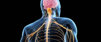
What is the sympathetic nervous system
This is part of the autonomic nervous system, which covers the upper lumbar and thoracic spinal cord, mesenteric nodes, cells of the sympathetic border trunk, and solar plexus. In fact, this section of the nervous system is responsible for the vital activity of cells and maintaining the functionality of the whole organism. In this way, a person is provided with an adequate perception of the world and the body’s reaction to the environment. The sympathetic and parasympathetic departments work together and are structural elements of the central nervous system.
The essence of interaction
In order for a person to adapt and adapt to any situation - external or internal threat, the sympathetic as well as the parasympathetic parts of the autonomic nervous system must interact closely. However, they have the exact opposite effect on the human body.
Parasympathetics are characterized by:
- lower blood pressure;
- reduce breathing rate;
- expand the lumen of blood vessels;
- constrict the pupils;
- adjust the concentration of glucose in the bloodstream;
- improve the digestive process;
- tone smooth muscles.
Protective reflexes also include the introduction of parasympathetic activity - sneezing, coughing, retching. It is inherent for the sympathetic department of the autonomic nervous system to increase the parameters of the cardiovascular system - pulse rate and blood pressure numbers, and increase metabolism.
A person learns that the sympathetic department predominates by feeling fever, tachycardia, restless sleep and fear of death, and sweating. If more parasympathetic activity is present, the changes will be different - cold, clammy skin, bradycardia, fainting, excessive salivation and shortness of breath. With balanced functioning of both departments, the activity of the heart, lungs, kidneys, and intestines corresponds to the age norm and the person feels healthy.
Structure
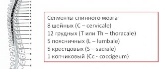
On both sides of the spine there is a sympathetic trunk, which is formed from two symmetrical rows of nerve ganglia. They are connected to each other using special bridges, forming a so-called “chain” with an unpaired coccygeal node at the end. This is an important element of the autonomic nervous system, which is characterized by autonomous operation. To ensure the required physical activity, the design distinguishes the following departments:
- cervical of 3 nodes;
- thoracic, which includes 9-12 nodes;
- area of the lumbar segment of 2-7 nodes;
- sacral, consisting of 4 nodes and one coccygeal.
From these sections, impulses move to the internal organs and support their physiological functionality. The following structural links are distinguished. In the cervical region, the nervous system controls the carotid arteries, in the thoracic region - the pulmonary and cardiac plexuses, and in the peritoneal region - the mesenteric, solar, hypogastric, and aortic plexuses. Thanks to postganglionic fibers (ganglia), there is a direct connection with the spinal nerves.
Morphology[ | ]
The sympathetic nervous system is topographically divided into central, located in the spinal cord, and peripheral, which includes numerous nerve branches and nodes connected to each other.
Central department[ | ]
The centers of the sympathetic system (Jacobson's spinal center) are located in the lateral horns of the thoracic and lumbar segments of the gray matter of the spinal cord in the thoracolumbar region (segments from the last cervical to the third lumbar inclusive).
Peripheral department[ | ]
The peripheral part of the sympathetic nervous system is formed by efferent and sensory neurons with their processes located in the paravertebral and prevertebral nodes remote from the spinal cord.
Sympathetic trunks[ | ]
Main article: Sympathetic trunk
There are sympathetic trunks on both sides of the spine. They look like chains formed by first-order nodes and stretch from the base of the skull to the coccyx, where they converge into the unpaired coccygeal node. The sympathetic trunk is divided into cervical, thoracic, lumbar and sacral sections. The nodes in each trunk chain are connected by internodal branches. In the lumbar and sacral regions, two chains are connected by transverse branches.
Sympathetic fibers[ | ]
Sympathetic fibers exit the spinal cord from the I-II thoracic to the II-IV lumbar segment. Along their course, sympathetic fibers are separated from motor somatic ones, and then, in the form of white connecting branches,
enter the thoracic and upper lumbar nodes of the border sympathetic trunk.
Gray communicating branches
and
visceral branches
depart from the nodes of the sympathetic trunk . The gray communicating branches connect to the spinal nerves and in their composition go to the smooth muscles of the blood vessels, the sebaceous and sweat glands, as well as to the skeletal muscles (they regulate its trophism). The visceral branches innervate the internal organs.
The sympathetic nervous system is characterized by a generalized influence
, while sympathetic fibers innervate all organs without exception.
Generalization, that is, expansion of the area of excitation
, is achieved due to the fact that preganglionic fibers in the nodes of the sympathetic trunk branch repeatedly
,
and the number of postganglionic fibers can be several tens of times greater than the number of preganglionic ones. This allows a relatively small number of central sympathetic neurons to provide innervation to all organs and tissues.
Ganglia of the second order[ | ]
In addition to the first-order nodes (vertebral ganglia), the peripheral part of the sympathetic nervous system also includes second-order nodes (prevertebral ganglia). They participate in the formation of plexuses (for example, celiac and solar).
Functions
The sympathetic system is an integral part of human anatomy, located closer to the spine, and is responsible for the proper functioning of internal organs. Controls the flow of blood through vessels and arteries, filling their branches with vital oxygen. Among the additional functions of this peripheral structure, doctors highlight:
- increasing the physiological abilities of muscles;
- decrease in the absorption and secretory capacity of the gastrointestinal tract;
- increased blood sugar and cholesterol;
- regulation of metabolic processes, metabolism;
- providing increased strength, frequency and rhythm of the heart;
- the flow of nerve impulses to the fibers of the spinal cord;
- dilated pupils;
- innervation of the lower extremities;
- increased blood pressure;
- release of fatty acids;
- decreased tone of smooth muscle fibers;
- adrenaline rush in the blood;
- increased sweating;
- stimulation of sensitive centers;
- dilation of the bronchi of the respiratory system;
- decreased saliva production.
Physiology[ | ]
General significance of the sympathetic nervous system[ | ]
The sympathetic nervous system is activated during stress reactions, which is why it is sometimes called the “fight or flight” system. Under the influence of this department, the rate of metabolic processes in organs and tissues increases, heart contractions intensify, the amount of oxygen supplied to the muscles increases, and digestion processes are inhibited.
Adaptive-trophic function of the sympathetic nervous system[ | ]
The sympathetic nervous system performs an adaptive-trophic function, that is, it ensures the body’s adaptation to changing environmental conditions by changing the level of metabolism in organs and tissues. The performance of the adaptive-trophic function by the sympathetic department was suggested by I.P. Pavlov when, during experiments on dogs, he discovered a branch of the sympathetic nerve going to the heart and, when excited, increasing heart contractions without changing their frequency. Subsequently, this idea was developed by Soviet physiologists L.A. Orbeli and A.G. Ginetsinsky, who discovered increased contractions of the tired skeletal muscle of a frog when the sympathetic nerve coming to it was irritated (the strength of muscle contractions increased, the excitability and contractility of the muscle increased). A similar experiment was repeated with mammalian muscles. The results of the experiments formed the basis of L. A. Orbeli’s theory of the adaptive-trophic function of the sympathetic department of the autonomic nervous system.
Innervation of individual organs and influence on their work[ | ]
1. Sympathetic fibers from the superior cervical ganglion (branches of the internal carotid nerve n.caroticus internus, forming the plexus caroticus internus around the carotid artery) innervate the pupillary dilator
, that is, under the influence of the sympathetic department of the autonomic nervous system,
the pupil dilates
. When the upper cervical ganglion is affected, there is a narrowing of the pupil on the side of the same name.
2. Sympathetic fibers arising from the superior cervical ganglion (external carotid nerves nn.carotici externi, forming the plexus caroticus externus around the carotid artery) innervate the salivary glands
.
Under the influence of this part of the nervous system, saliva is secreted in a small volume
.
When the sympathetic nerves in the glands are irritated, the blood supply is limited, so the consistency of the saliva is thick and viscous
.
Thus, the main function of the sympathetic nervous system in this case is the retention of salivation
(stress causes dry mouth).
3. Sympathetic fibers innervate the sweat glands of the skin in all areas of the body. Sweat glands are innervated by cholinergic nerve fibers - acetylcholine is released as a mediator at their endings. Under the influence of the sympathetic department of the autonomic nervous system, sweating increases
.
4. The sympathetic nerves innervating the heart (cardiacus cervicalis superior, n. cardiacus cervicalis medius, n. cardiacus cervicalis inferior, n. cardiaci thoracici) arise from the three upper cervical and five upper thoracic sympathetic nodes.
Two cardiac plexuses are formed from the sympathetic and parasympathetic nerves: the superficial, plexus cardiacus superficialis, located between the aortic arch and the bifurcation of the pulmonary trunk; and deep, plexus cardiacus profundus, located between the aortic arch and the tracheal bifurcation. The plexuses contain groups of ganglion cells and nerve ganglia. The branches of these plexuses then pass into a single intraorgan cardiac plexus.
The superior cardiac nerve is involved in the formation of the superficial and deep plexuses. The middle cardiac nerve, inferior cardiac nerve and thoracic cardiac nerves enter the deep cardiac plexus.
The impulses of these nerves increase heart contractions
and
speed up their rhythm
.
5. In most tissues, all vessels, except capillaries, are innervated by fibers of the sympathetic nervous system.
The sympathetic nervous system constricts blood vessels
and
increases blood pressure
, thereby removing blood from organs whose functions in a stressful situation are not necessary for the survival of the body, and, on the contrary, increases blood flow to organs that are vital and necessary during stress (for example, during stress, the skin turns pale because blood flow in it decreases in favor of skeletal muscles).
- The impulses of sympathetic nerves in the muscle tissue of small arteries and arterioles leads to an increase in vascular resistance and a decrease in blood flow.
- The impulses of sympathetic nerves in the smooth muscle tissue of large blood vessels, especially veins, leads to a decrease in their volume, which promotes the movement of blood towards the heart.
6. The sympathetic department has a complex effect on the respiratory tract and lungs. The main task it solves is to make breathing easier
during a stressful situation to more fully supply the body's cells with oxygen.
- The beginning of the respiratory tract - the nasal cavity - is innervated by sympathetic nerves, the preganglionic fibers of which arise from the upper thoracic segments of the spinal cord, and the postganglionic fibers originate in the superior cervical ganglion. Having reached the mucous membrane of the nasal cavity, the sympathetic nerves narrow the lumen of the blood vessels
and
reduce the secretion of mucus
.
- The larynx also has sympathetic innervation. Preganglionic fibers again begin in the upper thoracic segments of the spinal cord, and postganglionic fibers begin in the superior cervical ganglion. From the cervical ganglion to the larynx, sympathetic fibers go as part of the branches of the laryngopharyngeal nerves and as part of the nerve plexuses of the arteries. Under the influence of the sympathetic nervous system, the vessels of the larynx narrow
, and
the secretion of cells of the glandular epithelium
of the laryngeal mucosa is inhibited.
- Sympathetic nerves, the preganglionic fibers of which begin in the lower cervical and upper thoracic segments of the spinal cord, and the postganglionic fibers in the thoracic ganglia, innervate the tracheal mucosa and its smooth muscles. Under the influence of this section, the smooth muscles of the trachea relax
and
the secretion of the mucous glands slows down
, and
the blood vessels narrow
.
- As part of the pulmonary branches, sympathetic fibers come to the bronchial tree and lungs, where they innervate the mucous membrane and smooth muscles. The preganglionic fibers of these nerves begin in the upper thoracic segments of the spinal cord, and the postganglionic fibers begin in the upper thoracic ganglia. The action of the sympathetic department causes expansion of the lumen of the bronchi
and
a decrease in the secretion of the bronchial glands
.
7. Sympathetic fibers innervate smooth muscles in the walls of lymphatic vessels. They are concentrated mainly near the valves and in the places where smaller vessels transition into larger ones. Impulses from the sympathetic nervous system cause contractions of the walls of lymphatic vessels
and
an increase in lymph pressure
on the walls of blood vessels, which promotes the movement of lymph from the lymphatic capillaries to large ducts and, ultimately, to the veins of the bloodstream.
8. Sympathetic fibers innervate the walls of the hollow organs of the digestive tract. The impulses of the sympathetic department suppress the activity of the digestive organs
.
- Sympathetic fibers, the postganglionic neurons of which originate from the superior cervical ganglion, innervate the glands of the pharynx and, when excited, inhibit their secretion, and also cause contraction of the vascular walls
and provide
trophic innervation of the striated muscles
.
- Sympathetic fibers, the postganglionic fibers of which originate from the superior thoracic ganglia, innervate the glands, vascular walls and smooth muscles of the esophagus. In the walls of the esophagus, impulses from the sympathetic department constrict blood vessels, suppress the secretion of glands
and
inhibit the contraction of smooth muscles
, and also provide trophic innervation to the striated muscles of the upper third of the esophagus.
- Sympathetic nerves, the postganglionic fibers of which originate from the celiac plexus, innervate the smooth muscles, glands and vessels of the stomach wall. The impulses of the sympathetic department suppress the functions of the stomach: they inhibit the secretion of enzymes, hydrochloric acid and mucus; constrict blood vessels and suppress muscle contractions.
- The small intestine is innervated by sympathetic fibers, the postganglionic neurons of which arise from the superior mesenteric, as well as partially from the celiac and hepatic plexuses. Excitation of sympathetic fibers causes relaxation of smooth muscles, vasoconstriction and suppression of the secretion of glands of the small intestine.
- Efferent innervation of various parts of the colon is carried out by sympathetic fibers, the postganglionic neurons of which lie in the superior and inferior mesenteric, intermesenteric, superior and inferior pelvic plexuses. Excitation of the sympathetic department inhibits the peristalsis of the colon and the secretion of its glands, constricts blood vessels.
Sympathetic and parasympathetic nervous system
The interaction of both structures supports the vital functions of the whole organism; dysfunction of one of the departments leads to serious diseases of the respiratory, cardiovascular, and musculoskeletal systems. The effect is exerted through nerve tissues consisting of fibers that provide excitability of impulses and their redirection to internal organs. If one of the diseases predominates, the choice of high-quality medications is made by the doctor.
- Is it possible to get a haircut during the full moon?
- Abdominal vacuum is an exercise for weight loss. How to properly do a stomach vacuum at home
- Hearing aids for the elderly
Any person should understand the purpose of each department, what functions it provides to maintain health. The table below describes both systems, how they can manifest themselves, and what effect they can have on the body as a whole:
| Nervous sympathetic structure | Parasympathetic nervous structure | |
| Department name | Functions for the body | Functions for the body |
| Cervical region | Dilated pupils, decreased salivation | Constriction of pupils, control of saliva secretion |
| Thoracic region | Bronchial dilatation, decreased appetite, increased heart rate | Narrowing of the bronchi, decreased heart rate, increased digestion |
| Lumbar | Inhibition of intestinal motility, production of adrenaline | Possibility of gallbladder stimulation |
| Sacral section | Bladder relaxation | Bladder contraction |
Differences between the sympathetic and parasympathetic nervous systems
Sympathetic nerves and parasympathetic fibers can be located in a complex, but at the same time they provide different effects on the body. Before contacting your doctor for advice, it is recommended to find out the differences between the sympathetic and parasympathetic systems in structure, location and functionality in order to approximately understand the potential focus of pathology:
- Sympathetic nerves are located locally, while parasympathetic fibers are more discrete.
- Preganglionic sympathetic fibers are short and small, and parasympathetic fibers are often elongated.
- The nerve endings of the sympathetic are adrenergic, while the parasympathetic are cholinergic.
- The sympathetic system is characterized by white and gray connecting branches, but these are absent in the parasympathetic nervous system.
Embryology[ | ]
The ganglion plate is the embryonic source for the sympathetic system. It is divided into somites, which differentiate into the sympathetic and parasympathetic systems. The sympathetic level includes the cervical and thoracic somites.
During embryogenesis, the peripheral part of the sympathetic nervous system is formed as a result of the migration of sympathetic neuroblasts. Migration occurs along the fibers of the spinal cord segmentally. There are several “waves of migration”. As a result of the first wave, a “primary” sympathetic trunk is formed, represented in the neck by the superior (cranial), inferior (posterior), cervical and stellate ganglia. The second wave arises from the primary one, mainly from the dorsal sections. As a result, a “secondary” sympathetic trunk is formed.
What diseases are associated with the sympathetic system?
With increased excitability of the sympathetic nerves, nervous conditions develop that cannot always be eliminated by self-hypnosis. Unpleasant symptoms are reminiscent of themselves already in the primary form of the pathology and require immediate medical attention. The doctor recommends to beware of the following diagnoses and to consult your doctor in time for effective treatment:
- reflex sympathetic dystrophy syndrome;
- peripheral autonomic failure;
- Raynaud's phenomenon;
- nocturnal enuresis.
Treatment
If the sympathetic nerves are excited, it is necessary to contact the attending physician and promptly begin intensive therapy that can stabilize the general condition of the clinical patient. Pathology can occur under the influence of provoking factors, which must first be identified and eliminated. In order not to bring the situation to a critical limit, and to obtain a positive treatment result, it is recommended to pay attention to the following pharmacological groups:
- benzodiazepine tranquilizers (Phenazepam, Alprazolam);
- neuroleptics (Thioridazine, Periciazine, Azaleptin);
- antidepressants (Amitriptyline, Trazodone, Escitalopram, Maprotiline, Fluvoxamine);
- anticonvulsants (Carbamazepine, Pregabalin).