The spinal cord is a rather complex system that is responsible for many processes in the body and which is quite difficult to understand on your own. Basic knowledge can be obtained by studying anatomy at school, but when it comes to a more in-depth analysis, many incomprehensible issues arise.
Let's try to figure out what the spinal cord is, how it is structured, what functions it performs, and just understand why it is needed at all.
Spinal cord as part of the nervous system
The spinal cord is one of the components of the human nervous system. In Latin its name looks like medulla spinalis.
It is a thick cylindrical tube with a narrow channel located inside it. It is located in the spinal canal, or, more simply, inside the spine.
This organ has a rather complex structure and segmental structure. The main function of this organ is the transmission of various impulses and signals from the human brain to specific organs. In addition, it performs reflex activity, that is, it is responsible for human reflexes, both simple and more complex reflexes.
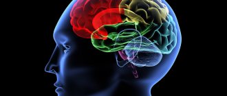
Human nervous system
The important role of the spinal cord in the human body
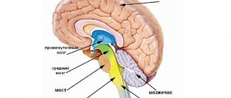
The brain is the most important organ of the human body. Without it, no movements, sensations or reactions would be possible. It is a kind of control center of the entire body, of all nerve endings. Without the reliable functioning of this organ, we would not be able to make a single movement or feel anyone’s touch.
Although the brain also plays an important role, its functions would not be so complete without the spinal cord. For example, in order to see what is happening around us, we need the work of the optic nerve, which obeys the brain. But only thanks to the guidance of the spinal cord, we can look in different directions by turning our pupils. The same is true with the ability to cry. Although we can experience negative emotions without the spinal cord, we cannot cry with its participation.
When some actions occur consciously, we need instructions coming from the brain. When a process becomes automatic, it occurs at the reflex level with the help of the spinal cord. Consequently, this small but very important organ requires our close attention and careful treatment!
The meaning of the spinal cord
There are only two main and most important functions:
- Reflex. To put it simply, a whole series of reflex arcs are closed on this organ. It is thanks to this that reflexes are carried out (the so-called spinal reflexes).
- Conductor. The organ in this case acts as a conductor. It conducts signals that come from various organs to the brain. It is through this organ that the brain receives all information and processes it. Everything works the same way in the opposite direction.
Spinal cord location
The organ is located in the spinal canal (located inside the human spine). This canal is quite long and practically reaches the lower vertebrae. In fact, this is a special canal, which is an oblong hole in which the spinal cord lies. From the sides it is protected by vertebrae, as well as intervertebral discs.
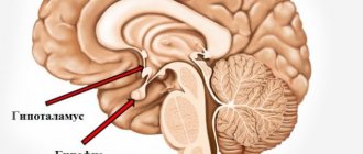
The organ is also located at the lower edge of the foramen magnum, where connections with the brain occur. It is in this place that there is a huge number of roots that directly connect to the human brain. This connection is called the left and right spinal nerves.
Spinal cord location
The bottom ends at the level of 1-11 vertebrae. Afterwards the organ turns into a thin terminal filament. In fact, it is still the spinal cord, because it contains nervous tissue.
Structure, functions and age-related characteristics of the spinal cord
The spinal cord in appearance is a long, cylindrical cord, flattened from front to back, with a narrow central canal inside. Externally, the spinal cord has three membranes - hard, arachnoid, and soft.
The spinal cord is located in the spinal canal and at the level of the lower edge of the foramen magnum passes into the brain. Below, the spinal cord ends at the level of the I-II lumbar vertebrae with a narrowing - the medullary cone, from which the terminal filament stretches down. In its upper sections it contains nervous tissue, and below the level of the second sacral vertebra it is a connective tissue formation, which is a continuation of all three membranes of the spinal cord. The terminal filament ends at the level of the body of the second coccygeal vertebra, fused with its periosteum. The filum terminale is surrounded by long roots of the lower spinal nerves, which form a bundle in the spinal canal called the cauda equina.
The length of the spinal cord in an adult is on average 43 cm (for men - 45, for women 41-42 cm), weight - about 34-38 g, which is approximately 2% of the mass of the brain.
In the cervical and lumbosacral sections of the spinal cord, two noticeable thickenings are found - cervical and lumbosacral. Their formation is explained by the accumulation in these parts of the brain of a large number of nerve cells and fibers innervating the upper and lower extremities.
The anterior median fissure is visible on the anterior surface of the spinal cord. The posterior median sulcus runs along the midline of the posterior surface of the brain. The anterior fissure and posterior sulcus are the boundaries that divide the spinal cord into right and left symmetrical halves.
On the anterior surface of the spinal cord, on each side of the median fissure, there runs the anterior lateral groove, which is the site of exit from the spinal cord of the anterior (motor) root. This groove also serves as a boundary on the surface of the spinal cord between the anterior and lateral cords. On the posterior surface of the spinal cord, on each half of it, there is a posterior lateral groove, the place where the dorsal (sensitive) root enters the spinal cord. This groove serves as the boundary between the lateral and posterior cords of the spinal cord.
The anterior roots of the spinal nerves consist of processes of motor (motor) nerve cells located in the anterior horn of the gray matter of the spinal cord.
The dorsal root is represented by a collection of central processes of pseudounipolar (sensitive) cells penetrating into the spinal cord, the bodies of which form the spinal ganglion, which lies at the junction of the dorsal root with the anterior one.
Along the entire length of the spinal cord, 31 pairs of spinal nerve roots arise from each side. The segment of the spinal cord corresponding to two pairs of spinal nerve roots (two anterior and two posterior) is called a spinal cord segment. The human spinal cord consists of 31 segments. There are 8 cervical, 12 thoracic, 5 lumbar, 5 sacral and 1 coccygeal segments of the spinal cord. The length of the spinal cord is significantly less than the length of the spinal column, therefore the serial number of the spinal cord segment and the level of their position, starting from the lower cervical region, do not correspond to the serial numbers of the vertebrae of the same name.
The spinal cord is made up of gray and white matter. Gray matter consists of nerve cell bodies and nerve fibers—the processes of nerve cells. The white matter is formed only by nerve fibers - processes of nerve cells of both the spinal cord itself and the brain. Gray matter occupies a central position in the spinal cord. The central canal runs through the center of the gray matter. Outside the gray matter is the white matter of the spinal cord.
In each half of the spinal cord, gray matter forms gray columns. The right and left gray pillars are connected by a transverse plate - a gray commissure, in the center of which the opening of the central channel is visible. Anterior to the central canal is the anterior commissure of the spinal cord, posteriorly is the posterior commissure. On a cross section of the spinal cord, the gray columns together with the gray commissure have the shape of the letter “H” or a butterfly with outstretched wings. The lateral projections of gray matter are called horns. There are paired, wider anterior horns and narrow, also paired posterior horns. The anterior horns of the spinal cord contain large nerve cells called motor neurons. Their long processes, neurites, form the bulk of the fibers of the anterior roots of the spinal nerves. The neurons located in each anterior horn form five large nuclei (nerve centers): two medial and two lateral, as well as a central nucleus. The processes of the cells of these nuclei are directed to the skeletal muscles. In the middle of the posterior horn is its own nucleus. It consists of so-called interneurons, the processes of which (axons) are sent to the anterior horn, and also pass through the anterior white commissure to the opposite side of the spinal cord. At the base of the dorsal horn there is another nucleus formed by large interneurons with highly branched dendrites. The nerve fibers (sensitive) of the dorsal roots, which are processes of nerve cells whose bodies are located in the intervertebral spinal nodes, end on the nerve cells of the nuclei of the dorsal horns.
The intermediate zone of gray matter of the spinal cord is located between the anterior and posterior horns. In this zone, from the VIII cervical to the II lumbar segment, there are projections of gray matter - the lateral horns. The lateral horns contain the centers of the sympathetic part of the autonomic nervous system in the form of groups of nerve cells united in the lateral intermediate substance. The axons of these cells pass through the anterior horn and exit the spinal cord as part of the anterior roots of the spinal nerves.
The white matter contains three paired cords. The anterior cord is located between the median fissure on the medial side and the anterior lateral groove on the lateral side (the exit point of the anterior roots). The posterior funiculus is located between the posterior median and posterior lateral grooves, the lateral funiculus is between the anterior and posterior lateral grooves. The white matter consists of nerve fibers along which nerve impulses travel either upward, towards the brain, or downwards, to the lower segments of the spinal cord. Deep in all cords, in close proximity to the gray matter, lie short intersegmental nerve fibers connecting adjacent segments of the spinal cord. These fibers establish a connection between the remaining segments, so these bundles are separated into their own segmental apparatus of the spinal cord. The fibers of the neurons of the spinal ganglia, penetrating the spinal cord as part of the dorsal roots, partially enter the dorsal horn, and partially continue their path in various directions. Some fibers end on interneurons of the dorsal horns of the opposite side or on neurons of the autonomic nervous system of the lateral horns. Other fibers are part of the posterior cords and rise upward to the brain. They belong to the ascending pathways of the spinal cord.
The conducting tracts of the spinal cord are located outward from its intersegmental (intrinsic) bundles. Impulses from sensory and interneurons of the spinal cord to the brain pass along the pathways in the ascending direction. In the descending direction, impulses follow from the overlying nerve cells of the brain to the motor neurons of the spinal cord.
The functional significance of the spinal cord lies in the fact that it conducts excitation in the ascending and descending directions and carries out a reflex function. The spinal cord connects the brain to the trunk and limbs. From the skin, muscles, tendons and joints, as well as from internal organs, afferent impulses arrive in an ascending direction to various parts of the brain. In the descending direction, the regulatory influences of the brain are transmitted to the working organs and systems of the human body. Afferent and efferent influences are transmitted along the spinal roots. When the spinal roots are damaged, movement disorders occur. The spinal cord carries out a regulatory function as a single unit with the brain. It is involved in the implementation of all complex motor and autonomic functions of the body, which can be the result of the reflex activity of the spinal cord itself or the overlying parts of the brain.
The spinal cord of a newborn is 14 cm long. The lower border of the brain is at the level of the lower edge of the second lumbar vertebra. By the age of two, the length of the spinal cord reaches 20 cm, and by the age of 10, compared with the neonatal period, it doubles. The thoracic segments of the spinal cord grow most rapidly. The weight of the spinal cord in a newborn is about 5.0 g (0.1% of body weight, in adults - 0.04%). In children of one year, the mass of the spinal cord is about 10 g. By three years, the mass of the spinal cord exceeds 13 g, by 7 years it is approximately 19 g, by 14 years - on average 22 g. In a newborn, the central canal is wider than in an adult. The decrease in its lumen occurs mainly during the 1st-2nd years, as well as in later age periods, when the mass of gray and white matter increases. The volume of gray matter of the spinal cord increases rapidly, especially due to its own bundles of the segmental apparatus, the formation of which occurs at an earlier time compared to the long pathways that form the connections of the spinal cord with the brain.
Topography and shape of the spinal cord
Let's look at the features of location (topography) and shape.
To do this, consider a number of features:
- The average length is 42-43 centimeters. In men, the length is often several centimeters longer, while in women, on the contrary, it is shorter.
- Weight 33-39 grams.
- There is a middle gap on the front part, it is clearly visible. You can notice that it seems to be growing into the organ. In essence, it creates a kind of partition that divides the brain into two sections.
- In the cervical and lumbosacral regions it is possible to
- mark two fairly serious thickenings. This is due to the fact that innervation of the upper and lower extremities occurs here. In simple terms, this is where the nerve endings from the limbs “attach” to the spinal cord, which
- Allows them to transmit the necessary signals.
- The spinal cord is topographically practically not connected with the vertebrae. The various sections are not located depending on a specific vertebra or several vertebrae.
The increase in volume in these areas is due to the fact that this is where the largest number of nerve cells are located, as well as fibers through which signals are transmitted from the limbs and back.
Despite the fact that the spine is a kind of “storage place” for the organ, the location of the nerve endings, especially in the lower part of the spine, does not correspond to specific vertebrae. This is due to the fact that the length of the spinal cord is less than the length of the human spine.
That is why it is necessary for doctors to know the exact location of each of the segments, because they will not be able to navigate along the spine.
Location in the spine
Structural and functional characteristics of the spinal cord.
Spin. the brain is a cord about 45 cm long in men and about 42 cm in women, has a segmental structure (31-33 segments); each of its sections is associated with a specific one. part of the body. Includes five sections: cervical, thoracic, lumbar, sacral and coccygeal. In the process of evolution, two thickenings were formed - cervical (segments innervating the upper limbs) and lumbosacral (segments innervating the lower limbs) as a result of increased. loads on these parts of the back. brain
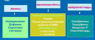
Neurons spin. brain
The total number of neurons is about 13 million. It is advisable to classify them according to several criteria:
• according to the NS department - neurons of the somatic and autonomic NS;
•as intended, i.e. according to the direction of information - efferent, afferent, intercalary;
• by influence - excitatory and inhibitory.
Efferent spin neurons. brain, related to the somatic nervous system, are effector, since they directly innervate the working organs - effectors (skeletal muscles), they are called motor neurons. Afferent neurons of the somatic NS are localized in the spinal ganglia and ganglia of the cranial nerves. Their processes, conducting afferent impulses from muscle, tendon and skin receptors, enter the corresponding spinal segments. brain and form synaptic. contacts, which can be excitatory and inhibitory. Intercalary (intermediate) neurons establish connections with spinal motor neurons. brain, with sensory neurons. They also provide. spin connection brain with the nuclei of the brain stem, and through them - with the cortex. brain They can be both exciting and inhibitory. Associative neurons form their own spin apparatus. brain, establishing connections between segments and within segments. Associative apparatus of spin. The brain is involved in coordinating posture, muscle tone, and movements of the limbs and torso. The reticular formation of the spinal cord consists of thin crossbars of the gray body, intersecting in different ways. directions, its neurons have numerous processes. The reticular formation is found at the level of the cervical segments between the anterior and posterior horns, and at the level of the upper thoracic segments - between the sides. and rear horns in a white tip adjacent to the gray one.
A collection of neurons forms various nerve centers
. In spin. The brain contains the centers for the regulation of most internal functions. organs and skeleton. muscles. Diff. centers are sympathetic. departments of the autonomic nervous system are localized in the center of the pupillary reflex, regulation of heart function, salivation, regulation of kidney function. The centers that regulate the functions of sweat glands and blood vessels, smooth muscles internally are located segmentally. organs, centers of pilomotor reflexes. Parasympathetic. innervation is received from the spinals. brain all the organs of the small pelvis: the bladder, part of the colon below its left bend, the genitals. Skeleton control centers. muscles are found in all parts of the back. brain and innervate the skeleton according to a segmental principle. muscles of the neck, diaphragm, upper limbs, trunk and lower limbs. In case of damage def. spin segments. brain or its wire. pathways develop specifically. engine disorders and sensitivity disorders.
Functions spin. brain
. There are conductive and reflex functions.
Conductor -
carried out with the help of descent. and sunrise. ways. Afferent information enters the spin. brain through the dorsal roots, efferent impulses and regulation of various functions. organs and tissues of the body are carried out through the anterior roots. Each root represents many nerves. fibers All afferent inputs to the spin. the brain carries information from 3 groups of receptors: 1) from skin receptors - pain, temperature, touch, pressure, vibration; 2) from proprioceptors - muscle, tendon, periosteum and joint membranes; 3) from internal receptors. organs - visceroreceptors. The value of afferent impulses entering the spin. brain: · participation in the coordination function of the central nervous system for the control of skeletal muscles. When afferent impulses are turned off from the working organ, its control becomes imperfect;
· participation in the processes of regulation of the functions of internal organs;
· maintaining central nervous system tone. When turned off afferent impulses decrease. total tonic activity of the central nervous system, inhibition of its activity;
· afferent impulses carry information about changes in the environment.
The hierarchy of the functions of the brain departments is such that its overlying departments control the underlying ones, and their own simpler reactions.
Characteristics of the spinal cord depending on age
Let's consider the features, depending on the age of the person:
- In a newly born child, the length of the organ is 13.5-14.5 centimeters.
- At 2 years, the length increases to 20 centimeters.
- At about 10 years old, the length can reach 29 centimeters.
- Growth ends in different ways, depending on the characteristics of a particular person’s body.
Let's look at the external features and changes depending on age:
- In infants, cervical and lumbar thickening is more noticeable than in adults. The same applies to the width of the central channel.
- The above features become almost invisible by the age of two.
- The volume of white matter grows many times faster than the volume of gray matter. This is due to the fact that the segmental apparatus is formed earlier than the pathways that connect the brain and spinal cord.
Otherwise, there are practically no age-related characteristics, because from birth the spinal cord performs almost all functions, as in an adult.
Human Spinal Cord Anatomy - Information:
The spinal cord, medulla spinalis (Greek myelos) , lies in the spinal canal and in adults is a long (45 cm in men and 41-42 cm in women), somewhat flattened from front to back cylindrical cord, which at the top (cranially) directly passes into medulla oblongata, and below (caudally) ends with a conical point, conus medullaris, at the level of the II lumbar vertebra. Knowledge of this fact is of practical importance (in order not to damage the spinal cord during a lumbar puncture for the purpose of taking cerebrospinal fluid or for the purpose of spinal anesthesia, it is necessary to insert a syringe needle between the spinous processes of the III and IV lumbar vertebrae). From the conus medullaris, the so-called filum terminale extends downwards, representing the atrophied lower part of the spinal cord, which below consists of a continuation of the membranes of the spinal cord and is attached to the II coccygeal vertebra.
The spinal cord along its length has two thickenings corresponding to the nerve roots of the upper and lower extremities: the upper one is called the cervical thickening, intumescentia cervicalis, and the lower one is called the lumbosacral thickening, intumescentia lumbosacralis. Of these thickenings, the lumbosacral one is more extensive, but the cervical one is more differentiated, which is associated with a more complex innervation of the hand as an organ of labor.
Formed as a result of thickening of the lateral walls of the spinal tube and passing along the midline by the anterior and posterior longitudinal grooves: deep fissura mediana anterior, and superficial, sulcus medianus posterior, the spinal cord is divided into two symmetrical halves - right and left; each of them, in turn, has a weakly defined longitudinal groove running along the line of entry of the posterior roots (sulcus posterolateralis) and along the line of exit of the anterior roots (sulcus anterolateralis). These grooves divide each half of the white matter of the spinal cord into three longitudinal cords: the anterior funiculus anterior, the lateral funiculus lateralis, and the posterior funiculus posterior. The posterior cord in the cervical and upper thoracic regions is further divided by an intermediate groove, sulcus intermedius posterior, into two bundles: fasciculus gracilis and fasciculus cuneatus. Both of these bundles, under the same names, pass at the top to the posterior side of the medulla oblongata.
On both sides, the spinal nerve roots emerge from the spinal cord in two longitudinal rows. Anterior root, radix ventralis s. anterior, exiting through the sulcus anterolateralis, consists of neurites of motor (centrifugal, or efferent) neurons, the cell bodies of which lie in the spinal cord, while the dorsal root, radix dorsalis s. posterior, part of the sulcus posterolateralis, contains processes of sensitive (centripetal, or afferent) neurons, the bodies of which lie in the spinal ganglia.
At some distance from the spinal cord, the motor root is adjacent to the sensory root and together they form the trunk of the spinal nerve, truncus n. spinalis, which neurologists distinguish under the name cord, funiculus. When the cord is inflamed (funiculitis), segmental disorders of both the motor and sensory spheres occur; in case of root disease (radiculitis), segmental disorders of one sphere are observed - either sensory or motor, and in case of inflammation of the branches of the nerve (neuritis), the disorders correspond to the zone of distribution of this nerve. The nerve trunk is usually very short, since upon exiting the intervertebral foramen the nerve splits into its main branches.
In the intervertebral foramina near the junction of both roots, the dorsal root has a thickening - the spinal node, ganglion spinale, containing false unipolar nerve cells (afferent neurons) with one process, which is then divided into two branches: one of them, the central one, goes as part of the dorsal root in the spinal cord, the other, peripheral, continues into the spinal nerve.
Thus, there are no synapses in the spinal ganglia, since only the cell bodies of afferent neurons lie here. This distinguishes the named nodes from the autonomic nodes of the peripheral nervous system, since in the latter intercalary and efferent neurons come into contact. The spinal ganglia of the sacral roots lie inside the sacral canal, and the ganglion of the coccygeal root lies inside the dura sac of the spinal cord. Due to the fact that the spinal cord is shorter than the spinal canal, the exit site of the nerve roots does not correspond to the level of the intervertebral foramina. To get to the latter, the roots are directed not only to the sides of the brain, but also downward, and the more vertically they extend from the spinal cord, the more vertical they are. In the lumbar part of the latter, the nerve roots descend to the corresponding intervertebral foramina parallel to the filum terminate, covering it and the conus medullaris with a thick bundle, which is called the cauda equina.
Internal structure of the spinal cord. The spinal cord consists of gray matter containing nerve cells and white matter made up of myelinated nerve fibers.
A. Gray matter, substantia grisea , is located inside the spinal cord and is surrounded on all sides by white matter. Gray matter forms two vertical columns located in the right and left halves of the spinal cord. In the middle of it is a narrow central canal, canalis centralis, of the spinal cord, running the entire length of the latter and containing cerebrospinal fluid.
The central canal is a remnant of the cavity of the primary neural tube. Therefore, at the top it communicates with the fourth ventricle of the brain, and in the region of the conus medullaris it ends with an extension - the terminal ventricle, ventriculus terminalis. The gray matter surrounding the central canal is called the intermediate, substantia intermedia centralis. Each column of gray matter has two columns: the anterior one, columna anterior, and the posterior one, columna posterior. On transverse sections of the spinal cord, these columns look like horns: anterior, widened, cornu anterius, and posterior, pointed, cornu posterius. Therefore, the general appearance of gray matter against a white background resembles the letter “H”.
Gray matter consists of nerve cells grouped into nuclei, the location of which generally corresponds to the segmental structure of the spinal cord and its primary three-membered reflex arc. The first, sensitive, neuron of this arch lies in the spinal ganglia, the peripheral process of which begins with receptors in organs and tissues, and the central one, as part of the dorsal sensory roots, penetrates through the sulcus posterolateralis into the spinal cord. Around the apex of the dorsal horn, a border zone of white matter is formed, which is a collection of central processes of the cells of the spinal ganglia ending in the spinal cord.
The cells of the dorsal horns form separate groups or nuclei that receive various types of sensitivity from the soma - somatic-sensitive nuclei. Among them are: the thoracic nucleus, nucleus thoracicus (columna thoracica), most pronounced in the thoracic segments of the brain; the gelatinous substance located at the top of the horn, substantia gelatinosa, as well as the so-called own nuclei, nuclei proprii. The cells embedded in the dorsal horn form the second, intercalary, neurons. The gray matter of the dorsal horns also contains scattered cells, the so-called tufted cells, the axons of which pass through the white matter in separate bundles of fibers. These fibers carry nerve impulses from certain nuclei of the spinal cord to its other segments or serve to communicate with third neurons of the reflex arc located in the anterior horns of the same segment. The processes of these cells, running from the posterior horns to the anterior ones, are located near the gray matter, along its periphery, forming a narrow border of white matter surrounding the gray on all sides. These are the spinal cord's own bundles, fasciculi proprii. As a result, irritation coming from a certain area of the body can be transmitted not only to the corresponding segment of the spinal cord, but also affect others. As a result, a simple reflex can involve a whole group of muscles in the response, providing a complex coordinated movement that remains, however, an unconditioned reflex.
The anterior horns contain third motor neurons, the axons of which, emerging from the spinal cord, form the anterior motor roots. These cells form the nuclei of the efferent somatic nerves that innervate the skeletal muscles - the somatic motor nuclei. The latter have the appearance of short columns and lie in the form of two groups - medial and lateral. The neurons of the medial group innervate the muscles that developed from the dorsal part of the myotomes (autochthonous muscles of the back), and the lateral group innervated the muscles that originated from the ventral part of the myotomes (ventrolateral muscles of the trunk and muscles of the limbs); The more distal the innervated muscles are, the more lateral the cells innervating them lie. The largest number of nuclei is contained in the anterior horns of the cervical thickening of the spinal cord, from where the upper limbs are innervated, which is determined by the participation of the latter in human labor activity. The latter, due to the complication of the movements of the hand as an organ of labor, has significantly more of these nuclei than in animals, including anthropoids.
Thus, the posterior and anterior horns of the gray matter are related to the innervation of the organs of animal life, especially the movement apparatus, in connection with the improvement of which the spinal cord developed in the process of evolution. The anterior and posterior horns in each half of the spinal cord are connected by an intermediate zone of gray matter, which in the thoracic and lumbar sections of the spinal cord, from the first thoracic to the second-third lumbar segments, is especially pronounced and appears in the form of a lateral horn, cornu laterale. As a result, in the named sections, the gray matter on a cross section takes on the appearance of a butterfly. The lateral horns contain cells that innervate the autonomic organs and are grouped into a nucleus called the columna intermediolateralis. The neurites of the cells of this nucleus emerge from the spinal cord as part of the anterior roots.
B. The white matter, substantia alba, of the spinal cord consists of nerve processes, which make up three systems of nerve fibers:
- Short bundles of associative fibers connecting parts of the spinal cord at different levels (afferent and interneurons).
- Long centripetal (sensitive, afferent).
- Long centrifugal (motor, efferent).
The first system (short fibers) belongs to the spinal cord proper apparatus, and the remaining two (long fibers) constitute the conductive apparatus of bilateral connections with the brain. The proper apparatus includes the gray matter of the spinal cord with the posterior and anterior roots and its own bundles of white matter (fasciculi proprii), bordering the gray in the form of a narrow strip. In terms of development, its own apparatus is a phylogenetically older formation and therefore retains primitive structural features - segmentation, which is why it is also called the segmental apparatus of the spinal cord, in contrast to the rest of the non-segmented apparatus of bilateral connections with the brain.
Thus, a neural segment is a transverse segment of the spinal cord and the associated right and left spinal nerves, which developed from one neurotome (neuromere). It consists of a horizontal layer of white and gray matter (posterior, anterior and lateral horns) containing neurons, the processes of which pass in one paired (right and left) spinal nerve and its roots.
There are 31 segments in the spinal cord, which are topographically divided into 8 cervical, 12 thoracic, 5 lumbar, 5 sacral and 1 coccygeal. A short reflex arc closes within the nerve segment. Since the spinal cord’s own segmental apparatus arose when there was no brain yet, its function is to carry out those reactions in response to external and internal stimuli that arose earlier in the process of evolution, i.e., innate reactions. The apparatus of bilateral connections with the brain is phylogenetically younger, since it arose only when the brain appeared. As the latter developed, the pathways connecting the spinal cord with the brain grew outward. This explains the fact that the white matter of the spinal cord seemed to surround the gray matter on all sides. Thanks to the conduction apparatus, the spinal cord's own apparatus is connected to the brain apparatus, which unites the work of the entire nervous system. Nerve fibers are grouped into bundles, and the bundles make up the cords visible to the naked eye: posterior, lateral and anterior. In the posterior cord, adjacent to the posterior (sensitive) horn, lie bundles of ascending nerve fibers; in the anterior cord, adjacent to the anterior (motor) horn, there are bundles of descending nerve fibers; finally, both are located in the lateral funiculus. In addition to the cords, the white matter is located in the white commissure, comissura alba, formed as a result of the intersection of fibers in front of the substantia intermedia centralis; There is no white commissure at the back.
The dorsal cords contain fibers of the dorsal roots of the spinal nerves, which are composed of two systems:
- Medially located thin bundle, fasciculus gracilis.
- Laterally located wedge-shaped bundle, fasciculus cuneatus. The thin and wedge-shaped bundles conduct from the corresponding parts of the body to the cerebral cortex conscious proprioceptive (muscular-articular sense) and cutaneous (sense of stereognosis - recognition of objects by touch) sensitivity related to determining the position of the body in space, as well as tactile sensitivity.
The lateral funiculi contain the following bundles:
A. Ascending.
To the hindbrain:
- tractus spinocerebellaris posterior, posterior spinocerebellar tract, located in the posterior part of the lateral cord along its periphery;
- tractus spinocerebellaris anterior, the anterior spinocerebellar tract, lies ventral to the previous one. Both spinocerebellar tracts conduct unconscious proprioceptive impulses (unconscious coordination of movements).
To the midbrain:
- tractus spinotectalis, spinocerebellaris tract, adjacent to the medial side and anterior part of the tractus spinocerebellaris anterior. To the diencephalon:
- tractus spinothalamics lateralis is adjacent to the tractus spinocerebellaris anterior on the medial side, immediately behind the tractus spinotectalis. It conducts temperature stimulation in the dorsal part of the tract, and painful stimulation in the ventral part;
- tractus spinothalamicus anterior s. ventralis is similar to the previous one, but is located anterior to the lateral one and is a pathway for conducting impulses of touch and touch (tactile sensitivity). According to the latest data, this tract is located in the anterior funiculus.
B. Descending.
From the cerebral cortex:
- lateral corticospinal (pyramidal) tract, tractus corticospinalis (pyramidalis) lateralis. This tract is the conscious efferent motor pathway.
From the midbrain:
- tractus rubrospinalis. It is an unconscious efferent motor pathway.
From the hindbrain:
- tractus olivospinalis, lies ventral to the tractus spinocerebellaris anterior, near the anterior cord. The anterior funiculi contain descending tracts.
From the cerebral cortex:
- The anterior corticospinal (pyramidal) tract, tractus corticospinalis (pyramidalis) anterior, forms a common pyramidal system with the lateral pyramidal fasciculus.
From the midbrain:
- ractus tectospinalis, lies medial to the pyramidal fascicle, limiting the fissura mediana anterior. Thanks to it, reflexive protective movements are carried out during visual and auditory stimulation - the visual-auditory reflex tract.
A number of bundles go to the anterior horns of the spinal cord from various nuclei of the medulla oblongata related to balance and coordination of movements, namely:
- from the nuclei of the vestibular nerve - tractus vestibulospinalis - lies on the border of the anterior and lateral cords;
- from formatio reticularis - tractus reticulospinalis anterior, lies in the middle part of the anterior cord;
- the bundles themselves, fasciculi proprii, are directly adjacent to the gray matter and belong to the spinal cord proper apparatus.
Features of the structure of the spinal cord
Now let's look at the structural features, one by one examining each segment separately that makes up the organ.
Spinal cord membranes
The spinal cord is located in a kind of canal, but at the same time it has protection, which also performs a huge number of functions.
The spinal membranes of the spinal cord, of which there are three in total:
- hard shell;
- arachnoid;
- soft shell.
All shells are interconnected, and below they fuse with the terminal filament.
Spinal cord membranes
White and gray matter
The spinal cord contains white and gray matter.
Let's try to figure out what it is:
- White matter is a complex system of pulpal and non-pulphate nerve fibers, as well as supporting nervous tissue.
- Gray matter is made up of nerve cells and their processes.
White and gray matter of the spinal cord
Spinal cord sections
There are five main sections of the spine, let’s look at them starting from the top:
- cervical;
- chest;
- lumbar;
- sacral;
- coccygeal
What is the spinal cord
The spinal cord, together with the brain, is part of the central nervous system of the human body. Both of them begin to form in the early stages of embryo development. Around the 20th day of pregnancy, certain folds form on the body of the embryo, which will eventually turn into what experts call the neural tube of the fetus. It represents the rudiment of the future central nervous system. During the development of the tube, its front part will turn into the brain, and the back will serve as the basis for creating the spinal cord.
Content:
- What is the spinal cord
- How does the spinal cord control the body?
- Functions of the spinal cord
- Consequences of central nervous system injury
Humanity has been studying the structure and role of the spinal cord since antiquity. For example, the ancient Greek physician Galen studied the central nervous system of animals and gladiators, analyzing how different types of injuries affect the functionality of the body. Modern scientists already know for sure that the spinal cord is a cylindrical bundle of nerve fibers that is connected to the brain and runs through the spine from the neck to the lower back. Scientists also know that in the body of an adult, this organ can reach 40-45 cm in length, 1-1.5 cm in width and weigh about 35 g. It has the shape of a thick-walled tube with a narrow channel inside, somewhat flattened in the anteroposterior direction, located inside the spinal canal, which is divided into segments (they correspond to the sections of the spine):
- 7 cervical;
- 12 breast;
- 5 lumbar;
- 5 sacral;
- 3-4 coccygeal.
Interestingly, in the adult body, the spinal cord does not occupy the entire cavity of the spinal canal, but only about 2/3 of its length. Experts explain this feature by the fact that the spinal column grows faster than the spinal cord, therefore, as a person grows, this part of the central nervous system takes up less and less space in the spinal canal. That is, in the body of adults, the spinal cord in its lower part reaches only the first lumbar vertebrae.
The spinal cord is covered by three meninges (protective membranes). The first, located closest to the spinal substance (internal), is called the pia mater of the spinal cord. It consists of a network of vessels that are responsible for the blood supply to the brain. On top of it is the middle arachnoid membrane, under which contains cerebrospinal fluid - cerebrospinal fluid. By the way, when a spinal cord puncture is performed, the cerebrospinal fluid is taken for analysis, that is, a needle is used to penetrate the space between the inner and middle membranes. The uppermost (outer) shell is hard. It protects the nerve roots leading to the intervertebral foramina.
If the spinal cord is cut across, you can see that its tube has an oval, almost round shape. The upper layer of this organ consists of white matter, under which lies the gray spinal substance. In cross section, it has the shape of a butterfly, from which horns of gray matter extend forward, backward and sideways. In the central part of the spinal substance is the intermediate nucleus of the gray matter. This part is the most important. She is responsible for collecting all the information and making decisions about the activation of one or another reaction of the body.
White matter is made up of bundles of nerve fibers called axons. They allow different levels of the central nervous system to exchange impulses. It is known that each axon bundle differs in the specificity of the signals transmitted. So, some of them (ascending) transmit information to the brain, while the rest (descending) are responsible for transmitting impulses from the brain to neurons in different muscles and glands located throughout the body. White matter axons are covered with an insulating myelin sheath, which facilitates the rapid and free transmission of impulses. Myelin is whitish in color, hence the name white matter.
The gray substance of the spinal cord is formed from nerve cells and their branched processes - dendrites. This substance is responsible for the perception and processing of information. For example, if we talk about the brain, it is the gray matter in it that gives a person the ability to think. If we talk about the spinal cord, the horns of the gray matter also process various information messages from the body. Moreover, each horn has its own function.
Functions of the spinal cord
Let's move on to looking at functions. For convenience, we will consider each separately.
Reflex and motor functions
This function is responsible for human reflexes. For example, if a person touches something very hot, he will reflexively withdraw his hand. This is a reflex or motor function. But let’s take a step-by-step look at how it’s all tripled and how it’s connected to the spinal cord.
It’s best to look at everything using an example, so let’s imagine a situation where a person touched a very hot object with his hand:
- When touched, the signal is primarily received by receptors that are located throughout the human body.
- The receptor transmits a signal to the nerve fiber.
- The signal is sent along the nerve fiber to the spinal cord.
- On the approach to the organ there is a spinal ganglion, where the body of the neuron is located. It was along its peripheral fiber that the impulse transmitted from the receptors was received.
- Now the impulse is transmitted along the central fiber to the posterior horns of the spinal cord. At this moment, a kind of switching of the impulse to another neuron occurs.
- The processes of the new neuron transmit the impulse to the anterior horns.
- Now the return journey begins, because the anterior horns transmit the impulse to the motor neurons. They are responsible for the movement of the upper limbs.
- Through these neurons, the impulse is transmitted directly to the hand, after which the person removes it (motor function).
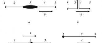
Reflex arc in the circuit
As a result of this whole process, a person withdraws his hand from the hot object and a reflex arc closes. The whole process takes a split second, so when touching any object, a person immediately feels its temperature, consistency and other features.
Conductor function
In this situation, the organ acts as a conductor. The conductor in this case is between the receptors and the brain. The receptors receive an impulse that is transmitted to the spinal cord and then to the brain. The information is analyzed there and transmitted back.
Thanks to this function, a person receives sensitivity, as well as a sense of himself in space. This has been proven many times, and this becomes especially evident in cases of serious spinal injuries.
Integrative function
This function is often forgotten, but it is no less important for a person than others. The integrative function manifests itself in reactions that cannot be classified as simple reflexes. In order for the body to respond, it is necessary to involve other parts of the human nervous system. This is how the spinal cord can form a connection between organs.
These include reflexes of chewing, swallowing, regulation of digestion, breathing and much more. In fact, this is an invisible function that ensures normal functioning.
Functions
Functional anatomy implies that, being part of the central nervous system, the spinal cord performs a reflex and conductive function. In the first case, the body controls the implementation of the simplest actions at the level of reactions contained in the subconscious. A striking example is the initiation of a motor function by withdrawing the hand if the surface is too hot. The limb does this before the person himself understands what happened. The second task of the organ is to transmit nerve impulses to the head section of the central nervous system, along the ascending and descending pathways.
Briefly about the main functions of the spinal cord
Reflex function
This main function of the organ is a response to external irritation. For example, the appearance of a reflex cough due to foreign objects and particles entering the respiratory tract, removing the hand from cactus spines or a source of danger. The impulse enters the spinal canal through motor neurons, which also trigger muscle contraction. This process does not require the involvement of the brain, and the motor reaction occurs without its participation. That is, a person does not even think about his action, often does not realize it.
Children's innate reflexes are checked after birth. They usually include the ability to suck milk, breathe, and jerk their legs. During development, acquired reflexes also appear, which help doctors identify the correct functioning of the arch elements and individual segments of the spinal cord. The test is carried out during a neurological examination. The main emphasis is on the plantar reflex, knee and abdominal. They allow you to check how healthy a person is at one time or another.
Conductor function
Treatment of spinal cord myelitis
Another important function of the spinal cord is conduction. It ensures the transmission of impulses from the skin, mucosal surface, internal organ to the brain and in the opposite direction. The white matter acts as a “conductor”. It is this that carries information about incoming impulses from the outside. Thanks to this ability, a person can characterize any object that surrounds him.
Cognition of the world is carried out through the transfer of information after touch to the brain. It is thanks to this function that a person understands that an object is slippery, smooth, rough or soft. With loss of sensitivity, the patient ceases to understand what is in front of him when touching an object. In addition, the brain receives data about the position of the body in space, tension in muscle tissue or irritation of pain receptors.
No ads 3
Spinal cord dysfunction
Impaired function can lead to serious consequences and often even death. Disorders often occur either due to injury or due to various diseases.
For example, due to dysfunction of the spinal cord, a person may lose sensitivity, in which case he may, for example, stop feeling temperature. In the worst case, the disorder can lead to uncontrollable actions of the limbs (or paralysis), disruption of the functioning of internal organs and the nervous system as a whole.
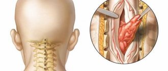
Spinal cord puncture
Puncture of cerebrospinal fluid (CSF) is a procedure that pursues diagnostic, anesthetic and therapeutic purposes. The procedure itself consists of an injection of an angle under the arachnoid membrane between the 3rd and 4th vertebrae, and then a certain amount of cerebrospinal fluid is extracted for research.
During the procedure, the brain itself is not affected, so there is no need to worry about violations. However, this procedure is quite serious and painful.
Spinal cord puncture