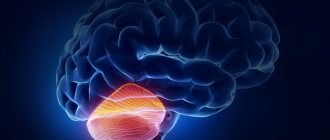
The cerebral cortex is represented by a uniform layer of gray matter 1.3-4.5 mm thick, consisting of more than 14 billion nerve cells. Due to the folding of the bark, its surface reaches large sizes - about 2200 cm2.
The thickness of the cortex consists of six layers of cells, which are distinguished by special staining and examination under a microscope. The cells of the layers vary in shape and size. From them, processes extend deep into the brain.
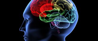
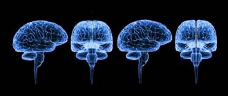
Structure of the cerebral cortex
It was found that different areas - fields of the cerebral cortex differ in structure and function. There are from 50 to 200 such fields (also called zones, or centers). There are no strict boundaries between the zones of the cerebral cortex. They constitute an apparatus that provides reception, processing of incoming signals and response to incoming signals.
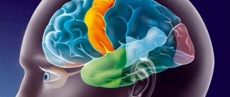
Areas of the cerebral cortex
In the posterior central gyrus, behind the central sulcus, there is an area of cutaneous and joint-muscular sensitivity . Here the signals that arise when touching our body, when it is exposed to cold or heat, or when it is painful are perceived and analyzed.
Areas of the cerebral cortex
In contrast to this zone, in the anterior central gyrus, in front of the central sulcus, the motor zone . It identifies areas that provide movement of the lower extremities, muscles of the trunk, arms, and head. When this area is irritated by electric current, contractions of the corresponding muscle groups occur. Injuries or other damage to the motor cortex lead to paralysis of the body muscles.
The temporal lobe contains the auditory area . The impulses arising in the receptors of the cochlea of the inner ear are received here and analyzed. Irritation of areas of the auditory zone causes sensations of sounds, and when they are affected by the disease, hearing is lost.
The visual zone is located in the cortex of the occipital lobes of the hemispheres. When it is irritated by electric current during brain surgery, a person experiences sensations of flashes of light and darkness. When it is affected by any disease, vision deteriorates and is lost.
Near the lateral sulcus there is a gustatory zone , where taste sensations are analyzed and formed based on signals arising in the receptors of the tongue. The olfactory zone is located in the so-called olfactory brain, at the base of the hemispheres. When these areas are irritated during surgery or during inflammation, people smell or taste something.
purely speech zone .
It is represented in the cortex of the temporal lobe, the inferior frontal gyrus on the left, and parts of the parietal lobe. Their diseases are accompanied by speech disorders.
Functions of the frontal lobe of the brain
The brain is a complex organ with billions of cells called neurons that work together. The frontal lobe works alongside other areas of the brain and controls the functions of the brain as a whole. Memory formation, for example, depends on many areas of the brain.
What's more, the brain can "repair" itself to compensate for damage. This does not mean that the frontal lobe can recover from all injuries, but other areas of the brain can change in response to head trauma.
The frontal lobes play a key role in future planning, including self-management and decision making. Some functions of the frontal lobe include:
Speech: Broca's area is an area in the frontal lobe that helps verbalize thoughts. Damage to this area affects the ability to speak and understand speech.
Motor skills: The frontal lobe cortex helps coordinate voluntary movements, including walking and running.
Comparing objects: The frontal lobe helps categorize objects and compare them.
From the editor: How neurasthenia is treated
Memory formation: Almost every region of the brain plays an important role in memory, so the frontal lobe is not unique, but it plays a key role in the formation of long-term memories.
Personality formation: The complex interaction of impulse control, memory, and other tasks helps shape the basic characteristics of a person. Damage to the frontal lobe can radically change personality.
Reward and Motivation: Most of the brain's dopamine-sensitive neurons are located in the frontal lobe.
Dopamine is a brain chemical that helps maintain feelings of reward and motivation.
Managing attention, including selective attention: When the frontal lobes are unable to control attention, attention deficit hyperactivity disorder (ADHD) may develop.
First and second signaling systems
The role of the cerebral cortex in improving the first signaling system and developing the second is invaluable. These concepts were developed by I.P. Pavlov. The signaling system as a whole is understood as the entire set of processes of the nervous system that carry out perception, processing of information and the response of the body. It connects the body with the outside world.
First signaling system
The first signaling system determines the perception of sensory-specific images through the senses. It is the basis for the formation of conditioned reflexes. This system exists in both animals and humans.
In the higher nervous activity of man, a superstructure has developed in the form of a second signaling system. It is peculiar only to humans and is manifested by verbal communication, speech, and concepts. With the advent of this signaling system, abstract thinking and generalization of countless signals from the first signaling system became possible. According to I.P. Pavlov, words turned into “signals of signals.”
Functions
In addition to the physiological division of the brain into lobes, the need arose to divide it into areas that are responsible for certain functions.
Frontal lobes
This is the so-called command center. What is the frontal lobe responsible for? It is a point of independence, self-awareness, and initiative. The defeat of these areas or the presence of pathologies in their functioning will affect a person’s attitude towards the world - almost everything will become indifferent to him, motivation will disappear, interest in current events will disappear, and laziness will appear.
The main functions of the frontal lobes are to control human behavior. It generates responses to social phenomena. When zones are violated, the limiter is deactivated, establishing a ban on certain actions, called uncultural.
The frontal lobes also allow you to analyze, plan, and learn new skills. Repeated repetition of the same sequences of movements over time becomes automatic and does not require effort or thought to perform them. Damage will force you to repeat monotonous movements every time as if for the first time, putting a lot of effort into it.
Perseveration is another consequence of deviations in the functioning of the frontal lobes. This is looping or repetition, such as repeating one phrase or word during a conversation
On the left side (for right-handed people) there are centers that are responsible for speech and attention.
These areas of the brain are also involved in coordinating and maintaining the body in an upright position when sitting and walking.
Temporal
They are located on the sides in the upper part of the brain, in the temple area. Thanks to them, the sound perceived by sound receptors turns into images, a person understands what he heard, certain sound vibrations are associated with images and are assigned to them. With the help of this part of the brain, people understand each other, their sound vibrations are filled with meaning, they choose the necessary words to describe certain phenomena.
Usually the left, non-dominant lobe is involved in determining the intonation of speech and reads emotions from facial expressions alone. Thanks to a small formation, the hippocampus, access to long-term memory is provided. Where it is located, on what medium and in what way our memories are recorded, this should be found out. The non-dominant part is used in visual memory, but the dominant part is used in verbal memory.
With problems with the temporal lobes, deviations in the functioning of the speech apparatus appear, in particular aphasia.
Parietal
MRI has shown that for left-handers and right-handers these lobes perform different functions, in fact directly opposite.
The left one gives us the ability to create a whole from fragments, that is, it helps to form a holistic picture of the world from small, at first glance, unrelated pieces.
They allow you not only to put together a mosaic from fragments, but also from letters into words, from a sequence of actions into a dance or technique, etc.
The non-dominant part allows you to perceive the world in three dimensions by processing information coming from the occipital lobes. Due to impairments, a person loses the ability to recognize faces, outline objects, and determine the distance to them and between them. These areas are also involved in the perception of pain and cold.
Occipital
Visual information processing center. They interpret the photons reflected from obstacles arriving at the biological light sensor - the retina - and form the resulting image, rotating it 180 degrees. Data about the size, color, shape, and material of an object are processed in separate centers and then recombined to form a single three-dimensional picture.
Editorial: Minimal brain dysfunction in children
METHODS FOR STUDYING THE FUNCTIONS OF THE CEREBRAL CORTEX
A significant number of methods are used in physiology to study the activity of the cerebral cortex. Some methods can only be used in so-called acute experiments, when the animal is under anesthesia and dies after the experiments; other methods make it possible to study for a long time. To study the functions of such a complex organ as the cortex, the greatest results are obtained by methods that allow research to be carried out over several months and even years.
Rice. 2 DIAGRAM OF THE COURSE OF NERVE FIBERS IN THE LARGE HEMISPHERES OF THE BRAIN. 1 - short associative fibers; 2 - long associative fibers; 3 - commissural fibers that communicate between both hemispheres of the pulp; 4 - centrifugal fibers
Let's get acquainted with some methods for studying the activity of the cerebral cortex.
Removal of individual sections of bark.
The essence of the method is that certain areas of the animal’s cortex are surgically removed. After the wound heals, when the animal recovers, the changes that have occurred in the animal’s behavior are observed. Based on the resulting disturbances, conclusions are drawn about the functions of the remote area of the cortex.
Electrical stimulation method
This method makes it possible, after opening the skull of an experimental animal or a person during brain surgery, to apply electrical stimulation to various points of the cortex. Thus, it is possible to establish the motor zone of the cortex and study its individual areas, the irritation of which causes the contraction of certain specific muscle groups. When studying the functions of the human cortex, this method turned out to be productive, since a person, when the cortex is irritated, is able to respond and communicate to the researcher the sensations that he experiences.
Chemical irritation method
To cause chemical irritation to the cerebral cortex, some poisons are used, most often strychnine.
To study the cortex, the property of strychnine to sharply increase the excitability of the nervous system was used. A small piece of filter paper is moistened with a solution of strychnine and applied to the area of the bark being examined. The excitability of the area of the cortex to which strychnine is applied increases sharply, which is reflected in the animal’s reactions. By studying these changes and knowing where the piece of paper moistened with strychnine solution is attached, they get an idea of the functions of this area.
Study of brain currents
The study of electrical phenomena in the brain first began in our country.
Much earlier than foreign authors, these studies were carried out by V. Ya. Danilevsky, I. M. Sechenov, N. E. Vvedensky, B. F. Verigo, V. V. Pravdich-Neminsky. In 1877, V. Ya. Danilevsky first published his research, which showed the presence of rhythmic electrical oscillations in the brain. He established a connection between the activity of the brain and the electrical fluctuations it observed. Soon after the work of V. Ya. Danilevsky, I. M. Sechenov in 1882, while studying electrical phenomena in the medulla oblongata, established the rhythmic nature of these phenomena and made a number of other observations.
In 1884, N. E. Vvedensky, applying to the cerebral cortex the technique he developed of listening to electrical muscle currents through a telephone receiver, grasped the rhythmic nature of electrical phenomena.
The modern method of electroencephalography, i.e., recording the biocurrents of the brain, allows, by applying special electrodes to the cerebral cortex or scalp during the experiment, to divert the currents of the cortex and record them. A recording of brain currents is called an encephalogram. Recording action currents during work and at rest, during sleep and during various other types of activity, as well as their further comparison, make it possible to draw certain conclusions. In Fig. 125 shows the electroencephalogram of a person during rest and work.