Anomaly in the structure of the brain (5th ventricle)
From time to time you have to deal with an atypical structure of the brain. Such cases are of particular clinical interest.
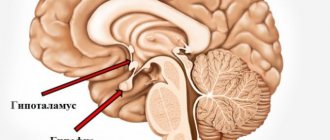
The fifth ventricle is a slit-like hypoechoic expansion located in the anterior midline of the brain, below the corpus callosum.
This is a longitudinal medullary fissure that begins to form at 12 gestational weeks from the lamina terminalis. By 6 gestational months, this expansion begins to close, merging into one septum. From 3 months to 6 months of infancy, the septum closes completely. Extremely rarely, the cavity does not close in adulthood, forming the 5th ventricle.
Some features:
1) In childhood, expansion of more than 10 mm may indicate a disease of the cerebrospinal fluid-dynamic system;
2) The presence of the 5th ventricle on ultrasound during pregnancy indicates the normal development of the child;
3) This condition should not be confused with cystic lesions; a traumatic lesion should be excluded (can develop in boxers);
4) The cavity can be connected to the rest of the liquor system (our case) or be closed, receiving liquor from neighboring cavities through the walls (more often);
5) The prognosis is favorable, it is a feature of the structure of the brain.
MRI of the brain, 22 years old:
Do you have anything to add?!
Related articles:
Lateral ventricles of the brain
The brain is a complex closed system protected by many structures and barriers. These protective supports carefully filter all material approaching the sinuous organ. However, such an energy-intensive system still needs to interact and maintain communication with the body, and the ventricles of the brain are one of the tools for ensuring such communication: these cavities contain cerebrospinal fluid, which supports the processes of metabolism, transport of hormones and removal of metabolic products. Anatomically, the ventricles of the brain are a derivative of the expansion of the central canal.
So, the answer to the question of what the cerebral ventricle is responsible for will be this: one of the main tasks of the cavities is the synthesis of cerebrospinal fluid. This cerebrospinal fluid serves as a shock absorber, that is, it provides mechanical protection for parts of the brain (protects against various types of injuries). Liquor, as a liquid, is in many ways similar to the structure of lymph. Like the latter, cerebrospinal fluid contains a huge amount of vitamins, hormones, minerals and brain nutrients (proteins, glucose, chlorine, sodium, potassium).
Different ventricles of the brain in an infant have different sizes.
Types of ventricles
Each part of the brain's central nervous system requires its own self-care, and therefore has its own storage facilities for spinal cerebrospinal fluid. Thus, the lateral stomachs (which include the first and second), third and fourth are distinguished. The entire ventricular organization has its own message system. Some (the fifth) are pathological formations.
Lateral ventricles - 1 and 2
The anatomy of the ventricle of the brain involves the structure of the anterior, inferior, posterior horn and the central part (body). These are the largest in the human brain and contain cerebrospinal fluid. The lateral ventricles are divided into the left - the first, and the right - the second. Thanks to the foramina of Monroe, the lateral cavities connect to the third ventricle of the brain.
The lateral ventricle of the brain and the nasal bulb as functional elements are closely interconnected, despite their relative anatomical distance. Their connection lies in the fact that between them there is, according to scientists, a short path along which pools of stem cells pass. Thus, the lateral stomach is a supplier of progenitor cells for other structures of the nervous system.
Speaking about this type of ventricles, it can be argued that the normal size of the ventricles of the brain in adults depends on their age, skull shape and somatotype.
In medicine, every cavity has its normal values. Lateral cavities are no exception. In newborns, the lateral ventricles of the brain normally have their own dimensions: the anterior horn is up to 2 mm, the central cavity is 4 mm. These dimensions are of great diagnostic importance when studying pathologies of the infant’s brain (hydrocephalus, a disease discussed below). One of the most effective methods for studying any cavity, including the cavities of the brain, is ultrasound. It can be used to determine both the pathological and normal size of the ventricles of the brain in children under one year of age.
3rd ventricle of the brain
The third cavity is located below the first two, and is located at the level of the intermediate section of the central nervous system between the visual tuberosities. The 3rd ventricle communicates with the first and second through the foramina of Monroe, and with the cavity below (4th ventricle) through the aqueduct.
Normally, the size of the third ventricle of the brain changes with the growth of the fetus: in a newborn – up to 3 mm; 3 months – 3.3mm; in a one-year-old child – up to 6 mm. In addition, an indicator of the normal development of cavities is their symmetry. This stomach is also filled with cerebrospinal fluid, but its structure differs from the lateral ones: the cavity has 6 walls. The third ventricle is in close contact with the thalamus.
4th ventricle of the brain
This structure, like the previous two, contains cerebrospinal fluid. It is located between the Sylvian water supply and the valve. The fluid in this cavity enters the subarachnoid space through several channels - two foramina of Luschko and one foramen of Magendie. The rhomboid fossa forms the bottom and is represented by the surfaces of the brain stem structures: the medulla oblongata and the pons. Also, the fourth ventricle of the brain provides the foundation for the 12th, 11th, 10th, 9th, 8th, 7th, and 5th pairs of cranial nerves. These branches innervate the tongue, some internal organs, the pharynx, facial muscles and facial skin.
5th ventricle of the brain
In medical practice, the name “fifth ventricle of the brain” is used, but this term is not correct. By definition, the stomachs of the brain are a set of cavities interconnected by a system of messages (channels) filled with spinal cerebrospinal fluid. In this case: the structure called the 5th ventricle does not communicate with the ventricular system, and the correct name would be “cavity of the septum pellucida.” From this follows the answer to the question of how many ventricles there are in the brain: four (2 lateral, third and fourth).
This hollow structure is located between layers of transparent partition. It, however, also contains cerebrospinal fluid, which enters the “stomach” through pores. In most cases, the size of this structure does not correlate with the frequency of pathology, however, there is evidence that in patients with schizophrenia, stress disorders and people who have suffered a traumatic brain injury, this part of the nervous system is enlarged.
Choroid plexuses of the ventricles of the brain
As noted, the function of the abdominal system is the production of cerebrospinal fluid. But how is this liquid formed? The only brain structure that provides the synthesis of cerebrospinal fluid is the choroid plexus. These are small villous formations belonging to vertebrates.
The choroid plexus is a derivative of the pia mater. They contain a huge number of vessels and carry a large number of nerve endings.
Ventricular diseases
In case of suspicion, an important method for determining the organic state of the cavities is puncture of the ventricles of the brain in newborns.
Diseases of the ventricles of the brain include:
Ventriculomegaly is a pathological expansion of cavities. Most often, such expansions occur in premature babies. The symptoms of this disease are varied and manifest themselves in the form of neurological and somatic symptoms.
Ventricular asymmetry (individual parts of the ventricles change in size). This pathology occurs due to an excessive amount of cerebral fluid. You should know that violation of the symmetry of cavities is not an independent disease - it is a consequence of another, more serious pathology, such as neuroinfections, massive contusion of the skull or tumor.
Hydrocephalus (fluid in the ventricles of the brain in newborns). This is a serious condition characterized by the excessive presence of cerebrospinal fluid in the gastric system of the brain. Such people are called hydrocephalus. The clinical manifestation of the disease is excessive volume of the child's head. The head becomes so large that it is impossible not to notice. In addition, the defining sign of pathology is the “sunset” symptom, when the eyes shift to the bottom. Instrumental diagnostic methods will show that the index of the lateral ventricles of the brain is higher than normal.
Pathological conditions of the choroid plexus occur against the background of both infectious diseases (tuberculosis, meningitis) and tumors of various locations. A common condition is cerebral vascular cyst. This disease can occur in both adults and children. The cause of cysts is often autoimmune disorders in the body.
Thus, the norm of the ventricles of the brain in newborns is an important component in the knowledge of a pediatrician or neonatologist, since knowledge of the norm allows one to determine pathology and find deviations in the early stages.
You can read more about the causes and symptoms of diseases of the cerebral cavity system in the article enlarged ventricles.
Causes of pathology
As a rule, this is a congenital disease. There are also acquired forms, but they are quite rare.
The causes of cerebral hydrocephalus in children are intrauterine infections, birth injuries, genetic disorders, and central nervous system defects.
Depending on the stage at which the disease developed, its causes vary:
- Fetal hydrocephalus in the vast majority of cases develops due to defects of the central nervous system. Infectious infections are to blame for the pathology of every fifth intrauterine hydrocephalus. Only a small proportion is due to genetic disorders.
- When the disease begins to develop immediately after birth, it is most likely due to infections in the womb. Such situations account for 75%. In 15% of cases, a newly born baby begins to suffer from dropsy due to a birth injury that led to meningitis or intracranial hemorrhage. The remaining 10% are malformations of either the spinal cord or brain, as well as disturbances in the functioning of blood vessels.
- In children aged one year and older, hydrocephalus is almost equally formed as a result of one of the following reasons: hemorrhage; brain tumor (spinal or brain); consequences of TBI; consequences of brain inflammation; genetic problems; malformations of cerebral vessels.
These generalized reasons also have their own classification.
So, among the infections that cause the disease, the following are common:
- rubella;
- cytomegalovirus;
- neurosyphilis;
- herpes virus type 1 or 2;
- toxoplasmosis;
- bacteria and viruses that cause meningitis;
- parotitis.
Defects responsible for the formation of dropsy:
- Narrowing of the canals connecting the cerebral ventricles.
- Arnold-Chiari syndrome (underdevelopment of the posterior fossa of the skull, due to which the brain structures located there do not fit into it).
- Anomalies of the cerebral venous system.
- Underdevelopment of the openings through which cerebrospinal fluid drains into the subarachnoid space.
- Dandy-Walker syndrome (anomaly of the development of the cerebellar spaces and cerebellum).
Also, among oncological diseases, hydrocephalus can be caused by:
- brain cancer;
- papilloma;
- tumor of the skull bones;
- brain ventricle tumor;
- vascular plexus meningioma;
- various forms of spinal cord oncology that impair fluid circulation.
Ventricles of the brain: structure, functions, diseases
The brain is the most complex organ in the human body, where the ventricles of the brain are considered one of the tools for interconnection with the body.
Their main function is the production and circulation of cerebrospinal fluid, due to which the transport of nutrients, hormones and the removal of metabolic products occurs.
Anatomically, the structure of the ventricular cavities looks like an expansion of the central canal.
What is a cerebral ventricle
Any ventricle of the brain is a special tank that connects with similar ones, and the final cavity joins the subarachnoid space and the central canal of the spinal cord.
Interacting with each other, they form a very complex system. These cavities are filled with moving cerebrospinal fluid, which protects the main parts of the nervous system from various mechanical damage and maintains intracranial pressure at a normal level. In addition, it is a component of the organ’s immunobiological protection.
The internal surfaces of these cavities are lined with ependymal cells. They also cover the spinal canal.
The apical portions of the ependymal surface have cilia that help move cerebrospinal fluid (cerebrospinal fluid, or cerebrospinal fluid). These same cells contribute to the production of myelin, a substance that is the main building material of the electrically insulating sheath that covers the axons of many neurons.
The volume of cerebrospinal fluid circulating in the system depends on the shape of the skull and the size of the brain. On average, the amount of fluid produced for an adult can reach 150 ml, and this substance is completely renewed every 6-8 hours.
The amount of cerebrospinal fluid produced per day reaches 400-600 ml. With age, the volume of cerebrospinal fluid may increase slightly: this depends on the amount of fluid absorption, its pressure and the state of the nervous system.
The fluid produced in the first and second ventricles, located in the left and right hemispheres, respectively, gradually moves through the interventricular foramina into the third cavity, from which it moves through the openings of the cerebral aqueduct into the fourth.
At the base of the last cistern there is a foramen of Magendie (communicating with the cerebellopontine cistern) and paired foramina of Luschka (connecting the terminal cavity with the subarachnoid space of the spinal cord and brain). It turns out that the main organ responsible for the functioning of the entire central nervous system is completely washed by cerebrospinal fluid.
Once in the subarachnoid space, the cerebrospinal fluid, with the help of specialized structures called arachnoid granulations, is slowly absorbed into the venous blood. Such a mechanism functions like valves that work in one direction: it allows fluid to pass into the circulatory system, but does not allow it to flow back into the subarachnoid space.
The number of ventricles in humans and their structure
The brain has several communicating cavities connected together. There are four in total, however, very often in medical circles they talk about the fifth ventricle in the brain. This term is used to refer to the cavity of the transparent septum.
However, despite the fact that the cavity is filled with cerebrospinal fluid, it is not connected to other ventricles. Therefore, the only correct answer to the question of how many ventricles are in the brain is: four (two lateral cavities, the third and fourth).
The first and second ventricles, located to the right and left relative to the central canal, are symmetrical lateral cavities located in different hemispheres just below the corpus callosum. The volume of any of them is approximately 25 ml, and they are considered the largest.
Each lateral cavity consists of a main body and canals branching from it - the anterior, inferior and posterior horns. One of these channels connects the lateral cavities with the third ventricle.
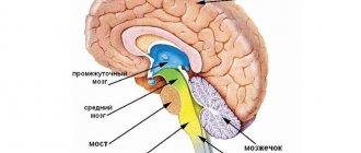
The third cavity (from the Latin “ventriculus tertius”) is shaped like a ring. It is located in the midline between the surfaces of the thalamus and the hypothalamus, and is connected inferiorly to the fourth ventricle by the aqueduct of Sylvius.
The fourth cavity is located slightly lower - between the elements of the hindbrain. Its base is called the rhomboid fossa and is formed by the posterior surface of the medulla oblongata and the pons.
The lateral surfaces of the fourth ventricle limit the superior cerebellar peduncles, and the entrance to the central canal of the spinal cord is located behind it. This is the smallest, but very important section of the system.
On the arches of the last two ventricles there are special vascular formations that produce most of the total volume of cerebrospinal fluid. Similar plexuses are present on the walls of two symmetrical ventricles.
Ependyma, consisting of ependymal formations, is a thin film that covers the surface of the central duct of the spinal cord and all ventricular cisterns. Almost the entire area of the ependyma is single-layered. Only in the third and fourth ventricles and the brain aqueduct connecting them can it have several layers.
Ependymocytes are elongated cells with a cilium at the free end. By the beating of these processes they move the cerebrospinal fluid. It is believed that ependymocytes can independently produce some protein compounds and absorb unnecessary components from the cerebrospinal fluid, which helps cleanse it of breakdown products formed during the metabolic process.
Functions of the ventricles of the brain
Each ventricle of the brain is responsible for the formation of cerebrospinal fluid and its accumulation. In addition, each of them is part of the fluid circulation system, which constantly moves along the cerebrospinal fluid pathways from the ventricles and enters the subarachnoid space of the brain and spinal cord.
The composition of cerebrospinal fluid is significantly different from any other fluid in the human body. However, this does not give reason to consider it a secretion of ependymocytes, since it contains only cellular elements of blood, electrolytes, proteins and water.
The liquor-forming system forms about 70% of the necessary fluid. The rest penetrates the walls of the capillary system and the ventricular ependyma. The circulation and outflow of cerebrospinal fluid is due to its constant production. The movement itself is passive and occurs due to the pulsation of large cerebral vessels, as well as due to respiratory and muscle movements.
Absorption of cerebrospinal fluid occurs along the perineural nerve sheaths, through the ependymal layer and capillaries of the arachnoid and pia mater.
Liquor is a substrate that stabilizes brain tissue and ensures full neuronal activity by maintaining optimal concentrations of essential substances and acid-base balance.
This substance is necessary for the functioning of the brain systems, since it not only protects them from contact with the skull and accidental impacts, but also delivers the hormones produced to the central nervous system.
To summarize, let us formulate the main functions of the ventricles of the human brain:
- production of cerebrospinal fluid;
- ensuring continuous movement of cerebrospinal fluid.
Ventricular diseases
The brain, like all other internal human organs, is prone to various diseases. Pathological processes affecting parts of the central nervous system and the ventricles, including, require immediate medical intervention.
In pathological conditions developing in the organ cavities, the patient’s condition rapidly deteriorates because the brain does not receive the required amount of oxygen and nutrients. In most cases, the cause of ventricular diseases is inflammatory processes resulting from infections, injuries or neoplasms.
Hydrocephalus
Hydrocephalus is a disease characterized by excessive accumulation of fluid in the ventricular system of the brain. The phenomenon in which difficulties arise in its movement from the site of secretion to the subarachnoid space is called occlusive hydrocephalus.
If the accumulation of fluid occurs due to a violation of the absorption of cerebrospinal fluid into the circulatory system, then this pathology is called aresorptive hydrocephalus.
Hydrocele of the brain can be congenital or acquired. The congenital form of the disease is usually detected in childhood. The causes of the acquired form of hydrocephalus are often infectious processes (for example, meningitis, encephalitis, ventriculitis), neoplasms, vascular pathologies, injuries and developmental defects.
Dropsy can occur at any age. This condition is dangerous to health and requires immediate treatment.
Hydroencephalopathy
Another common pathological condition due to which the ventricles in the brain may suffer is hydroencephalopathy. In this pathological condition, two diseases are combined at once - hydrocephalus and encephalopathy.
As a result of impaired circulation of cerebrospinal fluid, its volume in the ventricles increases, intracranial pressure increases, and because of this, brain function is disrupted. This process is quite serious and without proper control and treatment leads to disability.
Ventriculomegaly
When the right or left ventricles of the brain become enlarged, a disease called ventriculomegaly is diagnosed. It leads to disturbances in the functioning of the central nervous system, neurological abnormalities and can provoke the development of cerebral palsy. This pathology is most often detected during pregnancy at a period of 17 to 33 weeks (the optimal period for detecting pathology is 24-26 weeks).
A similar pathology often occurs in adults, but ventriculomegaly does not pose any danger to a mature organism.
Ventricular asymmetry
Changes in the size of the ventricles can occur under the influence of excessive production of cerebrospinal fluid. This pathology never occurs on its own. Most often, the appearance of asymmetry is accompanied by more serious diseases, for example, neuroinfection, traumatic brain injury, or a tumor in the brain.
Hypotensive syndrome
A rare phenomenon, usually a complication after therapeutic or diagnostic procedures. Most often it develops after a puncture and leakage of cerebrospinal fluid through the hole from the needle.
Other causes of this pathology may be the formation of cerebrospinal fluid fistulas, disruption of the water-salt balance in the body, and hypotension.
Clinical manifestations of low intracranial pressure: the appearance of migraine, apathy, tachycardia, general loss of strength. With a further decrease in the volume of cerebrospinal fluid, pallor of the skin, cyanosis of the nasolabial triangle, and breathing problems appear.
Finally
The ventricular system of the brain is complex in its structure. Despite the fact that the ventricles are only small cavities, their importance for the full functioning of human internal organs is invaluable.
The ventricles are the most important brain structures that ensure the normal functioning of the nervous system, without which the life of the body is impossible.
It should be noted that any pathological processes leading to disruption of brain structures require immediate treatment.
The membranes and spaces of the brain.
The meninges of the brain are a direct continuation of the meninges of the spinal cord, but have a number of features (Fig. 20.).
The pia mater is closely adjacent to the substance of the brain, extending into all the grooves and crevices of its surface. In some places, the vessels of the pia mater are very developed and form choroid plexuses. The choroid plexuses consist of a large number of leaf-shaped processes; each process contains a small artery or arteriole, which turns into a capillary plexus. The tortuous capillaries form tubercles in the epithelium called villi. The epithelium covering the choroid plexus develops from the ependyma. This is cuboidal epithelium located on a thin layer of connective tissue originating from the soft and arachnoid membranes.
The choroid plexuses (they are located in each of the four ventricles of the brain) are producers of cerebrospinal fluid or cerebrospinal fluid. The latter is formed by extravasation from the choroid plexuses.
The arachnoid membrane of the brain has a structure similar to the spinal cord. It is very thin, transparent, devoid of blood vessels. It is covered with endothelium on the outside and inside.
Between the arachnoid and soft membranes there is a subarachnoid
(subarachnoid) space, 120-140 µm wide, crossed by jumpers and containing cerebrospinal fluid.
Above the large fissures and grooves of the brain, the arachnoid membrane forms containers called cisterns. These include:
1. cerebellar-cerebral (cisterna cerebellomedullaris)
, lying between the cerebellum and medulla oblongata;
2. cistern of the lateral sulcus (cisterna fossa lateralis)
- in the area of the same gap;
3. cistern of the optic chiasm (cistern chiasmatis)
- at the base of the brain anterior to the chiasm;
4. interpeduncular cistern (cisterna interpeduncullaris)
between the cerebral peduncles) - anterior to the posterior perforated substance;
5. cistern of the great cerebral vein (cisterna venae cerebri magnae)
located in the area of the transverse fissure, in the circumference of the great vein of the brain;
6. bridge tank (cisterna pontis)
located on the ventral surface at the transition of the midbrain into the pons;
7. cistern of the corpus callosum (cisterna corporis collosi)
located above the corpus callosum.
The subarachnoid spaces of the brain and spinal cord communicate with each other at the junction of the spinal cord and the brain.
The cerebrospinal fluid, formed in the ventricles of the brain by their choroid plexuses, flows into the subarachnoid space. From the lateral ventricles, through the right and left interventricular foramina, cerebrospinal fluid enters the third ventricle, then through the cerebral aqueduct into the fourth ventricle, and from it through the azygos foramen (Magendie) in the posterior wall and the paired lateral aperture (Lyushko) into the cerebellocerebral cistern subarachnoid space.
In the lateral (I and II), III and IV ventricles of the brain there are choroid plexuses (plexus chorioideus)
, which are capillary-rich structures protruding into the lumen of the ventricles.
Hydrostatic pressure in the capillaries of the choroid plexuses is increased, which facilitates the formation of fluid, and in the venous sinuses, where arachnoid granulations are located, it is decreased. This facilitates the outflow of fluid into the venous sinuses.
Outside the arachnoid membrane, the brain, like the spinal cord, is covered by the dura mater.
The hard shell in the area of the skull gives rise to special outgrowths - processes that go deep between the individual parts of the brain.
1. Falx cerebri – separates the cerebral hemispheres.
2. tentorium cerebellum – separates the cerebral hemispheres from the cerebellum.
3. Tentural notch – the brain stem passes through it.
4. Falx cerebellum – separates the cerebellar hemispheres.
5. Sella diaphragm – in the area of the sella turcica.
6. Trigeminal cavity – it contains the trigeminal nerve ganglion.
In some places, the layers of the dura mater split and form sinuses (sinuses), through which venous blood flows freely from the brain. The sinuses are triangular in shape, do not collapse, and lack valves. In them, venous blood flows from the brain through the veins, which then enters the internal jugular veins.
The dura mater contains the following sinuses:
1. Superior sagittal sinus (sinus sagittalis superior)
(unpaired) runs along the entire outer (upper) edge of the falx cerebri and flows into the transverse sinus.
2. Inferior sagittal sinus (sinus sagittalis inferior)
(unpaired) is located on the lower edge of the falx cerebri, flows into the straight sinus.
3. Direct sinus (sinus rectus)
(unpaired) located at the junction of the falx cerebellum and the tentorium cerebellum. It connects the inferior and superior sagittal sinuses and empties into the transverse sinus.
4. Occipital sinus (sinus occipitalis)
(unpaired) lies at the base of the falx cerebellum along the internal occipital crest. At the posterior edge of the foramen magnum, the sinus divides into two branches, each of which flows into the sigmoid sinus of the corresponding side. The superior end of the occipital sinus communicates with the transverse sinus.
5. Transverse sinus (sinus transversus)
(unpaired) lies at the base of the tentorium cerebellum. The superior sagittal, occipital and straight sinuses flow into it - this is a sinus drainage located in the area of the internal occipital protrusion. The transverse sinus continues to the right and left into the sigmoid sinus on its side.
6. Sigmoid sinus (sinus sigmoideus)
(paired), located in the groove of the same name in the temporal bone, in the area of the jugular foramen it passes into the internal jugular vein.
7. Cavernous sinus (sinus petrosus inferior)
(paired) located on the sides of the sella turcica. Both cavernous sinuses are connected to each other by the anterior and posterior intercavernous sinuses.
8. Sphenoparietalis sinus
(paired) runs along the free posterior edge of the lesser wing of the sphenoid bone and flows into the cavernous.
Rice. 20. Shells and spaces of the brain.
Ventricles of the human brain
The human brain is made up of an amazing number of neurons - about 25 billion of them, and this is not the limit. Neuronal cell bodies are collectively called gray matter because they have a gray tint.
The arachnoid membrane protects the cerebrospinal fluid circulating inside it. It acts as a shock absorber that will protect the organ from impact.
The brain mass of a man is higher than that of a woman. However, the opinion that a woman’s brain is inferior in development to a man’s is erroneous. The average weight of the male brain is about 1375 g, the female brain is about 1245 g, which is 2% of the weight of the entire body. By the way, brain weight and human intelligence are not interrelated. If, for example, you weigh the brain of a person suffering from hydrocephalus, it will be larger than usual. At the same time, mental abilities are significantly lower.
The brain consists of neurons - cells capable of receiving and transmitting bioelectric impulses. They are supplemented by glia, which help neurons function.
The ventricles of the brain are cavities inside the brain. It is the lateral ventricles of the brain that produce cerebrospinal fluid. If the lateral ventricles of the brain are impaired, hydrocephalus may develop.
How does the brain work?
Before moving on to considering the functions of the ventricles, let us recall the location of some parts of the brain and their significance for the body. This will make it easier to understand how this whole complex system works.
The brain is finite
It is impossible to briefly describe the structure of such a complex and important organ. The telencephalon runs from the back of the head to the forehead. It consists of large hemispheres - right and left. It has many grooves and convolutions. The structure of this organ is closely related to its development.
Conscious human activity is associated with the functioning of the cerebral cortex. Scientists distinguish three types of bark:
- Ancient.
- The old one.
- New one. The rest of the cortex, which was the last to develop during human evolution.
Hemispheres and their structure
The hemispheres are a complex system that consists of several levels. They have different parts:
- frontal;
- parietal;
- temporal;
- occipital
In addition to the lobes, there is also a cortex and subcortex. The hemispheres work together, they complement each other, performing a set of tasks. There is an interesting pattern - each part of the hemispheres is responsible for its own functions.
Bark
It is difficult to imagine that the cortex, which provides the main characteristics of consciousness and intelligence, is only 3 mm thick. This thinnest layer reliably covers both hemispheres. It is composed of the same nerve cells and their processes, which are located vertically.
The layering of the crust is horizontal. It consists of 6 layers. The cortex contains many vertical nerve bundles with long processes. There are more than 10 billion nerve cells here.
The cortex is assigned various functions that are differentiated between its different sections:
- temporal – smell, hearing;
- occipital – vision;
- parietal – taste, touch;
- frontal – complex thinking, movement, speech.
It affects the brain structure. Each of its neurons (we remind you that there are about 25 billion of them in this organ) creates about 10 thousand connections with other neurons.
In the hemispheres themselves there are basal ganglia - these are large clusters that consist of gray matter. It is the basal ganglia that transmit information. Between the cortex and the basal ganglia are the processes of neurons - the white matter.
It is the nerve fibers that form the white matter; they connect the cortex and those formations that are under it. The subcortex contains the subcortical nuclei.
The telencephalon is responsible for physiological processes in the body, as well as intelligence.
Intermediate brain
It consists of 2 parts:
- ventral (hypothalamus);
- dorsal (metathalamus, thalamus, epithalamus).
It is the thalamus that receives stimuli and sends them to the hemispheres. This is a reliable and always busy intermediary. Its second name is the visual thalamus. The thalamus ensures successful adaptation to an ever-changing environment. The limbic system reliably connects it to the cerebellum.
The hypothalamus is a subcortical center that regulates all autonomic functions. It affects through the nervous system and glands. The hypothalamus ensures the normal functioning of individual endocrine glands and participates in metabolism, which is so important for the body. The hypothalamus is responsible for the processes of sleep and wakefulness, eating and drinking.
Below it is the pituitary gland. It is the pituitary gland that provides thermoregulation, the functioning of the cardiovascular and digestive systems.
hindbrain
It consists of:
- front axle;
- the cerebellum behind it.
The bridge visually resembles a thick white cushion. It consists of a dorsal surface, which is covered by the cerebellum, and a ventral surface, the structure of which is fibrous. The bridge is located above the medulla oblongata.
Cerebellum
It is often called the second brain. This department is located behind the bridge. It covers almost the entire surface of the posterior cranial fossa.
The large hemispheres hang directly above it, separated only by a transverse gap. Below, the cerebellum is adjacent to the medulla oblongata. There are 2 hemispheres, the lower and upper surface, the worm.
The cerebellum has many slits along its entire surface, between which one can find convolutions (ridges of the medulla).
The cerebellum consists of two types of substance:
- Gray. It is located on the periphery and forms the cortex.
- White. It is located in the area under the bark.
The white matter penetrates into all convolutions, literally penetrating them. It can be easily recognized by its characteristic white stripes. In the white matter there are inclusions of gray - the nucleus. Their interweaving in cross-section visually resembles an ordinary branched tree. It is the cerebellum that is responsible for coordinating movements.
Midbrain
It is located from the anterior region of the bridge to the optic tracts and papillary bodies. There are many nuclei (tubercles of the quadrigeminal). The midbrain is responsible for the functioning of latent vision and the orienting reflex (it ensures that the body turns to where the noise is heard).
Ventricles
The ventricles of the brain are cavities connected to the subarachnoid space, as well as the spinal cord canal. If you're wondering where cerebrospinal fluid is produced and stored, it happens in the ventricles. Inside they are covered with ependyma.
Ependyma is a membrane that lines the surface of the ventricles from the inside. It can also be found inside the spinal canal and all cavities of the central nervous system.
Types of ventricles
Ventricles are divided into the following types:
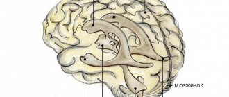
- Lateral. Inside these large cavities there is cerebrospinal fluid. The lateral ventricle of the brain is large in size. This is explained by the fact that quite a lot of fluid is produced, because not only the brain, but also the spinal cord needs it. The left ventricle of the brain is called the first, the right - the second. The lateral ventricles communicate with the third ventricle through foramina. They are symmetrically located. From each lateral ventricle departs the anterior horn, the posterior horns of the lateral ventricles, the lower, and the body.
- Third. Its location is between the visual tuberosities. It has the shape of a ring. The walls of the third ventricle are filled with gray matter. There are many autonomic subcortical centers here. The third ventricle communicates with the midbrain and lateral ventricles.
- Fourth. Its location is between the cerebellum and the medulla oblongata. This is the remnant of the cavity of the brain bladder, which is located behind. The shape of the fourth ventricle resembles a tent with a roof and a bottom. Its bottom is diamond-shaped, which is why it is sometimes called a diamond-shaped fossa. The spinal cord canal opens into this fossa from behind.
The shape of the lateral ventricles resembles the letter C. They synthesize cerebrospinal fluid, which must then circulate in the spinal cord and brain.
If the cerebrospinal fluid does not drain correctly from the ventricles, the person may be diagnosed with hydrocephalus. In severe cases, it is noticeable even in the anatomical structure of the skull, which is deformed due to strong internal pressure. Excess liquid tightly fills the entire space. It can change the functioning of not only the ventricles, but also the entire brain. Excessive amounts of cerebrospinal fluid can cause a stroke.
Diseases
The ventricles are susceptible to a number of diseases. The most common among them is the above-mentioned hydrocephalus. With this disease, the cerebral ventricles can grow to pathologically large sizes. In this case, the head hurts, a feeling of pressure appears, coordination may be impaired, nausea and vomiting appear. In severe cases, it is difficult for a person to even move. This can lead to disability and even death.
The appearance of the mentioned signs may indicate congenital or acquired hydrocephalus. Its consequences are detrimental to the brain and the body as a whole. Blood circulation may be impaired due to constant compression of soft tissues, and there is a risk of hemorrhage.
The doctor must determine the cause of hydrocephalus. It can be congenital or acquired. The latter type occurs with a tumor, cyst, injury, etc. All departments suffer. It is important to understand that the development of pathology will gradually worsen the patient’s condition, and irreversible changes will occur in the nerve fibers.
The symptoms of this pathology are associated with the fact that more cerebrospinal fluid is produced than necessary. This substance quickly accumulates in the cavities, and since there is a decrease in outflow, the cerebrospinal fluid does not drain away as it should normally. The accumulated cerebrospinal fluid can be in the ventricles and stretch them, it compresses the vascular walls, impairing blood circulation. Neurons do not receive nutrition and quickly die. It is impossible to restore them later.
Hydrocephalus often affects newborns, but it can appear at almost any age, although it is much less common in adults. Proper circulation of cerebrospinal fluid can be established with proper treatment. The only exception is severe congenital cases. During pregnancy, ultrasound can reveal possible hydrocephalus in the baby.
If during pregnancy a woman indulges in bad habits and does not follow proper nutrition, this entails an increased risk of fetal hydrocephalus. Asymmetric development of the ventricles is also possible.
To diagnose pathologies in the functioning of the ventricles, MRI and CT are used. These methods help to identify abnormal processes at a very early stage. With adequate treatment, the patient's condition can improve. Even a complete recovery is possible.
The ventricles may be subject to other pathological conditions. For example, their asymmetry has a negative impact. Tomography can reveal it. Asymmetry is caused by vascular dysfunction or degenerative processes.
Pathological changes can also be caused by tumors and inflammation.
If there is an increased volume of cerebrospinal fluid, this can happen not only due to its excessive production, but also due to the fact that there is no normal outflow of fluid. This may be the result of the appearance of neoplasms, hematomas, or blood clots.
With diseases of the ventricles, the patient is concerned about serious health problems. The brain suffers from a deficiency of nutrients, oxygen, and hormones. In this case, the protective function of the cerebrospinal fluid is disrupted, poisoning of the body begins, and intracranial pressure increases.
Conclusion
The ventricles are interconnected with many organs and systems; human health as a whole depends on their condition. If an MRI or CT scan reveals their expansion, you should immediately consult a doctor. Timely treatment will help you return to a full life.


