Structure and functions of the ventricles of the brain
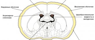
The brain has four chambers. They produce and circulate in the cerebrospinal fluid. Its main job is to transport nutrients to brain cells, remove waste products from the brain, and absorb head impacts.
Two lateral ventricles (ventriculi lateralis) are located in the cerebral hemispheres - the first on the left, the second on the right. The normal depth of the sinuses is from 1 to 4 mm. An increase in the capacity of the organ to 5 mm leads to a noticeable change in shape: the lateral C-shaped curvature disappears, the defect acquires a rounded shape.
In this case we are talking about the initial stage of asymmetry of the lateral ventricles. Many people wonder what it is and how dangerous is this pathology?
Anatomy of the ventricular system of the brain
The ventricles of the brain are a cerebral system of cavities communicating with each other, the subarachnoid space and the spinal canal. Their inner surface is lined with ependyma. Under this layer are the choroid plexuses, which produce cerebrospinal fluid.
The lateral (or lateral) ventricles are the most voluminous. They are localized on both sides of the midline and have pairs of anterior, posterior and lower horns that are symmetrical relative to each other. The lateral ventricular cavities communicate with each other with the third ventricle, located between the thalami, through the foramen of Monroe. Between the cerebellum and the brain stem is the fourth ventricle. From it, the cerebrospinal fluid enters the subarachnoid space through the foramina of Luschka (paired) and Magendie (unpaired).
Causes of ventricular dilatation
An increase in the size of the lateral ventricles is formed as a result of impaired circulation of the cerebrospinal fluid. This picture emerges when:
- overproduction of cerebrospinal fluid;
- impaired adsorption of cerebrospinal fluid;
- difficulty in the outflow of cerebrospinal fluid.
Changes in liquor dynamics due to overproduction and slower reabsorption of cerebrospinal fluid occur as a result of irritation of the ventricular choroidal plexuses and the arachnoid membrane of the brain by a pathological process (most often as a result of neuroinfection).
Difficulty in the outflow of cerebrospinal fluid is caused by blockage of the cerebrospinal fluid pathways by neoplasms and cysts.
The main reasons for the expansion of the lateral ventricles are:
- neuroinfections (meningitis, meningoencephalitis);
- skull injuries;
- brain tumors;
- idiopathic hydrocephalus;
- formed hematomas;
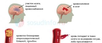
- hemorrhagic stroke;
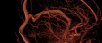
- thrombosis of cerebral vessels;
- atypical embryonic anlage of the ventricular system.
Clinical signs
Manifestations of asymmetry of the lateral ventricles in an adult are often absent. In this case, such a structure of cerebral structures is not considered a pathology; it is revealed as a finding during neuroimaging. Along with this, a pronounced disturbance of liquor dynamics leads to the following clinical symptoms:
- headaches;
- feeling of heaviness and fullness in the head;
- dizziness;
- nausea;
- vomiting that does not bring relief;
- anxiety-phobic syndrome;
- apathy.
In addition, the clinical picture of ventricular asymmetry is complemented by symptoms of the disease that forms the basis of this condition. Paresis, pathology of cranial nerves, sensitivity disorders, cognitive decline, and cerebellar disorders may occur.
Causes of asymmetry of the lateral ventricles
The expansion (expansion) of caverns is associated with the accumulation of liquors in them. There may be two reasons for this: excessive production of fluid or disruption of its outflow.
The following diseases can provoke an increase in the amount of cerebrospinal fluid:
- Hydrocephalus, in which the absorption of cerebral fluid is impaired.
- Changes in the central nervous system - schizophrenia, bipolar disorder.
Inadequate circulation of cerebrospinal fluid occurs when mechanical compression of the exit ducts occurs.
This phenomenon is most often caused by damaging factors:
- Traumatic brain injuries;
- Malformation of the Sylvian canal (a narrow canal connecting the third and fourth ventricles);
- Brain hemorrhages;
- Intracranial tumors, cysts, papillomas, hematomas;
- Meningitis;
- Thrombosis of cerebral veins.
Lateroventriculoasymmetry of the brain often occurs in newborns, more often in premature infants.
Principle of the method
What is neurosonography? In fact, it has nothing to do with sleep. For some reason, many parents think that this is a functional diagnostic method that is used to study sleep. This is a false misconception. Neurosonography is a routine ultrasound performed on a child for... The result of the study is a neurosonogram, and the safety of the method is so high that neurosonography of newborns has become a mandatory method for regular examination of children in clinics.
NSG of the brain is not used in adults for one compelling anatomical reason: their skull bones fuse, as a result of which the skull becomes a single bone ball that protects the internal structures of the central nervous system. In children under one year of age, the skull is structured differently: between the bones of the skull there are open spaces called fontanelles. It is the zone of the fontanel that is passable for the acoustic signal, and ultrasound can be done through this zone.
In fact, this study is a regular Doppler ultrasound (ultrasound), which is performed through the fontanelles. Of course, there are methods of transcranial research, but you can see much better through the fontanelles. In the eighties and nineties of the last century, it was generally believed that the bones of the skull were not capable of conducting ultrasound and no studies were carried out. However, due to the constant improvement of ultrasound equipment for ultrasound in general and NSG in particular, in recent years it has become possible to perform NSG through the squama of the temporal bone, which is very thin in children.
About the testimony
What indications exist for this study? First of all, it is so safe that it has become a screening method. This shows effectiveness and popularity. At what age can this study be carried out? At the present stage of development of medical science, not only neurosonography of newborns is carried out, but also regular repetition of studies. Additional indications for NSG are as follows:
- obstetrics by cesarean section, rapid labor, or prematurity;
- when there is a delay in the child’s development, from a month or more;
- when the child has a neurological pathology (paresis, muscle wasting);
- a child's head injury occurred during childbirth or is suspected of it;
- in case of threatening fetal asphyxia;
- in case of Rh-conflict pregnancy;
- for various infectious diseases of the mother, especially in the first trimester;
- if there is a suspicion of increased intracranial pressure in the baby;
Of course, there are other indications. Since this study is safe, informative and carried out very quickly, any suspicion by a neurologist of the presence of focal neurological symptoms allows NSG to be performed on the child. The standards for conducting this study allow for the presence of a pediatric neurologist or neurosurgeon who can help decipher the results of the study.
An important fact is that this study does not require any special preparation and can be performed on a child under one year of age at least every month.
This study can be done if there is a suspicion of increased intracranial pressure, if a baby develops convulsive seizures, or meningism.
Symptoms of enlarged ventricles
The mechanism of development of pathology has common features, regardless of the reasons. Fluid accumulates in the ventricles, which expand and contract brain tissue. The pressure in the cavities themselves also increases.
Patients may complain of sudden headaches, nausea and vomiting, a feeling of distraction in the eyeballs, hearing and vision problems. Performance gradually decreases, memory impairment, apathy, and drowsiness appear.
All these are symptoms of pathology of the spinal system. If the abnormality is not detected and treated early, it becomes a chronic condition.
How it manifests itself
The main function of the ventricles is to secrete cerebrospinal fluid, as well as ensure its normal circulation in the subarachnoid space. If the balance of exchange and production of cerebrospinal fluid is disturbed, then stagnation is formed and, as a result, the walls of the cavities are stretched. The same slight expansion of the lateral segments may be a normal variant, but their asymmetry and enlargement of individual parts (for example, only the horn) will be a sign of the development of pathology.
Enlarged ventricles of the brain in an infant can be diagnosed with a congenital disease such as ventriculomegaly. It varies in severity:
- Slight expansion of the ventricles of the brain up to 11-12 mm, with no significant symptoms. It manifests itself in the child’s behavior: he becomes more excitable and irritable.
- Increasing the depth of the ventricles up to 15 mm. Most often, the pathology is accompanied by asymmetry and impaired blood supply to the affected area, which entails the appearance of seizures, an increase in head size and a lag in mental and physical development.
- Ventricular dilatation up to 20 mm is characterized by irreversible changes in brain structures and is often accompanied by Down syndrome and cerebral palsy in infants.
In adulthood, an increase in ventricular volume is manifested by the following symptoms:
- Gait disturbance, with the child walking “on tiptoes” or vice versa, focusing only on the heels.
- The appearance of visual disorders, such as squint, insufficient focus of the gaze, as well as double images when trying to see small details.
- Tremor of arms and legs.
- Behavioral disorders that manifest themselves in excessive lethargy and drowsiness, while it is difficult to captivate the child with any activity.
- The appearance of headaches due to increased intracranial pressure, sometimes nausea and even vomiting may occur.
- Dizziness.
- Frequent regurgitation, loss of appetite. Some newborns are able to refuse breastfeeding.
Diagnostics
Lateral ventricular asymmetry is detected using the following procedures:
- Ultrasound of the brain is the most informative examination;
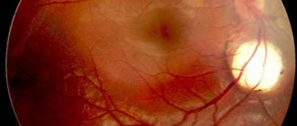
- Examination of the fundus, detection of swelling of the eye disc, spasms, hemorrhages;
- Neurosonography;
- Magnetic resonance imaging and computed tomography of the brain.
If the above methods give conflicting results, lumbar puncture is recommended. Analysis of the cerebrospinal fluid reveals the cause of the curvature of the lateral sinuses.
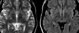
What is the MRI normal for the lateral ventricles in an adult?
The dimensions depend on the cause of the pathology. An asymmetrical arrangement of cerebral structures is observed in neoplasms, intracranial hypertension, and hydrocephalus. Some differences in the measured values depend on the type of examination - native or contrast magnetic resonance imaging.
Tomographic indicators of the cerebral ventricles
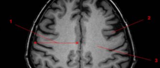
MRI of the head shows the lateral ventricles as anechoic zones (density below surrounding structures). Three-dimensional modeling allows us to distinguish the anatomical structures of the lesion:
- Triangle (atrium);
- Body;
- inferior temporal horn;
- Occipital (back) part;
- Frontal (front) zone
The size of the ventricular spaces increases with age. In the gestational period, the value is several tenths of a millimeter. In a mature child, the ventricular spaces are slit-like. MRI determines the width of the space in the area of the foramen of Monroe, where the norm is 2 mm.
The expansion in adults begins posteriorly, so any examination requires clear visualization of the occipital structures.
Pathological dilatation is diagnosed when the diameter of the anterior horns, determined on coronal sections, increases over five millimeters.
Magnetic resonance scanning can accurately determine when the lateral ventricles are dilated and when disposition occurs.
Symptoms of asymmetry
Clinical manifestations of lateral ventricular asymmetry may differ depending on the level of intracranial pressure. The main symptom in this case is pain, which accompanies many diseases, including brain pathologies.
Among the clinical manifestations characteristic of the condition are the following:
- headache;
- nausea and vomiting syndrome;
- increase in skull size;
- animated postural reflexes;
- constant anxiety syndrome;
- restlessness, tearfulness;
- weakened grasping and swallowing reflexes;
- divergence of the sagittal suture;
- increase and increase in fontanelle tension;
- decreased muscle tone;
- tremor of the upper extremities;
- swelling of the optic disc of the eye;
- insomnia;
- loss of appetite;
- anemia;
- the occurrence of hallucinations;
- the appearance of a veil, “flies” before the eyes.
The above symptoms are more typical for pathology occurring in newborns and young children. In adults, asymmetry of the lateral ventricles is rarely accompanied by severe symptoms and is more often diagnosed accidentally during an ultrasound examination to detect other diseases.
A child who has asymmetry of the lateral ventricles refuses the breast, becomes restless, and cries constantly.