Structures associated with the limbic system are located in the inner part of the temporal lobe of the brain: gyrus hippocampalis, which goes rostrally into the uncus and includes the amygdala and hippocampus, are involved in the regulation of the functions of the autonomic nervous system and the affective sphere. These areas of the brain are also credited with being responsible for drive and motivation, memory, and learning.
The limbic system of the brain is known to be associated with the emotional sphere of a person; its damage in schizophrenia is quite acceptable, given the mood swings, manic states, depression and agitation observed in this disease.
It is possible that in schizophrenia, not all components of the limbic system are affected equally, but more subtle morphological changes may occur in the structure of the neurons included in this system. Some, but distinct, of these changes have been found in some cases in patients suffering from bipolar affective disorder.
The results of many studies in schizophrenia show a decrease in the density of neurons in the hypothalamus, amygdala-hippocampus complex and parahippocampal gyrus.
The dimensions of the parahippocampal gyrus and entorhinal cortex, according to K. Prasad et al. (2004), find a certain correlation with the severity of delusions and psychotic symptoms in schizophrenia. According to researchers, these brain structures play an important role in regulating processes related to memory.
A reduction in size and change in the shape of neurons was found in the hippocampus, hippocampal gyrus and entorhinal cortex (Bogerts B., 1993).
Definition
The limbic system consists of a number of brain structures responsible for processing feelings and emotions to lead to new memories and bodily changes. It processes these stimuli as electrical signals throughout the central nervous system, allowing memory formation as well as autonomic and behavioral changes. Ultimately, the limbic system communicates with the endocrine system to allow the release of hormones so that the body can properly respond to presented stimuli.
How it works?
The limbic system spans multiple regions of the brain, and the full functionality of the system remains to be fully understood. Despite this, scientists have identified many structures that are important to the limbic system, such as the limbic lobe, hippocampal formation, amygdala, thalamus, and hypothalamus. These structures are able to communicate with each other, stimulating the individual cells of the structures, known as neurons. Eventually, connections are established between neurons that enable rapid processing from the initial stimulus to the final physical change.
Overview of the Nervous System
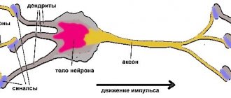
The nervous system is made up of individual nerve cells known as neurons, which are specialized in their ability to interact with electrical signals at speeds of up to 120 meters per second. These high speeds are critical to the functioning of the nervous system because it allows a single neuron to send information to and from the brain almost instantly.
- Dendrites, which convert and receive electrical signals
- cell body (or soma) that contains the nucleus and processes signal
- An axon that transmits a signal to the next neuron
At rest, neurons have a membrane potential, which is determined by the concentration of ions on either side of the neuron's membrane. When a stimulus is received, neurons create a rapid and temporary change in this potential, now called an action potential. These action potentials quickly change the polarization of the neuron, allowing signals to be transmitted between neurons.
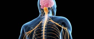
Neurons form the entire nervous system, which is divided into two subsystems: the central nervous system and the peripheral nervous system. The central nervous system (CNS) includes the brain and spinal cord, and the peripheral nervous system (PNS) includes all other neurons distributed throughout the body. When discussing the limbic system we will primarily focus on the CNS brain, and when discussing the hypothalamus we will primarily focus on the PNS.
Limbic system
The scheme is designed for 2-3 months. Children under 10 years old - ½ dose.
The limbic system is the culprit of our positive and negative emotions, as well as obsessive negative thoughts. After getting to know this part of the brain, you will know what to do if you are sad, how to cope with anger or a bad mood.
The limbic system (from Latin limbus - border, edge) is a collection of a number of brain structures located on both sides of the thalamus, directly under the telencephalon. It envelops the upper part of the brain stem, like a belt, and forms its edge (limb). This is not a separate system, but a collection of structures from the telencephalon, diencephalon (diencephalon) and midbrain (mesencephalon).
The limbic system is based in the cerebral cortex, the organ that is responsible for speech, logic, the ability to analyze - for everything that distinguishes us from animals, and becomes both a big plus and a problem if there is no understanding of where the unexpected outbreak comes from anger or negative thoughts that poison your life.
How does the fight or flight response lead to neurosis?
The deep limbic system regulates our emotional state: at different times we feel relaxed or tense. The anterior part of the hypothalamus sends signals to the body through the PSNS, the parasympathetic nervous system, which is responsible for relaxation. Activation of the posterior hypothalamus triggers the fight-or-flight response. We inherited this reaction from our ancestors, when survival was a physical matter and was taken literally.
The “fight or flight” principle is a “highly motivated response” that occurs instantly when the hypothalamus is activated. As soon as we see or feel a physical or emotional threat, changes immediately occur in the body: the heart begins to beat faster, breathing becomes faster, blood pressure rises, limbs become cold due to the outflow of blood to large muscles, so that a person on a physical level can either repel the enemy’s attack or run away from him. At the same time, the pupils dilate to see better.
The above defense reaction was designed by nature in case of physical threat. Today the world has become much safer, but our brain still scans the surrounding reality in search of potential threats. In the absence of physical threats, the brain successfully finds social threats, preventing you from relaxing and fully enjoying life. Even the lack of a smile from your opponent can increase cortisol levels. It is no coincidence that the limbic system is also called the “emotional brain.”
The Emotional Brain and Big Problems
Responsible for our survival, preservation and self-defense, managing social behavior, maternal care and education, participating in the regulation of the functions of internal organs, smell, instinctive behavior and experiences, our limbic system strives for safety and constancy. Often it is this limbic love for routine that gives side effects:
Adverse habits The brain constantly filters people according to the “friend or foe” principle, pre-emptively tuning negatively to the entire world around us. A person is in stress mode 24/7 without even realizing it. Naturally, the body begins to look for affordable ways to obtain hormones of happiness and safety. Which? The simplest and easiest! Eating, smoking, alcohol - these actions directly affect dopamine and serotonin levels, briefly distracting us from stress. We become absolutely passive, lazy and find many excuses not to act.
Mirroring Undesirable Reactions Under the influence of the limbic system, we repeat the reaction to similar situations over and over again, creating strong neural connections. Many people have “mirror” neurons since childhood, formed by observing the behavior of loved ones and their reaction to stress. And even realizing that the scenario is not the most attractive, we still reproduce it, accumulating negativity.
Inaction and depression Focusing on the negative and a depressive state often lead to inaction, patiently waiting “by the sea for the weather”, and this is a mistake - this way you can fall out of life for a long period.
Frontal lobes and behavioral responses
The frontal lobes, part of the limbic system, arose in humans through evolutionary means. The more an early man could share food among his community, the more likely it was that the community would be able to survive. Only thanks to the frontal lobe is a person able to refuse food, sharing it with his neighbor and thereby maintaining relationships within society. In women, the frontal lobes began to emerge for the specific purpose of sharing food. Men received this area evolutionarily, as a gift. Without the assigned tasks that lie on the shoulders of women, men began to use the frontal lobes in a wide variety of ways (thinking, building, showing dominance). The frontal lobe supported social connections in ancient hominids. Those who were unable to share food were eaten themselves or expelled. Therefore, in just a few million years, as a result of social selection, the frontal lobes of the brain grew significantly and one day became the basis of the mind.
We no longer need to “survive”, spend the bulk of our lives in search of food and defend ourselves from predators. The majority of the world's population is not in danger. And the greater activity of the limbic system is directed towards social moments.
Currently, the frontal lobes of the brain control cognitive abilities, coordination of movements, and expression of emotions.
The left frontal lobe is responsible for movement and positive feelings, and the right frontal lobe is responsible for nonverbal abilities and negative emotions. Knowing how to activate your left frontal lobe can make a huge difference in your mood and life in general.
The emotional brain does not differentiate between threats to our body and threats to our ego. Therefore, we begin to defend ourselves without even understanding the situation. When someone hurts our feelings, we release adrenaline, which stimulates blood flow to the large muscles, instantly focusing our thoughts to protect ourselves from the threat.
The limbic system, with the participation of the cerebral cortex, makes us critical of new people and situations, information (we remember that the emotional brain loves stability). For the same reason, we see all the vicious circles in other people, and prefer to point out their mistakes, but do not notice our own imperfections (because stability and old neural connections take their toll).
When the activity of the limbic system increases, and the system is in an overexcited state for some time, this leads to exhaustion and inhibition of the work of all its structures. And then even the simplest and most harmless things will be perceived through negativity.
A simple example: a conversation between a conditionally normal person and a person with a hyperactive limbic system (already in a negative mood). The latter will interpret almost everything said in a negative way, and his characteristic fear will be the fear that he is not being told something or is being lied to. In harmless speech patterns, he can hear irony or insult, the so-called. “reading between the lines” effect. If such a mood continues in a person long enough, it causes a reaction of rejection from society and a desire to retire from everything that causes pain.
Dependence of mood on the season
Light levels are extremely important for people suffering from seasonal affective disorder. People with this disorder often feel more depressed in the winter when daylight hours are shorter. Seasonal affective disorder affects particularly large numbers of people in the northwestern United States and northern Europe, where there are often cloudy skies and short days in winter. Short daylight hours and the cold season stimulate the activity of the limbic system, the frontal cortex reduces its functionality, and people involuntarily experience anger and aggression. In women, the limbic system is more active, so they feel weather changes especially acutely.
Most people who become depressed prefer to shut themselves out from the world, preferring to be indoors with low light. However, low light levels change brain chemistry. The brain receives a signal from the retina to determine whether it is dark or light. The retina will then send this information to the pineal gland. If it is dark, the pineal gland will secrete melatonin, which causes sleepiness and inactivity. It's no wonder that people become more depressed in winter (due to little daylight - little serotonin, little dopamine, high melatonin).
At the same time, it is important to understand that the limbic system is stronger than the cerebral cortex (logic, awareness and control): at the moment when the limbic becomes hyperactive, the cortex is unable to control it, especially in such situations everything is bypassed: the impulse goes to the hypothalamus, he, in turn, instructs the pituitary gland to secrete certain hormones. So if you're feeling depressed, try to stay in brightly lit areas as much as possible. One way to treat mood disorder is to use a full-spectrum lamp. Of course, natural light is more effective, but if you live in an area where light is scarce in winter, then at least try this lamp.
Tonsils as part of the limbic system
Negative thoughts are part of a vicious circle. The limbic gives a signal - it causes bad thoughts - bad thoughts cause activation of the amygdala (the main guard of the brain) - the amygdala partially releases the excitement into the limbic - the limbic becomes even more activated.
The amygdala processes emotions, including fear. This area of the brain is hyperactive during times of stress, fear, anxiety, or a panic attack. The amygdala is connected to the prefrontal cortex (PFC), the area of the brain that regulates thoughts and is responsible for focus, concentration and attention. If the amygdala is very active, it can change the functioning of the PFC, and therefore we cannot concentrate, concentrate, anxiety takes over.
The amygdala region consists of three almond-shaped groups of neurons closely connected in the temporal lobes of the brain. It also plays a major role in the formation and storage of memories associated with strong emotional events, especially those involving fear and anxiety. The amygdala is also responsible for memory consolidation (short and long term). Neurotransmitters and hormones, especially those produced by the adrenal glands, are closely related to the function of the amygdala.
In moments of prolonged anxiety, negativity, and irritability, do not try to pull yourself together and dull your emotions with willpower. Firstly, it most likely will not work, and secondly, the limbic system will control the cerebral cortex and start a powerful chain reaction. Some of the signals will still pass through the hypothalamus, turning into hormones, after which they will accumulate in a sufficient area of the cerebral cortex, gradually leading you to a nervous breakdown.
In those moments when you feel mentally ill, you don’t need to prove to your brain that everything is fine and you feel amazing. Just take it for granted and go ahead and calm the limbic system.
How not to drown in negativity?
Daily work on your own perception of the world will help you avoid drowning in negativity. Constant practice of extremely simple techniques will improve the functioning of the limbic system. Allow 3-4 weeks to restore the functions of the hyperactive limbic system.
Try to talk it out It is really important to pour out the stream of confused and oppressive thoughts - this way you will have a much better chance of understanding the problems. The longer the negativity accumulates inside, the more serious the consequences will be. You can chat with friends, a loved one, parents, a psychologist, or you can simply write your thoughts on a piece of paper.
Controlling Negativity Control your negative thoughts - this will help you control your life. When you know how the brain works, you will find ways to direct your actions towards something positive. The limbic system can only be calmed by catching negative thoughts and transforming them into positive antagonistic thoughts. For example, “I’m fat and ugly,” but (we translate the thought into a positive direction) “But I’m the life of the party, I find a great common language with men and I cook like a god.”
You become what you think because thoughts shape actions and the brain interprets thoughts as directives or commands. Your body reacts to the thoughts that pass through your mind. By understanding how powerful your brain can be, you can make a conscious effort to positively influence it.
A good technique for tracking negative thoughts and turning them into positive ones is using a rubber band on your wrist. When a stream of negative thoughts appears, simply pull the elastic band back as much as possible and click it on your hand until it hurts. Pavlov's reflex will work. The pain will stop the flow of negativity and you will be able to consciously switch to the positive thoughts you need.
Focus on the good Notice 5-10 times a day 3-5 positive things (in the appearance of others, the environment, the current situation).
Smile “Pull up” your smile for at least 5 minutes. When you smile, you physically activate the parts of your brain associated with positive emotions. There are neural pathways that connect facial muscles to important parts of the brain. The movement you show in your face can affect the signals you send to your brain, activating your frontal lobe. A simple smile can activate your left lobe, which processes positive emotions, and a negatively expressed emotion can activate your right lobe, which processes negative emotions.
Kiss and hug. You can even ask your partner to give you a relaxing massage. Actions that help the production of oxytocin are necessary. Good music, delicious food, pleasant smells, close people - all this will help. Sometimes, in order to overcome bad thoughts and emotions, you need to distract yourself and try to look at what is happening less seriously. Humor develops neuroplasticity in the brain and is a great treatment for anxiety. When you're sad, humor can quickly move you from one state to another. You should not watch emotional films - they will only make you want to cry. It’s better to watch comedies - they distract from bad thoughts.
Breathe correctly Practice diaphragmatic breathing - it helps to shift from the SNS to the PSNS. The slowing of the heart rate that occurs with this type of breathing leads to inhibition of nervous reactions and activates neurochemical systems that reduce the activity of the amygdala. By focusing on your breathing, you can free your mind from anxious thoughts. Videos on YouTube will help you learn diaphragmatic breathing.
Many world cultures have breathing-based relaxation techniques (meditation, prayer) - they activate the parasympathetic nervous system, although there was no trace of any scientific research at that time.
Also, in moments of fear and anxiety, you can use exercises to activate the parasympathetic system.
Stand up straight, your feet should be shoulder-width apart. Bend forward and try to reach your toes with your hands, feel how the blood rushes down and the muscles stretch. Slowly straighten up, raise your arms to the sides and up in the shape of the letter V. Inhale deeply from your belly. Continuing to be in this position, hold your breath for 10 seconds, then slowly lower your arms. Take a deep breath, exhale as much as you can. Do this ten more times.
Physical activity 30 minutes of cardio training. The left hemisphere of the brain is responsible not only for the formation of positive emotions, but also for physical activity. Exercising helps reduce the level of depression, but laziness and passivity, on the contrary, only aggravate depression.
Why do you need to breathe into a bag during a panic attack?
We take from 9 to 16 inhalations and exhalations per minute at rest. When stressed or having a panic attack, we already take up to 30 breaths per minute. Since the cardiovascular system combines the respiratory and circulatory systems, rapid breathing provokes an increase in heart rate, and a person feels anxious. Slowing your breathing and simultaneously slowing your heart rate is calming and relaxing.
With hyperventilation (frequent shallow breathing), physiological changes occur in the human brain. Excessive inhalation of oxygen reduces the level of carbon dioxide in the blood. CO₂ helps maintain an optimal level of acid-base balance (pH) in the blood. A decrease in pH levels causes nerve cells to become more excitable, and a person may feel anxious. Bodily sensations coupled with uncontrollable anxiety can even cause a panic attack.
Too much reduction in CO₂ levels in the blood leads to alkalosis, a condition in which the acid-base balance is disturbed (the blood contains an increased amount of alkali). Because of this, the blood vessels narrow, and much less oxygen reaches the tissues and organs. Paradox: the more air a person inhales, the less tissue and organs receive it.
Hypercapnic acidosis leads to dizziness, lightheadedness, cerebral vasoconstriction (hence the feeling of unreality) and peripheral vasoconstriction (this explains the feeling of trembling in the limbs). A person prone to panic attacks may overreact to such bodily sensations and breathe even more frequently.
Conclusion: if you feel severe stress with hyperventilation or a panic attack - breathe into the bag (inhale - exhale) to restore carbon dioxide levels. Try to breathe with your belly!
Biochemistry of stress
Anemia, hypothyroidism, hormonal deficiencies, ineffective digestion, vitamin D deficiency, dysbiosis, toxic load, insulin resistance, impaired absorption of amino acids, which are the building blocks for the same neurotransmitters - all this can lead to permanent stress. But in most cases, stress is a lack of awareness, a lack of understanding of neuroprocesses, and an obsession with the negative.
Anxiety is also associated with elevated levels of corticotropin-releasing factor (CRF). It causes the adrenal glands to release "stress" hormones called glucocorticoids. These “stress hormones” include cortisol and norepinephrine. If CRF is high during anxiety or a stressful situation, the result is that the body releases excess amounts of stress hormones, which leads to feelings of tension and fear - the heart begins to beat faster, breathing becomes more rapid, and the stress response becomes more intense.
In a recent study, the receptors that release CRF were blocked in mice. And then, despite the extreme conditions that created extreme stress in which the animals were placed, they secreted much less of the hormone and tolerated the state of stress more easily.
High CRF means a priori there is a lot of glutamate in the brain. Studies have shown that glutamate receptor antagonists are effective in treating anxiety; they help alleviate fear, anxiety, and have a calming effect. In anxiety, the ratio between glutamate (an excitatory neurotransmitter) and gamma-aminobutyric acid, also known as GABA and GABA (an inhibitory neurotransmitter), is unbalanced. Treatment may involve decreasing glutamate (through glutamate-inhibiting drugs) or increasing GABA levels.
How glutamate makes us tense and GABA makes us calm
Glutamic acid (glutamate) is the most common excitatory neurotransmitter in the vertebrate nervous system, in neurons of the cerebellum and spinal cord. Glutamate enhances and accelerates the transmission of impulses in the nervous system and brain; makes it easier to remember, learn, understand, and react faster; gives more anxiety, stress, irritability.
For the most part, glutamate's main job is to carry an impulse to the muscles. After that, glutamate, with the help of the enzyme glutamate decarboxylase, is converted into GABA, an inhibitory neurotransmitter, which ensures normal brain function (excitation is extinguished). However, as a result of failures in enzymatic systems, an imbalance can occur when there is excess glutamate in the brain.
An excess of glutamate is actually poison for the body. Increased membrane conductivity of nerve cells, depletion of cellular resources, and, as a result, death of neurons. Aggressive behavior, poor memory, inability to control one’s desires, deviant behavior in its purest form - these are the characteristic “side” effects of an excess of glutamate.
Symptoms of excess glutamate in the brain:
- hyperactivity (inability to concentrate on some activity or control one’s behavior. Attention jumps from subject to subject, while more than one task is not completed)
- insomnia
- anxiety
- headache
- blood pressure surges
- the occurrence of stereotypies that are observed in children with autism
- thinking becomes sluggish (thoughts, words, melodies of songs are obsessively spinning in your head, which prevent you from concentrating on something really important)
- internal dialogues that a person cannot stop by force of will
- imbalance of neurotransmitter balance
Glutamic acid is a necessary source for the synthesis of GABA. Excess glutamate can occur due to a lack of the enzyme glutamate decarboxylase, which converts glutamate into GABA, and with high CRF, stress, and cortisol, glutamate literally “paralyzes” brain function.
A lack of vitamin B6 (pyridoxine), with the direct participation of which glutamate decarboxylase works, as well as high eosinophils in the blood may indirectly indicate a high level of glutamate.
Increased glutamate is a direct correlation to the high rate of reproduction of streptococci - bacteria that cause inflammatory diseases (sore throat, scarlet fever, erysipelas) and autoimmune damage to the kidneys, joints, and heart valves. People with imbalances who have high levels of glutamic acid in their bodies, including children with autism, are prone to streptococcal infections.
In 1939, Sigmund Freud said: “Psychology has its limits. Biochemistry will one day provide a solution to mental illness." And he turned out to be right.
If there is an excess of glutamate, additional intake of gamma-aminobutyric acid and regular physical activity are recommended, during which the level of glutamate concentration drops and GABA naturally increases.
This also applies to hyperactive children. Exercise helps burn off adrenaline, so exercising when you're stressed can help balance your neurotransmitters.
Important supplements for children with hyperactivity: B6, B12 (B9 is added after taking them for two weeks), Omega-3 (3-4 g), GABA (1-2 g), selenium 200 mcg, magnesium glycinate (700-800 mg ).
nutraceuticals for anxiety
Let's take a closer look at some of the additives from the scheme.
Kava-kava Second name for Piper methysticum. Kava kava has long been used in folk medicine to treat anxiety, depression, insomnia, and has a mild analgesic effect. A clinical study has shown that kava kava reduces excessive activity in the amygdala and limbic system in general. Non-addictive, it has been approved for use in the fight against insomnia and anxiety in Germany and Switzerland.
Some time ago, kava kava was not delivered to Russia (but was sold on its territory!). The persecution against her is not justified; a negative effect was noted when taking 2 g per kg of body weight, and this dosage is 15 times higher than what doctors prescribed in Germany. However, pregnant women and children are still not recommended to take this drug.
St. John's wort St. John's wort has long been used to relieve depression, anxiety and sleep disorders. It works primarily by increasing serotonin levels in the brain. According to a Pakistani study on rats, St. John's wort has a significant effect on stress-related hormones and neurotransmitters and reduces activity in the amygdala and limbic system in general.
Omega-3 Omega enhances the number of serotonin receptors and increases serotonin levels in the brain. EPA increases the release of serotonin, and DKG affects serotonin receptors by causing cell membrane fluidity.
Magnesium threonate This is the only form of magnesium that crosses the blood-brain barrier and has calming properties.
B vitamins Without B vitamins, the normal functioning of the nervous system becomes impossible. Vitamins B1, B6 and B12 are neurotropic: they are often used in the treatment of neurological disorders, and their deficiency causes damage to peripheral nerves. Each of these vitamins is involved in methylation, and deficiency leads to depression and various disorders.
L-theanine Theanine calms the brain, promotes recovery from depression, improves cognitive functions of the body, and increases concentration. Acts more as a relaxant.
Electrolyte concentrate is necessary to eliminate the imbalance of microelements, primarily magnesium, calcium, potassium and sodium, which affect the functioning of glutamate receptors and the transmission of signals through nerve cells. When there is a lack of potassium, a lot of sodium enters the channels in the brain, causing overactivity and anxiety.
An alternative from the pharmacy is Regidron, but this is for a very last resort, the drug contains a large amount of dextrose.
Structures, connections and functions of the limbic system
Limbic lobe
The dentate cortex is the inner part of the brain that is located superior (above) and dorsal (behind) the corpus callosum. It consists of an anterior (frontal) and posterior (posterior) division, which both receive information from the thalamus and hippocampus formation.
The main function of this area is to regulate both autonomic function and conscious function. Autonomic functions involve involuntary changes, such as increasing heart rate to a threat in the environment, while conscious responses involve voluntary choices, such as deciding to respond to a threat (such as taking a step back). Conscious response is also important in controlling emotions. In other words, rather than perceiving the emotion itself (such as feeling pain), the cingulate cortex controls what a person thinks about the emotion (such as how you feel about pain).
The parahippocampal gyrus consists of several distinct areas in the medial temporal lobe. The most significant of these areas includes the entorhinal cortex, which the parahippocampal gyrus uses to send information from the cortex to the hippocampal formation.
Hippocampal formation
The hippocampus is a horn-like structure that surrounds the lateral ventricles. The main function is to process new information and form long-term memories, which are then stored in other areas of the brain. If the damaged person is unable to create new long-term memories. However, a person can remember long-term memories that have already been stored.
The hippocampus most often receives information from the entorhinal cortex of the parahippocampal gyrus. When received, information is usually mediated through execution. An efficient pathway sends signals using fibers throughout the subiculum of the specific complex and into the dentate gyrus, where fibers then extend from the dentate gyrus to CA3 axons. Schaffer collaterals then connect to dendrites on CA1 axons. CA1 axons contain fibers that extend from the hippocampus to subsequent brain regions.
amygdala
The amygdala supports two main information processing pathways: dorsal and ventral. Both routes send information to the hypothalamus, while the ventral route also sends information to the thalamus.
thalamus
*Note: Smell is not processed by the thalamus. Smell is received by the olfactory bulbs and is directly sent for emotional processing and memory formation.
Hyopthalamus
The hypothalamus is also located in the diencephalon, which is inferior to the thalamus and both inferior and lateral to the third ventricle. The main function is homeostatic regulation, as the hypothalamus directly controls the autonomic nervous system (ANS). The ANS is part of the PNS, which controls the involuntary actions and rhythms of internal organs, including heart rate, blood pressure, and digestive movements. The ANS covers two subsystems – the Sympathetic Nervous System (“Fight or Flight”) and the Parasympathetic Nervous System (“Rest and Digest”).
The hypothalamus is the central structure that must be stimulated to create physical changes in the body. This is because the hypothalamus is directly connected to the pituitary gland and thus immediately influences the hormones released by the pituitary gland. This connection is a crosstalk between the nervous system and the endocrine system, translating neural stimuli received by the brain into hormonal changes throughout the body.
Alternatively, the hypothalamus may release neurosecretory cells into blood vessels that penetrate and activate the anterior pituitary gland. The anterior pituitary gland then increases or decreases the hormones distributed in the blood depending on whether neurosecretory cells from the hypothalamus are directed to stimulate or inhibit a particular hormone.
The part of the pituitary gland with which the hypothalamus interacts - anterior or posterior - depends on the signal received by the brain and the needs of the body.
Hippocampus
In a study by C. McDonald et al. (2006) revealed a decrease in the volume of the right and left hippocampus, 2.47 ml and 2.50 ml, versus 2.54 ml and 2.61 ml in healthy individuals, which corresponds to approximately a decrease in the volume of this brain structure by approximately 6%.
Some authors note that a decrease in the volume of the hippocampus, as part of the limbic system, is noticeable after the first psychotic episode, however, according to other researchers, these changes are recorded before the manifestation of schizophrenia and progress after its onset. Note that in relatives of patients with schizophrenia, a decrease in the volume of the hippocampus and amygdala can also be detected.
In schizophrenia, the functioning of the amygdala is impaired; this fact is discovered when studying patients with schizophrenia using the method of evoked potentials (P300).
In the hippocampus of patients with schizophrenia, with a decrease in the volume of neurons and “sparseness” of their mutual arrangement, an increase in the number of pathologically altered myelinated axons with thinned myelin sheaths, swollen perioxal glial processes and wrinkled axons was found. The proportion of these fibers in the total number of myelinated axons and their numerical density in schizophrenia is 2 times greater than in the control group. At the same time, it is known that pathological and reparative changes in axons depend on the reaction of the microglial cells surrounding them (Kolomeets N.S., 2007).
Peculiar memory impairments, primarily working memory, in schizophrenia, affecting learning difficulties in persons suffering from this disease, may indicate the involvement of the hippocampus in the pathological process.
According to Shenton et al. (2001), the volumes of the medial temporal lobes, which usually include the hippocampus and amygdala, are significantly reduced in size. These changes were noted in 70% of patients with schizophrenia.
Bibliography
Show hide
