More useful information about other diseases starting with the letter “K” here: Causalgia, Brain cyst, Cluster headache, Tick-borne encephalitis, Kozhevnikov epilepsy, Colloid cyst of the third ventricle, Coma, Compressive myelopathy, Balo concentric sclerosis, Radicular syndrome, Corticobasal degeneration, Craniovertebral anomalies, Craniospinal tumor, Craniopharyngioma, Myasthenia gravis crises, Hemorrhage into the ventricles of the brain.
What does hemorrhage into the ventricles of the brain mean?
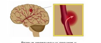
Hemorrhage into the ventricles of the brain is a disease that is one of the forms of malfunction of the circulatory system of the brain. The presented disease is most often diagnosed in modern medicine. Neurologists say that the disease is caused by arterial hypertension (the treatment of which has been neglected for a long time), bad habits (smoking, alcoholic beverages, drugs) and the use of pharmacological agents that can lead to poor blood clotting. To establish a conclusion, the doctor conducts a detailed conversation with the patient, finds out existing complaints and problems, and also prescribes a certain list of tests for a final affirmative diagnosis. Therapeutic treatment helps eliminate the formed edema, stabilize lung function and normalize blood pressure.
Why is intraventricular hemorrhage dangerous in a child?
Hemorrhage inside the brain is a type of acute cerebrovascular accident. In adults, pathology develops with hypertension, secondary hypertension, alcohol abuse, and blood clotting disorders.
In children, ventricular hemorrhage develops due to underdevelopment of the vascular wall. Any external pressure on the weak arteries of the brain provokes rupture.
In old age, hemorrhage into the ventricles causes death in approximately 40-50% of cases. Persistent arterial hypertension with unstable relief by drugs and the occurrence of a hypertensive crisis are dangerous factors in the development of the nosology.
Hemorrhage in the lateral ventricle has a favorable course. Gradual spread of blood, filling of the third ventricle is characterized by an unfavorable prognosis. Neurological disorders are rapidly increasing. A rapid and sharp increase in the amount of blood provokes death. A similar situation occurs when blood instantly breaks through from the lateral ventricular zones into adjacent anatomical structures. Before death, severe neurological symptoms appear. Mortality is even higher when the fourth ventricle is filled.
Physiological ventricular spaces are filled with cerebrospinal fluid. The leakage of any fluid creates external pressure, creating conditions for intracranial hypertension. Maintaining the condition for several hours provokes irreversible changes and death of the brain parenchyma.
Brief information
In neurology, a similar disease of hemorrhagic type is classified as stroke. The anomaly has different names. According to surveys and average statistical information received from practicing doctors, the described deviation from the norm ranks first in the number of deaths of patients around the globe. Based on publicly available information, such a process can cause death within 48 hours of onset in 60% of cases. The risk increases significantly during the recovery period after a stroke. People who survive a critical attack have a 90% chance of bleeding.
Basically, the largest number of recorded pathologies is observed in the population of retirement age, whose age has exceeded the mark of 50 years. However, in addition to this, danger threatens not only people with arterial hypertension, but also any other abnormalities that have absolutely nothing to do with the level of pressure.
Types of hemorrhage
The compilation of classes of the described disease was created in the late 90s, as a result of which the list was included in international medical documents on the classification of diseases. Based on the source in the form of ICD-10, the presented pathological process is divided into several forms:
- Subependymal (SEC).
- SEC with damage to the extreme parts.
- SEC with progression to areas and substance of the brain.
In addition, there are additional differences between IVH, which differ in location:
- Changes in the lateral structures - begins from nearby soft cavities and manifests itself as a gradual filling of the entire volume with further leakage into other areas. In the case of the formation of a surplus, the size of the main organ begins to increase, as a result of which symptoms of a neurological nature progress (if only one object is filled, the specialists’ prognosis is positive, since this process is similar to a parenchymal disorder).
- Location in the third ventricle - the medial zones of parenchymal disorders become the culprit.
- The focus of inflammation in the 4th lobe originates from the dorsal perimeter of the brainstem or cerebellum.
Intraventricular hemorrhage
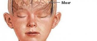
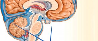
The brain has a very complex structure. In addition to gray and white matter, the brain contains structures filled with a special fluid called cerebrospinal fluid. The internal structures of the brain filled with cerebrospinal fluid are called the ventricles of the brain; there are only four of them: two lateral, a third and a fourth. Liquor is formed in the ventricles of the brain, flows through them, then enters the intrathecal spaces of the brain and spinal cord and is absorbed there into the circulatory system.
Due to the immaturity of the entire brain, premature babies are at risk of rupture of fragile cerebral vessels and the development of hemorrhage in the brain, and most often this hemorrhage occurs in the ventricles of the brain.
Intraventricular hemorrhage (IVH) is bleeding into the ventricles of the brain.
The younger the baby's gestational age, the higher the incidence of IVH. The incidence of IVH is especially high in children born before 28 weeks of gestation. Typically, IVH develops within the first 3 days after birth.
IVH is diagnosed using brain ultrasound (neurosonography - NSG). There are three degrees of IVH.
Stage 1 IVH is a small hemorrhage. If the child’s condition has stabilized and no progression of hemorrhage is observed, then grade 1 IVH becomes harmless; it, as a rule, does not lead to complications and does not require treatment. A child with stage 1 IVH undergoes neurosonography several times to make sure that the hemorrhage does not progress and then enters the resolution stage. IVH of the 1st degree does not have an adverse effect on the development of the child.
IVH of 2 and, especially, 3 degrees are already more significant hemorrhages that can affect both the child’s condition and lead to complications. With IVH of the 2nd degree, there is an outpouring of blood into the ventricle; with IVH of the 3rd degree, hemorrhage also occurs in the brain substance surrounding the ventricle (periventricular region). The development of IVH of degrees 2 and 3 can worsen the general condition of the child, worsen breathing, lead to apnea (stopping breathing) and impaired cardiac function. It is also possible to develop seizures. Treatment is carried out taking into account all developing symptoms.
With massive outflow of blood into the ventricles of the brain, the risk of the main complication— occlusive hydrocephalus— . Unfortunately, there are currently no treatments that can prevent this complication.
Occlusive hydrocephalus (dropsy of the brain) is characterized by progressive expansion of the ventricles of the brain as a result of impaired outflow of cerebrospinal fluid. Blockage of the cerebrospinal fluid outflow tract occurs due to the proliferation of certain cells in the walls of the ventricles under the irritating influence of blood and its breakdown products. Thus, cerebrospinal fluid continues to form in the ventricles of the brain, but since the outflow tract is blocked, the cerebrospinal fluid accumulates more and more in the ventricles, leading to their progressive expansion and increased intracranial pressure. In such cases, neurosurgical assistance is required. At the beginning, ventricular punctures (punctures of the cerebral ventricle) can be performed to remove excess cerebrospinal fluid; temporary drains (tubes for draining cerebrospinal fluid) can also be installed into the cerebral ventricle. When the child’s condition allows, then the main stage of neurosurgical treatment is carried out, which should ensure constant drainage of cerebrospinal fluid from the ventricles.
The prognosis for the development of a child with IVH of degrees 2 and 3 is determined by the degree of damage to the brain substance and the presence of occlusive hydrocephalus. With grade 2 IVH without hydrocephalus, there is a high probability of subsequent favorable development of the child. With IVH of the 3rd degree, as well as if occlusive hydrocephalus develops, the risk of impaired psychomotor development of the child, the formation of cerebral palsy, and epilepsy increases.
Kryuchko Daria Sergeevna
Head of the Department for Analysis and Coordination of Work to Improve the Provision of Neonatological Care
FSBI "Scientific Center of Obstetrics, Gynecology and Perinatology named after Academician V. I. Kulakov of the Ministry of Health of the Russian Federation,
Director of the Directorate of Social Projects of the Foundation for Socio-Cultural Initiatives.
Scientific editor of the journal “Neonatology. News, opinions, training."
Doctor of Medical Sciences.
19.01.2019
Main factors of appearance and symptoms
There are two stages of bleeding - primary and secondary. PIH is associated with arterial problems in the body or amyloidosis in the cerebral vessels, so they are diagnosed in rare situations. Based on some data, such an anomaly occurs once in 300 patients. The second version of FFM is formed due to the incorrect use of certain medications (for example, antiplatelet substances or fibrinolytic drugs), the presence of a neoplasm capable of breakthrough, or cancerous tumors.
With such an illness, the patient experiences a rapid increase in depression of thinking. Usually, a coma appears after a stroke in the first hours. Only with a gradual progression of the disease and, most importantly, a minimum amount of fluid released, the consciousness of the individual remains stable for a long time and decreases gradually.
Often, bleeding occurs along with:
- meningeal manifestations and gag reflexes.
- Extreme sweating.
- Trembling, similar in nature to chills.
- Change in skin color.
- Abnormal accumulations of blood cells in the vascular ducts, occurring in the face, torso, arms and legs.
- An initial decrease in body temperature, and as a result a rapid jump reaching above 40 degrees.
A typical symptom of such a disorder in the body is a decrease in the tone of muscle tissue, which manifests itself in the form of hormetonic syndrome or an irreversible stage of coma. In the first method, an attack of increased muscle activity in the inflamed area begins. This symptom appears due to external conditions. In the second, deformations develop exclusively in the extensor layers. The patient takes a supine position and arches his back, while tilting his head. The upper limbs are clasped and the forearms are facing inward. Also, it may be accompanied by paresis in the opposite extremities from the pathology, the formation of reflexes in the tendons, the presence of anomalies and lack of action in the abdominal region, disruption of the performance of the pelvic region and its organs.
During the disease, which is localized in the third compartment, difficulties arise in breathing and in the functioning of the circulatory system, and hormetonia syndrome has a bilateral effect. With a deviation located in the fourth ventricle, hiccups and problems with swallowing saliva are caused, uncontrolled movements are not observed, and signs of hormetonia are less noticeable. If the described process continues for a long time in a patient, then an increase in the amount of fluid begins, which provokes an increase in VD, swelling and compression of the nerve areas responsible for the life support of the entire body. The supplement favors disorders of the heart and vascular system.
Diagnostic techniques
To establish a correct conclusion, experts will need to conduct an evaluative check of the person’s medical history, that is, find out the presence of blood diseases, previous hemorrhagic strokes, what medications the patient took that contributed to poor clotting, etc. In addition, doctors conduct a standard examination and prescribe additional tests to create a full picture.
You need to understand that if an anomaly is likely to develop, you should urgently go to the nearest medical center to provide professional clinical care. Also, experts do not exclude the possibility that the patient will have to undergo resuscitation measures on the way to the hospital. In the inpatient building, doctors write out directions for the following procedures:
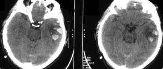
- MRI scan or computed tomogram, depending on the presence of contraindications.
- Donation of blood cells for analysis, during which there will be a report on the number of platelets in the body.
- Coagulogram examination.
- Electrocardiogram.
- Check blood pressure readings.
If doctors do not have the opportunity to perform magnetic resonance or computer diagnostics on a person due to a coma, then echo-encephalography is prescribed. At certain points, it may be necessary to perform a lumbar puncture in order to distinguish hemorrhage, which provokes the entry of fluid into the spinal substance, from an ischemic stroke.
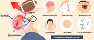
Symptoms of cerebral hemorrhage in adults
The key symptoms that occur during a cerebral hemorrhage were given to us by neurologist Polina Petrosyan. According to her, intracerebral hemorrhage is characterized by an acute onset, with a rapid development of the clinical picture:
- sudden severe headache;
- nausea and vomiting;
- dizziness.
Depending on the volume and location of the hemorrhage, the following may occur:
- one-sided weakness in the limbs (hemiparesis) up to a complete lack of movement in them (hemiplegia);
- restriction of eyeball movement;
- speech impairment - both its reproduction and understanding;
- difficulty swallowing and breathing;
- disturbance of consciousness up to coma.
Much of the severity of symptoms depends on what part of the brain is damaged, how much blood leaks out, and how large the vessel is.
Treatment Options
The first point for medical staff is the prompt provision of first emergency aid and the immediate implementation of basic level treatment - stabilization of the functions of the heart and lungs, as well as monitoring of blood pressure levels and regulation of the constant flow of internal processes of the body.
In addition, doctors prescribe therapy to get rid of symptoms: intravenous injections that have an anticonvulsant effect, as well as help resolve swelling and normalize blood pressure (in addition, a drug can be used to stop vomiting). To fully stop bleeding, a treatment plan is currently being developed by the most qualified scientists. The pathogenic option involves maintaining optimal blood pressure levels and pumping out excess blood through surgery. Such procedures include:
- The use of agents that have a neurotrophic effect and increase metabolism.
- Taking antioxidants (including vitamin E) and substances that block calcium ducts.
The surgical procedure is selected on an individual basis.