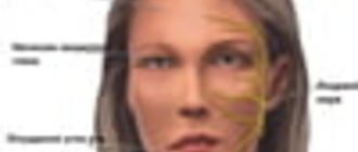
The main symptom of the disease is asymmetry and immobility of one part of the face (this is usually shown in the photo of facial nerve paresis). The patient cannot close one eye, smile, or speak normally. If you seek qualified medical help, you can completely get rid of the problem.
There are three forms of paresis:
- Peripheral paresis of the facial nerve. It begins with severe pain in the parotid area, behind the ear. The inflammatory process affects only one half of the face; swelling of the nerve fibers occurs and their compression in the canal through which they pass. The muscles of the affected side become flaccid, hypotonicity is observed.
- Central paresis of the facial nerve. A rare form of the disease caused by pathology of neurons in the brain. Associated with inflammation of the muscles located in the lower part of the face. The eyes and forehead remain in their normal physiological position, so the patient can wrinkle and blink freely. There are no changes in taste. During palpation, the doctor notices that the muscles at the bottom of the face are very tense.
- Congenital paresis of the facial nerve (10% of cases). Requires differentiation from Mobius syndrome. With paresis of the facial nerve in newborns, lesions of other nerve branches are also recorded. If the disease is mild or moderate, the prognosis is favorable; if the disease is severe, surgery is recommended for the child.
Causes
The causes of facial paresis in a child and an adult may be different. Most often, the problem is provoked by severe hypothermia of the parotid area or head. The disease is also caused by:
- respiratory diseases of the upper respiratory tract;
- nerve damage during otitis media or surgery;
- head injuries;
- herpes virus;
- tuberculosis;
- mumps;
- syphilis;
- polio.
All of these diseases contribute to the occurrence of a strong inflammatory process.
Another factor that triggers the pathological process is a violation of the blood supply to the face, which is noted in hypertensive crisis, ischemic stroke, multiple sclerosis, and diabetes mellitus. Sometimes the sensitive and motor functions of the facial nerve are disrupted during dental procedures.
What causes facial paralysis?
The occurrence of Bell's palsy can be facilitated by various factors that disrupt the conductivity of the nerves of the facial muscles, which leads to their immobilization and disruption of blood flow to the face.
These factors are:
- Suffered a stroke.
- Frequent infectious diseases, for example: herpes, colds, flu.
- Suffered a concussion.
- Diabetes.
- Malignant neoplasms.
- Old age, as a result, deteriorates the elasticity of facial muscles and impairs blood flow throughout the body.
- Vascular thrombosis.
- Abuse of “beauty injections”.
- HIV.
- Facial injuries or injuries caused from outside.
Healthy people and even children can be at risk for developing pathologies. Among other factors, facial paralysis can even be triggered by a stressful or emotional situation, intoxication, or such a nuisance as an unsuccessful turn of the head, as a result of which one of the nerves may be pinched.
It's scary to hear that facial paralysis can occur from simple stress. However, there is a logical explanation for this: when a person is under stress for a long time, his body weakens, the functionality of muscles and organs decreases, due to which the body becomes susceptible to various infections and diseases, one of which can be Bell's palsy .
causes of facial paralysis
Symptoms of facial paresis
The branches of the facial nerve are responsible for motor function. They provide muscle mobility and make facial expressions lively. If the nerve impulse does not pass, facial movements become impossible. There are three stages of facial nerve paresis:
- Acute (lasts up to two weeks).
- Subacute (lasts up to one month).
- Chronic (illness lasts more than a month).
On the affected side of the face there are:
- drooping corner of the mouth;
- smoothing the nasolabial fold;
- a wide eyelid, when closing the eye, lagophthalmos is observed (a visible strip of light-colored sclera remains noticeable);
- loss or deterioration of taste sensations on the first third of the surface of the tongue;
- inability to stretch lips;
- leakage of food from the half-open half of the mouth;
- lacrimation (tears begin to flow while eating);
- worsening of hearing in the first days of illness (discomfort with loud sounds);
- pain behind the ear;
- inability to wrinkle the forehead much.
Paresis can occur through three stages:
- Mild degree. Facial asymmetry is slightly expressed. There is a slight distortion of the lips due to inflammation, difficulty when trying to completely close the eyes and frown the eyebrows.
- Moderate severity. The eyelid does not close completely (lagophthalmos); only minor movements are possible in the upper part of the face. The patient cannot puff out his cheek or perform various movements with his lips.
- The degree is difficult. Facial asymmetry is very pronounced. The mouth is distorted, the eye on the side of the paresis almost does not close. The patient cannot perform even the simplest movements that require the participation of facial muscles.
If you experience similar symptoms,
consult your doctor . It is easier to prevent a disease than to deal with the consequences.
Prerequisites for the occurrence of neuritis
Causes of Bella's Palsy:
- Genetics. One of the reasons for the development of Bella syndrome is a narrow canal of the facial nerve, which is inherited.
- Alcoholism and drug abuse.
- Necrosis of facial tissue due to severe hypothermia.
- Oncology.
- Plastic surgery.
The following prerequisites will help determine the development of pathology:
- Noticeable weakening of the facial muscles, inability to control one or another muscle.
- Difficulty closing the eyes completely. This pathology is called lagophthalmos.
- Increased sensitivity to loud noises.
- Dulling of taste buds.
- Unreasonable tearing of the eyes or, conversely, their excessive dryness and redness.
- There is noticeable facial asymmetry.
- Paralysis of parts of the face or individual muscles.
- Swelling of the face.
- Difficulty controlling salivation.
- Jaw dropping.
Paralysis of the facial nerves, or so-called Bella syndrome, is characterized by slow progression, thanks to which it is possible to determine the development of the pathology and take the necessary treatment measures in a timely manner.
Diagnostics
The diagnosis of facial paresis is made taking into account the existing symptoms. Additionally, the patient is referred for consultation to an otolaryngologist to exclude the presence of pathology in the ear. Tests and diagnostic procedures are also prescribed to identify the cause of paresis.
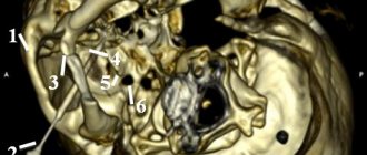
It is important to make sure that the disease is not associated with an abscess or tumor of the face. Electroneurography is used to measure the speed of nerve impulses passing through peripheral fibers. It makes it possible to accurately determine the localization of the source of inflammation, the severity and degree of the pathological process.
Treatment of facial nerve paresis
Whether it will be possible to completely get rid of facial nerve paresis depends on whether the patient sought medical help in time. If the process becomes chronic, it is impossible to restore the innervation of the nerve 100%. This means that the patient will forever be left with an asymmetrical face, which looks unnatural.
Treatment for facial paresis usually takes about six months. All this time it is necessary to take various medications, do massage, special gymnastics, and undergo physical therapy sessions.
Treatment of facial nerve paresis with medications
Before starting treatment for acute facial paresis, the cause of the disease is identified. Afterwards, measures are taken to relieve swelling, eliminate the inflammatory process and activate the process of nerve cell regeneration.
Drug treatment of facial nerve paresis includes the use of:
- Analgesics in the form of tablets or injections. “Baralgin”, “Ketorol”, “Spazgan” are suitable.
- Decongestant drugs - Furosemide, Triampur.
- Corticosteroids. If moderate or severe paresis is diagnosed, Prednisolone is used. It allows you to relieve inflammation and swelling in a minimally short period of time.
- Vasodilator drugs - nicotinic acid, Complamin.
- Sedatives (if there is a high level of patient anxiety) - “Sibazon”, “Relanium”. They help the patient calm down and relax, thereby reducing muscle tone.
- Vitamin-mineral complexes (B vitamins are especially necessary).
- Artificial tears – if the eyes are affected. Their use makes it possible to exclude the addition of a secondary infection and drying out of the mucous membranes.
- Other medications aimed at eliminating pronounced symptoms.
Surgical treatment of facial nerve paresis
Surgical treatment of paresis is carried out if a complete nerve rupture is diagnosed. This happens with congenital anomalies or injuries. The operation can achieve positive results only if it is performed within a year from the onset of the disease. If you wait longer, the facial muscles will atrophy and the restored nerve will no longer be able to control them.
In case of rupture, the nerve is sutured. If we are talking about a congenital anomaly, autotransplantation is performed - the graft is taken from the patient’s leg and placed in the desired place on the face. Then the branches of the nerve from the healthy side are sewn to it. As a result, the facial expressions of the operated person are controlled by one nerve. The scar remains only behind the ear. There are no other changes to the skin associated with the operation.
Physiotherapeutic treatment of facial nerve paresis
During the first week, the disease can be treated with Sollux. This is a special lamp designed for light therapy. Later, the patient is prescribed UHF sessions, paraffin therapy and phonophoresis.
Acupuncture and homeopathy in the treatment of facial paresis
You cannot hope for the success of homeopathy in all cases. If improvements are not observed within a month, it is wiser to turn to traditional methods of treating paresis. Otherwise, the disease can severely disfigure the face, and it will no longer be possible to change anything. It is important not to exceed the dosage of homeopathic medicines, because they may contain extracts of poisonous plants.
The homeopathic drug Gelsemium is most often used to treat paresis. However, you should not prescribe it yourself. Consult a specialist at your nearest homeopathy center
.
Acupuncture can also achieve good results.
Psychotherapy for paresis
A distorted face negatively affects the emotional state of many patients. They develop depression, their mood worsens, and their self-esteem decreases.
If sedative medications prescribed by a doctor do not help improve the current situation, consultation with a qualified psychotherapist is recommended. It is also important that the patient remains at home during the acute period of illness.
Traditional methods of treating facial nerve paresis
Treatment of facial nerve paresis at home can be carried out using folk recipes. But they should complement drug therapy and not be used in isolation.
- The affected side of the face should be rubbed with fir oil every evening.
- Heat sand or salt. Place in fabric bags and apply to the inflamed part of the face.
- Mix tinctures of hawthorn, calendula, motherwort and peony in equal proportions. Add 25 ml of Corvalol and 3 tbsp to 50 ml of the resulting mixture. natural honey. Take a teaspoon shortly before bed for 3 months. Afterwards, take a month's break and repeat the course.
Gymnastics for facial nerve paresis
A very important stage in the process of restoring the functioning of a damaged nerve is gymnastics. The patient needs to do the following exercises every day:
- Frown your eyebrows, count to 5, relax your face. Repeat at any time as often as possible.
- Puff out your cheeks and hold your breath. To increase the effectiveness of the exercise, you can press on your puffed out cheeks with your palms, thus creating resistance.
- Curl your lips into a tube and pull them forward.
- Open your eyes wide and close your eyes tightly.
A set of exercise therapy exercises for facial nerve paresis is usually selected by the attending physician on an individual basis. This takes into account the form of pathology and the severity of symptoms.
Massage for paresis
Massage helps improve muscle function with facial nerve paresis. a medical massage center near you
. You can start sessions 5-7 days after the first symptoms of the disease. Since massage has its own characteristics, it is better if it is done by an experienced massage therapist.
- First you need to stretch your neck muscles. To do this, tilt the head in different directions and rotate. They need to be done very slowly and carefully (10 times for each movement).
- Start the massage from the neck and back of the head. This allows you to prepare the lymphatic vessels to supply an additional portion of lymph from the facial part of the head.
- You need to massage both the healthy and diseased sides of the face.
- Perform all movements along the lymph outflow lines.
- Combine stroking with light vibration.
- Pay maximum attention to the mastoid process, neck, collar area, and face.
- For muscle soreness, the massage should be light and superficial.
- Useful movements are from the middle of the chin, forehead and nose to the parotid glands.
- There is no need to massage the areas around the lymph nodes (they may become inflamed).
- Place your thumb behind your cheek and gently stretch the muscles.
- At the end of the session, re-massage the muscles of the neck and back of the head. This allows you to accelerate the outflow of lymph to the main vessels.
- You should always finish with neck exercises.
The massage should be repeated until all symptoms of facial nerve paresis disappear.
Social program to help patients with paresis of facial muscles
Correction of the consequences of facial nerve damage using APTOS thread technologies
Purpose:
Social rehabilitation of patients with paresis of the facial muscles due to damage to the facial nerve.
Tasks:
Performing thread lifting procedures for patients with the described pathology free of charge. Popularization of the company’s social initiative among the medical community in order to increase the number of specialists providing free assistance in a given area.
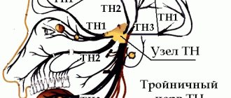
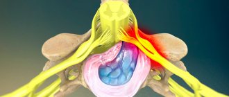
Facial nerve
(nervus facialis)
, the seventh of twelve cranial nerves, is responsible for the innervation of the facial muscles.
Also part of the facial nerve is the intermediate nerve responsible for the innervation of the lacrimal gland, stapedius muscle and taste sensitivity of the two anterior thirds of the tongue. Facial nerve paralysis is a disease characterized, most often, by a unilateral impairment of facial muscle facial activity. Impaired motor activity can vary from muscle weakness to complete paralysis with the appearance of severe facial asymmetry.
Frequency of occurrence of pathology: 20 people per 100,000 population.
The main pathogenetic link of the disease is a violation of the conduction of nerve impulses along the fibers of the facial nerve. Depending on the location of the source of primary damage, central and peripheral paralysis of the facial nerve are distinguished.
Peripheral facial paralysis
Damage to the facial nerve is possible both due to diseases, injuries and their consequences, and due to iatrogenic causes. The latter includes damage to the facial nerve during surgical interventions (neurosurgeries, maxillofacial and plastic surgeries)
Damage to the motor portion of the facial nerve leads to peripheral paralysis of the innervated muscles - the so-called. peripheral paralysis n.facialis. In this case, facial asymmetry develops, noticeable at rest and sharply increasing with facial movements. Half of the face on the affected side is motionless. When trying to wrinkle the skin of the forehead into folds on this side, the skin of the forehead does not gather, and the patient is unable to close his eyes.
When you try to close your eyes, the eyeball on the affected side turns upward (Bell's sign) and a strip of sclera becomes visible through the gaping palpebral fissure (hare's eye, lagophthalmos). In the case of moderate paresis of the orbicularis oculi muscle, the patient is usually able to close both eyes, but cannot close the eye on the affected side, while leaving the eye on the healthy side open (eyelid dyskinesia, or Revillot's sign). It should be noted that during sleep the eye closes better (relaxation of the muscle that lifts the upper eyelid).
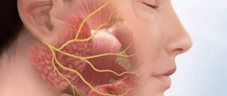
When the cheeks are puffed out, the air comes out through the paralyzed corner of the mouth, the cheek on the same side “sails” (sail symptom). The nasolabial fold on the side of the muscle paralysis is smoothed, the corner of the mouth is lowered. Passive lifting of the corners of the patient's mouth with fingers leads to the fact that the corner of the mouth on the side of the lesion of the facial nerve rises higher due to decreased muscle tone (Russetsky's symptom). When you try to bare your teeth on the side of the paralyzed orbicularis oris muscle, they remain covered with your lips. In this regard, the asymmetry of the oral fissure is roughly expressed; the oral fissure is somewhat reminiscent of a tennis racket, with the handle turned towards the affected side (racket symptom).
A patient with paralysis of the facial muscles caused by damage to the facial nerve experiences difficulty while eating; food constantly falls behind the cheek and has to be removed from there with the tongue. Sometimes there is biting of the mucous membrane of the cheek on the side of paralysis. Liquid food and saliva may leak from the corner of the mouth on the affected side. The patient also experiences a certain awkwardness when talking. It is difficult for him to whistle or blow out a candle.
Due to paralysis of the orbicularis oculi muscle (paretic lower eyelid), the tear does not completely enter the lacrimal canal and flows out - the impression of increased lacrimation is created.
Damage to the facial nerve in the pyramid of the temporal bone
- Proximal to the chorda tympani (chorda tympani) – peripheral paralysis of the facial nerve, lack of taste sensitivity in the anterior 2/3 of the tongue. Patients often experience dry mouth due to a disorder in the secretion of the submandibular and sublingual salivary glands.
- Proximal to the stapedius nerve (n.stapedius) - peripheral paralysis of the facial nerve, lack of taste sensitivity in the anterior 2/3 of the tongue. Patients often experience dry mouth due to a disorder in the secretion of the submandibular and sublingual salivary glands, hyperacusis - abnormally fine hearing and special sensitivity to low tones
- Proximal to the relatively large petrosal nerve (n.petrosus major) - peripheral paralysis of the facial nerve, lack of taste sensitivity in the anterior 2/3 of the tongue. Patients often experience dry mouth due to a disorder in the secretion of the submandibular and sublingual salivary glands; often nerve deafness due to combined damage to the vestibular-cochlear nerve (n.vestibulocochleari)s; only when it is absent - hyperacusis; lack of tear production – xerophthalmia.
Damage to the facial nerve in the cranial cavity
The above symptoms. Bilateral facial paralysis (basal meningitis) is common. In most cases, other nerves are also affected and there are general cerebral symptoms.
Damage to the facial nerve nucleus
The nuclei may suffer from degenerative diseases (progressive bulbar palsy, syringobulbia), dyscirculatory and inflammatory processes, tumors of the pons or hemorrhages in the pons. Clinically, damage to the nucleus of the facial nerve is manifested by its peripheral paralysis.
Central facial palsy
When the pathological focus is localized in the cerebral cortex or along the corticonuclear pathways related to the facial nerve system, central paralysis of the facial nerve develops. In this case, central paralysis or, more often, paresis develops on the side opposite to the pathological focus, only in the muscles of the lower part of the face, the innervation of which is provided through the lower part of the nucleus of the facial nerve. Paresis of the facial muscles of the central type is usually combined with hemiparesis.
With a purely limited focus in the cortical projection zone of the facial nerve, a lag in the corner of the mouth on the opposite half of the face in relation to the pathological focus is detected only with arbitrary baring of the teeth. This asymmetry is completely leveled out during emotionally expressive reactions (when laughing, crying), because the reflex ring of these reactions closes at the level of the limbic-subcortical-reticular complex. In this regard, despite the existence of supranuclear palsy, the facial muscles are capable of involuntary movements in the form of a clonic tic or tonic facial spasm, since the connections of the facial nerve with the extrapyramidal system are preserved. A combination of isolated supranuclear palsy and attacks of Jacksonian epilepsy is possible.
Treatment
Conservative therapy is reduced to medication (corticosteroids, vitamins) and physiotherapeutic treatment. If paralysis is caused by an infectious disease, then specific pathogenetic therapy (antibacterial, antiviral) is prescribed.
In case of iatrogenic or traumatic damage to the facial nerve, surgical reconstructive options (cross-plasty, facial nerve reconstruction) can be quite effective. Unfortunately, these operations do not always bring the desired result. The cost of such operations is quite high, and not all patients can afford such interventions.
Social characteristics of patients
As a rule, due to pronounced facial asymmetry, patients with consequences of damage to the facial nerve are difficult to socially rehabilitate. Most of these patients are characterized by a secluded lifestyle and social maladjustment, which in turn lead to depressive states. During consultation, many of the patients in this group ask to restore facial symmetry, relegating facial activity to the background.
APTOS methods for correction of facial asymmetry due to damage to the facial nerve
Thread lifting methods are successfully used to correct age-related facial changes resulting from gravitational tissue ptosis. A variety of methods makes it possible to eliminate both the initial signs of tissue ptosis and pronounced changes, including facial asymmetries. Aptos thread lifting methods can eliminate facial asymmetry even in case of damage to the facial nerve due to static suspension (suspension) of the soft tissues of the face using threads with notches. The effectiveness of the methods is, as a rule, very high, and the result lasts even longer than after a similar procedure in patients with age-related facial changes, since there is no facial activity on the affected side.
Thanks to APTOS methods, we have been helping patients with the consequences of facial nerve injuries free of charge for more than 10 years. For this reason, thread lifting procedures have been performed in more than 300 cases in Georgia and Russia.
In order to get into the program, the patient must contact the Aptos hotline or find a specialist in Aptos methods in the region of your residence.
For the free procedure you will need:
- neurologist's report confirming the diagnosis
- electroneuromyography results
- consent to photographing and publishing your photographs in scientific periodicals.
APTOS: NOTHING IS IMPOSSIBLE!
Danger
Paresis of the facial nerve, provided that treatment is started in a timely manner, cannot cause harm to health and psyche. If the disease becomes chronic, the asymmetry remains forever. This means that the patient will not be able to lead his previous lifestyle. He will feel complex about his appearance and will begin to avoid contact with friends.
Among the most common complications of paresis:
- Contracture of facial muscles (muscles tighten on one side of the face and become inelastic).
- Muscle atrophy (muscles weaken, their volume decreases).
- Involuntary twitching of facial muscles.
- Keratitis, conjunctivitis (due to the fact that the patient cannot completely close the eye, the cornea, the inner lining of the eyelid, becomes inflamed).
- Facial synkinesis (nervous excitement mistakenly spreads from one area of the face to another, which is why, for example, tears may flow while chewing food).