A characteristic feature of the human brain is the incredible size of the cortex and complex folding. The cortex is the most developed area of the brain, responsible for non-reflexive activity (memory, perception, cognition, thinking, etc.).
The formation of cortical-subcortical structures occurs during embryonic development, providing the possibility of placing the cortex in a limited volume of the cranium. Convolutions (giri) and grooves (sulci) make up its folded surface. Pathological changes in the size or folds of the cortex lead to severe mental disability and intractable epilepsy. Consequently, cortical expansion and folding are considered key processes in brain evolution.
Fissures and convolutions: formation and functions
The grooves and gyri in neuroanatomy that give the brain its wrinkled appearance serve two critical functions. They help increase the surface area of the cortex, which allows more neurons to pack into it and enhance the brain's ability to process information. The sulci and convolutions of the brain form divisions, creating boundaries between the lobes of the brain, dividing it into two hemispheres.
Main grooves:
- The interhemispheric fissure is a deep groove in the center of the brain that contains the corpus callosum.
- The Sylvian fissure (lateral sulcus) separates the parietal and frontal lobes.
- Roland's fissure (central sulcus), separating the fusiform gyrus and the hippocampal gyrus on the inferior surface of the temporal lobes.
- Parieto-occipital - separates the parietal and occipital lobes.
- The calcarine fissure (spur-like groove or prominent fissure) is located in the occipital lobes and divides the visual cortex.
The main convolutions of the brain:
- The angular gyrus of the parietal lobe helps in processing auditory and visual recognition.
- Broca's gyrus (Broca's center) is an area of the brain located in the left frontal lobe in most people that controls functions related to speech production.
- The cingulate gyrus, an arched fold located above the corpus callosum, is a component of the limbic system and processes sensory input regarding emotions and regulates aggressive behavior.
- The fusiform gyrus is located in the temporal and occipital lobes and consists of lateral and medial parts. It is thought to play a role in word and face recognition.
- The hippocampal gyrus lies on the inner surface of the temporal lobe, which borders the hippocampus. Plays an important role for memory.
- The lingual gyrus in the occipital lobe is involved in visual processing. It is limited by the collateral groove and calcarine fissure. In front it is in contact with the pararpopampal gyrus, and together they form the medial part of the fusiform gyrus.
As the embryo develops, gyri and sulci form with the appearance of depressions on the surface of the cortex. Not all gyri develop at the same time. The primary form is formed starting from the 10th week of pregnancy (in humans), then secondary and tertiary forms develop. The most prominent groove is the lateral one. It is followed by the central one, separating the motor cortex (precentral gyrus) from the somatosensory cortex (postcentral gyrus). Most of the cortical sulci and gyri of the brain, the anatomy of which begins to take shape between 24 and 38 weeks of pregnancy, continue to grow and develop after the newborn is born.
The early state of the brain has a strong influence on the final level of gyrification. In particular, there is an inverse relationship between cortical thickness and gyrification. Brain regions with low thickness values have a higher level of gyrification. The opposite is also true: brain regions with a high thickness value (for example, thickening of the hippocampal gyrus cortex) have a low level of gyrification.
Brain: functions
You may be interested in: “Indapamide”: instructions for use and analogues
In fact, there are a huge number of functions of the human brain, and more than one article can be written about them. In the list below, all functions are combined into separate groups:
- processing information coming from outside;
- planning and decision making;
- carrying out movements;
- emotions;
- memorization and memory;
- attention;
- speech;
- intelligence and thinking.
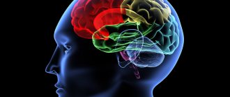
Lobes of the brain and their functions
Each hemisphere is divided into four lobes: frontal, parietal, temporal and occipital. Most brain functions rely on different regions throughout the brain working together, but each lobe performs the bulk of relatively specific functions.
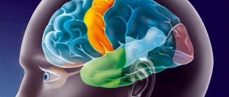
The frontal lobe is located in the most anterior region of the cerebral cortex, separated from the parietal lobe by the central sulcus, and from the temporal lobe by the lateral sulcus. The region typically contains the most important executive functions for a person, including emotion regulation, planning, reasoning, and problem solving.
The parietal lobe is responsible for the integration of sensory information, including contact, temperature, pressure, and pain. Because of the processing that occurs in the parietal lobe, it is possible to distinguish between the touch of two objects at nearby points (rather than as a single object). This process is called two-point.
The temporal lobe also contains areas involved in sensory processing, especially important for hearing, language recognition, and memory formation. The primary auditory cortex receives audio information through the ears and secondary areas and processes the data so that a person understands what he hears (words, laughing, crying, etc.). The medial (closer to the center of the brain) part contains the hippocampus, an area important for memory, learning, and perception of emotions. Certain areas of the temporal lobe process complex visual information, including faces and scenes.
Parts of the brain
As noted above, the structure of the brain is truly complex. To simplify its study, depending on the functions performed and the characteristics of intrauterine development, the brain is divided into the following parts:
- the forebrain (telecephalon), which consists of the cerebral hemispheres;
- the diencephalon (diencephalon), which includes the thalamus and surrounding structures;
- the midbrain (mesencephalon), consisting of the quadrigeminal cord and cerebral peduncles;
- the hindbrain (metencephalon), which includes the pons and cerebellum;
- medulla oblongata (myelencephalon).
You may be interested in: Venous and arterial blood: features, description and differences
Cellular mechanisms leading to expansion and folding of the cerebral cortex
The structure of the human brain distinguishes it from other mammals, and for this reason may explain its unique mental abilities compared to other animals. The number of folds in the cortex may correlate with some specific cognitive, sensory, and motor abilities. Although there is no clear explanation of how the unique division of the human brain into sulci and convolutions occurs. Today there is progress in understanding the extremely complex processes in the brain, the cortex of which is built with so many grooves and convolutions. Even though all cells have the same DNA, different neural stem cells are formed. It is their work with various properties that creates the basic structure of the brain, consisting of neurons and glial cells.
Telencephalic neuroepithelium
Brain growth occurs through two types of stem cells—neural stem cells and neural progenitors. Both of these forms form neurons, which become permanent in the brain, as well as intermediate cells that create the building material for building the brain. Four different types of stem cells determine the structure of the cortex.
During early embryonic development, expansion of the rostral domain of the neural tube leads to the appearance of two telencephalic vesicles. The dorsal half of these vesicles is molecularly defined as the primordium of the cerebral cortex. At this stage, the cortical primordium consists exclusively of a monolayer of neuroepithelial progenitor cells. They are highly polarized and attached to each other by tight junctions at the level of the apical domain (inner surface of the telencephalic vesicle) and move the cell nucleus between the apical (apical) and basal (lower) side of the neuroepithelium in coordination with the cell cycle.
- basal-directed movement during G1 phase;
- basal position during S phase;
- apically directed movement during G2 phase;
- mitosis on the apical surface.
The cycling movement is known as interkinetic nuclear migration and is completely asynchronous between neuroepithelial cells, giving the neuroepithelium a pseudostratified appearance. Cells undergo only symmetrical self-aggressive divisions, with each division generating two daughter cells, hence exponentially increasing their number. Because they are the fundamental progenitor cells of the cerebral cortex, the size of their association determines the number of derived neurogenic progenitor cells and the final number of cortical neurons, and therefore it has a fundamental influence on the size of the mature cerebral cortex. An increase in quantity leads to an expansion of surface area and the formation of neuroepithelium.
Spread and neurogenesis
Immediately before the onset of neurogenesis, neuroepithelial progenitor cells begin to lose tight junctions and acquire features typical of glial cells (including expression of brain lipid-binding protein, vimentin, and Pax6), thereby becoming apical radial glial cells (ARGCs). They also undergo interkinetic nuclear migration, divide at the apical surface of the developing cortex, and at this early stage also undergo self-reinforcing divisions.
However, they gradually begin to divide asymmetrically to generate one similar cell plus another cell. These new cells accumulate in the basal part of the cortical primordium, while the cell bodies of the ARGC remain on the apical side, forming the ventricular zone (VZ). With the accumulation of cells above the GC, the ARGK process is prolonged, remaining attached to the basal plate, and is now called radial glia. Asymmetric ARGK divisions generate one ARGK plus one neuron or one intermediate progenitor cell. Intermediate progenitor cells (secondary progenitor cells without apical-basal polarity) do not undergo interkinetic nuclear migration, divide in a layer located in the ventricular zone, the subventricular zone (SVZ), and all express a transcription factor (Tbr2).
However, due to the fact that each neuron itself consumes during mitosis, their relative quantity compared to ARGK is quite low. Intermediate progenitor cells in the cerebral cortex generate the majority of cortical excitatory neurons. As neurogenesis progresses, the need for ARGK expansion/renewal decreases and the production of neurons increases. In addition to the expanded ventricular zone, the subventricular zone thickens, populated in abundance by basal precursors, especially in the later stages of neurogenesis. As a result, the SVZ splits into internal and external parts. The outer portion contains a wide variety of progenitor cell types with high developmental potential, which is a key factor for the expansion and formation of the cortex.
Medial surface
The sulcus of the corpus callosum is located most medially, which then passes into the sulcus of the hippocampus, which borders the hippocampus itself. Next to the callosal sulcus are the subparietal and callosal-marginal sulci. The rhinal sulcus runs parallel to the hippocampus.
The recesses of the brain listed above limit a specific system, which is called limbic. It, in turn, consists of the cingulate and hippocampal gyri.
In addition to the limbic system itself, on the inner surface of the brain there are also structures that continue their course from the outer part of the cerebral cortex. In this way, the parieto-occipital groove extends, behind which the precuneus is located (a gyrus resembling a trapezoid in shape). Next to this depression there is also a calcarine groove, which extends from the back of the head and forward all the way to the corpus callosum. Between the two recesses mentioned above is the sphenoid gyrus.
Cerebral cortex
The cerebral cortex forms a thin layer of gray matter responsible for higher mental function. On the superficial part of the cortex you can visually see grooves, which is why all parts of the brain have a folded surface. The central organ of each person has a different shape of grooves, depth and length, thus creating an individual pattern.
Important Autism - causes, symptoms and types of disease
Studies of brain structures have made it possible to determine the most ancient cortical layer and the evolutionary development of the organ through histological analysis. The bark is divided into several types:
- Archipallium - the oldest part of the cortex, regulates emotions and instincts;
- Paleopallium is the younger part of the cortex, responsible for vegetative regulation and maintains the physiological balance of the whole organism;
- Neocortex is a new area of the cortex that forms the upper layer of the cerebral hemispheres;
- Mesocortex - consists of an intermediate old and new cortex.
All areas of the cortex are in close interaction with each other, as well as with subcortical structures. The subcortex includes the following structures:
- The thalamus (visual thalamus) is a collection of large masses of gray matter. The thalamus contains sensory and motor nuclei, and nerve fibers allow it to be connected to many parts of the cortex. The visual thalamus is connected to the limbic system (hippocampus) and is involved in the formation of emotions and spatial memory;
- The basal ganglia (nuclei) are a collection of white matter in the thickness of gray matter. The layer is located on the side of the thalamus, near the base of the hemispheres. The basal ganglia carry out higher processes of nervous activity, the active phase of work occurs during the daytime, and stops during sleep. Neurons in the nuclei are activated during mental work of the organ (concentration), and produce electrochemical impulses;
- Brainstem nuclei - regulate the mechanisms of redistribution of muscle tone, and are responsible for maintaining balance;
- The spinal cord is located in the spinal canal and has a cavity filled with cerebrospinal fluid. It is presented in the form of a long cord and provides communication between the cerebrum and the periphery. The spinal cord is divided into segments and performs reflex activities. Information flows through the spinal canal to the brain.
The hierarchy of these structures in relation to the cortex is lower, but each performs important functions and, in case of violations, independent self-government is triggered. The subcortical region is represented by a complex of various formations that are involved in the regulation of behavioral reactions.
Structure and functions of the cerebellum
The cerebellum is located above the brain stem and is connected to its parts by 3 pairs of peduncles. The cerebellum has 2 small hemispheres covered by the cerebellar cortex. The main functional significance of the cerebellum is to maintain body balance, regulate and coordinate body movements, giving them smoothness, accuracy and proportionality. The cerebellum programs the automatic execution of movements, which is made possible by its connections with the spinal cord, brainstem and cerebral cortex. For example, when walking and running, the cerebellum controls the position and movement of the torso and arms in accordance with the movements of the legs and the movement of the body's center of gravity. When writing, it is responsible for maintaining optimal posture and coordinating the movements of the head, eyes and hands. The cerebellum plays an important role in performing rapid sequential and simultaneous movements, such as the movements of the hands of a pianist or typist.
Bottom surface
The lower, or basal, surface of the brain is formed by parts of the frontal, temporal and occipital lobes. However, in addition to these structures, the so-called olfactory brain is also located on the basal surface. It consists of the olfactory sulcus, surrounded by the straight gyrus and orbital sulci.
The temporal lobe, based on the brain, contains the inferior temporal and occipitotemporal sulci, between which the gyrus of the same name is located. The lingular gyrus is also detailed nearby.
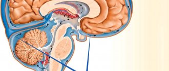
Structure and functions of the diencephalon
Anterior to the brain stem, between the midbrain and the cerebral hemispheres, is the diencephalon. The upper part of the diencephalon is called the thalamus or thalamus optic, the lower part is the hypothalamus.
The meaning of the thalamus The thalamus, a paired ovoid-shaped formation, is a collector of all types of sensitivity from all parts of the body and sensory organs. From here this information is transmitted to the cerebral cortex. Certain areas of the thalamus are important components of the limbic system of the brain, which controls the psycho-emotional behavior of a person, while others are involved in ensuring memory processes. There is evidence that the thalamus is involved in the perception of pain. Destruction of certain areas of the thalamus can lead to a decrease in anxiety, tension, aggressiveness, elimination of obsessive thoughts, as well as a sharp decrease in motor activity.
The significance of the hypothalamus The significance of the hypothalamus is associated primarily with the regulation of the activity of internal organs. The nuclei of the hypothalamus produce special substances - neurohormones, which enter the pituitary gland, and from it into the blood.
The pituitary gland is an endocrine gland, closely related in structure and location to the hypothalamus. The single hypothalamic-pituitary system of the diencephalon controls the work of other endocrine glands and, with their help, regulates the functions of the body. This system controls the state of water-salt balance, metabolism and energy, the functioning of the immune system, thermoregulation, reproductive function of the body, etc. There is evidence that the hypothalamus contains specific pleasure centers that play an important role in the formation of motivations and emotional forms of behavior . In the area of the hypothalamus there are areas of the optic nerves through which information is transmitted from the retina of the eye.
The significance of the pineal gland The diencephalon also includes the pineal gland, or pineal gland, an endocrine gland that influences the work of other endocrine glands and is involved in the regulation of the seasonal rhythms of the body.