How is SCT research carried out?
The x-ray technician positions the patient in a special way, depending on the examination being performed. Next, the gantry (the device on which the tube and receiving sensors are installed) begins to rotate around the scanned area and makes slices of the area under study with certain parameters of power and tube passage step. When scanning the chest and abdominal organs, you will need to hold your breath several times, which the x-ray technician will warn you about. The scanning procedure lasts 2-4 minutes.
CT scan of the chest: what will it show and how will it help the patient?
Computed tomography of the chest (CT CT) is one of the modern diagnostic methods. When and for what purpose may you need to do a CT scan of the chest? What does it include?
These and other questions are answered by Yulia Aleksandrovna Rutskaya, Deputy Chief Physician for Radiation Diagnostics, radiologist of the highest qualification category “Clinic Expert” Kursk.
— Yulia Aleksandrovna, how often do patients come to you for a CT scan of the chest?
“People come to our clinic for this service every day. Computed tomography of the chest organs accounts for on average more than a third of all examinations per day. Now, during the coronavirus epidemic, these numbers are, of course, much higher.
— What does a chest CT scan show?
- Anything that goes into the chest. First of all, the study allows us to study the condition of the lungs. We can assess the location of the vessels and bronchi, their patency, and find out whether there are neoplasms, inflammatory processes, or congenital pathologies in the lungs. Also, with the help of this study, you can see the esophagus, dilatation (expansion) of the chambers of the heart and large vessels, and the condition of the mediastinal lymph nodes.
In addition, we see a frame formed by the ribs, the thoracic spine, and the sternum. These bony structures are assessed for destructive changes.
— What diseases can be identified or excluded during this study?
— The main pathologies that can be detected are pneumonia (pneumonia) of various natures, including viral, tuberculosis, the detection of which is of social importance, and lung cancer. Interstitial lung diseases are also detected, diffusely affecting the lung tissue; accumulation of fluid in the pleural cavity; tumors and metastases in the lungs, bones and mediastinum (for example, thymoma, lymphoma, dermoid tumors, cysts, esophageal tumors); abnormalities of bone structure.
Read materials on the topic:
Tuberculosis: a disease that knows no boundaries What is a dermoid cyst? COVID-19 and more: what are the features of viral pneumonia?
— Yulia Aleksandrovna, please tell us how a CT scan of the chest organs is done
— First of all, the patient undergoes a registration procedure in our clinic. This takes 5-10 minutes. Then the laboratory assistant accompanies him to the locker room, where the patient undresses to the waist. A mandatory requirement is to remove all metal jewelry. The patient lies on a special tomograph table on his back. The tomograph itself is a ring-shaped device that rotates smoothly around the patient and takes pictures.
At the command of the radiologist, the patient inhales and holds his breath 2-3 times for a certain time (from 10 to 30 seconds). Next comes the processing of information received by the radiologist from the tomograph computer. The doctor previews the images over the next 10-15 minutes. If no changes are detected, the result is reported to the patient immediately, but the specialist needs time to describe the images. The conclusion is sent to the patient via email within a few hours, or he can pick it up from the clinic himself the next day.
Read material on the topic:
Electronic media for recording MRI studies: marketing ploy or benefit to patients?
— What is the purpose and how is a CT scan of the chest organs with contrast performed? What is special about this type of research?
— Such a study is carried out, for example, if there is a suspicion of a blood clot in the pulmonary artery. This condition is life-threatening; contrasting is required to clarify the diagnosis. In addition, we perform CT of the chest with contrast when there are space-occupying formations in the mediastinum or roots of the lungs. Contrasting allows you to study in detail the localization and spread of the tumor, assess its nature, and the location of the vessels in it.
The contrast agent is an iodine-containing substance that is injected using a special syringe-flask at the beginning of the procedure.
— Is preparation required for this study? If so, how to prepare for a chest CT scan?
— No special preparation is required for traditional CT examination of the OGK. If a contrast study is necessary, the patient must first undergo blood tests to assess the kidney's glomerular filtration rate and creatinine level. The doctor also finds out whether the patient has a history of severe allergic reactions, bronchial asthma, or individual iodine intolerance.
— In what cases is CT scanning of the chest organs contraindicated?
— Absolute contraindications are pregnancy and excess weight (more than 150 kg). A contrast study is not performed in cases of severe renal failure or individual iodine intolerance; it is used with caution in cases of diabetes mellitus and thyroid pathologies.
— Is it possible to do a CT scan of the chest organs for children?
— It is possible, at any age, only if there are direct indications and no contraindications. I would like to note that this requires a referral from a specialist with justification for conducting this particular study.
— And if an adult wants to sign up for a CT scan of the chest organs, does he need to take a referral from a doctor?
— It is desirable because from the referral the radiologist receives preliminary information about the expected diagnosis and, based on it, decides how best to conduct the study, and also determines further tactics for managing the patient.
Interviewed by Sevilya Ibraimova
If you need a CT scan of the chest, you can sign up for the study here ATTENTION: the service is not available in all cities
The editors recommend:
What does MRI of the mediastinum show? How to live with chronic obstructive pulmonary disease without losing quality of life? Formula of Love by Alexander Abdulov
For reference:
Rutskaya Yulia Alexandrovna
Graduate of the medical faculty of Kursk State Medical University in 2002. In 2004, she completed an internship in radiology. Currently, she is the deputy chief physician for radiation diagnostics, a radiologist of the highest qualification category at the Expert Clinic Kursk. Receives at the address: st. Karl Liebknechta, 7
What can SKT do?
Computed tomography is simply irreplaceable in diagnosing the conditions of any bone structures, the condition of the lungs, and mediastinum in humans. SCT is also widely used in the study of abdominal organs (liver, spleen, gallbladder, pancreas), adrenal glands, kidneys, and pelvic organs. With the help of CT, it became possible to accurately detect any stones (for example, in the gall bladder, kidneys) and calcifications (for example, those changes that accompany tuberculosis, sclerosis).
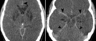
Spiral computed tomography is leading in the diagnosis of complex bone injuries , multiple injuries (for example, road accidents) of bones and internal organs, in the diagnosis of complex or multiple injuries of the skull and acute injuries and hemorrhages of the brain.
SCT is also widely used by ENT doctors for a detailed assessment of the condition of the paranasal sinuses, auditory canals and temporal bones, since no other diagnostic method can compare with the accuracy and “depth” of computed tomography.
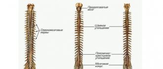
In studies of the spine using SCT, it is possible to determine the smallest damage in the bone structure, pathological calcifications in the ligamentous apparatus; to diagnose the condition of the intervertebral discs, it is best to use MRI.
The advisability of prescribing SCT in each individual case should be determined by the attending physician, who will subsequently interpret the results obtained from CT taking into account the entire picture and characteristics of the patient.
Who should not undergo SCT or MSCT
Diagnosis using computed tomography is painless, but requires a person to remain absolutely still for about 5-15 minutes (in total), while the direct time of x-ray radiation in general is no more than 10 seconds. During the procedure, at the doctor’s command, you will have to hold your breath one or more times.
CT machine platforms are not designed to weigh more than 150 kg
Contraindications to computed tomography of the heart and blood vessels:
- sustained tachycardia with heart rate above 75 beats/min;
- high coronary calcium index;
- extrasystole, other forms of arrhythmias;
- pregnancy, lactation period;
- claustrophobia, mental illness;
- allergy to iodine;
- pathologies of the thyroid gland;
- decompensated diabetes mellitus;
- severe renal or liver failure;
- myeloma.
SCT or MSCT is not recommended for use in children under 14 years of age without substantial justification.
On a note. How often can a spiral or multispiral computer study be done? The SanPin instruction limits the total radiation exposure to internal organs received from all types of x-ray examinations per year to no more than 5 mSv. Therefore, possible options for the number of procedures are from 1 to 3(5), which are carried out no more than once every 2-3 months.
To normalize the pulse in case of severe anxiety or if there is a tendency to claustrophobia (fear of closed spaces), a person will be asked to take an oral sedative or be given an injection of a powerful sedative.
What does SKT “dislike”?
First of all, these are movements . Any movements during scanning of the study area distort the resulting images, of course, reducing their information content for the doctor. If in a series of images obtained, even just 1-2 scans are distorted by motion artifacts, then subsequent 3D reconstructions will already have defects and their information content for the doctor, of course, will be reduced.
Secondly, metal. Metal is to X-rays what a mountain is to the sun—it leaves a “shadow.” A large amount of metal in the examination area can significantly distort the resulting images. But if the metal is located in any place other than the area being examined, it cannot in any way affect the quality of the scans.
What do you need to have with you for SCT?
Before the study, you must give your existing medical documentation to the x-ray technician or radiologist (a referral from the attending physician, which indicates the preliminary diagnosis, study area, clinical task, specific comments), data from previous studies (which directly relate to the reason for prescribing SCT to you) and other medical documentation, after analyzing which the radiologist will be able to formulate the most accurate diagnostic conclusion on SCT, taking into account the data of your medical. cards and recommendations of the attending physician. If you do not provide medical documentation in full, then the radiologist will evaluate only the directly obtained SCT scans, without taking into account previous injuries and diseases, any possible consequences that they could entail, and without taking into account possible concomitant pathologies.
Remember, there is no absolute and universal method of medical diagnosis: MRI will never replace SCT, SCT will never replace ultrasound, ultrasound will never replace radiography. All these methods complement each other and sometimes, data from previous studies have the decisive word in the correct interpretation of the resulting picture by a radiologist during computed tomography.
Make an appointment
Make an appointment by calling:
+38 (044) 393 0933
+38 (067) 230 2432
+38 (050) 332 3339
Who deciphers the results and when they can be obtained.
The resulting SCT images must be examined by a radiologist (a doctor who specializes in CT, MRI, X-ray studies and has the appropriate certificate). It is necessary to “read” all the scans, study the provided medical information. documentation, compare it with the obtained CT image, describe the study, draw a diagnostic conclusion, shoot film with scans. On average it takes about 1-3 hours. At ACMD, we undertake to prepare the results for delivery by the end of the Clinic’s working day.
After the examination, you receive films with scans and a diagnostic report, which will be signed by a radiologist. Also, the diagnostic report may contain recommendations regarding your further actions (which doctor to contact with these results, what types of further examination are recommended, and so on).
Consultations and questions regarding your SCT study.
A radiologist is not a clinician (a doctor who treats you) and, as such, cannot provide any advice or recommendations regarding your treatment or diagnostic procedures.
To make objective comments on an SCT study, it is necessary to fully know the patient’s objective data, the picture of his previous studies, the characteristics of his current condition, anamnestic data, and much more.
You can ask all your questions to your attending physician or physician, whose consultation will be recommended to you by a radiologist.
If you still have any specific questions about computed tomography, you can ask them in the specialized MRI and SCT diagnostics section on our Forum.
Check out comprehensive body check packages with up to 30% discount
Contrast techniques for SCT.
When examining the abdominal organs, the patient will be asked to drink 500–1000 ml. drinking water with a contrast agent dissolved in it in order to “separate” the stomach and intestines from nearby organs and tissues, as well as to ensure that intestinal loops do not create additional “interference” for the radiologist during the study. This is the so-called oral contrast.
The same technique of intravenous administration of a contrast agent is very often used in order to “highlight” areas with pathological changes, to study the blood flow of tumor formations, or specifically to study blood vessels.
How to prepare
In most cases, no special steps are required before performing a CT scan. The exceptions are studies of the kidneys, liver, pelvis and abdominal cavity. Preparation for them includes:
- diet correction - within a few days you need to give up food and drinks that cause flatulence;
- cleansing the intestines - the doctor may prescribe laxatives or an enema.
If the procedure involves the use of a contrast agent, a test is performed in advance to check for an allergic reaction.
Is the SCT procedure painful and is it harmful? What are the contraindications? Where to get a CT scan in Kyiv?
The SCT examination is absolutely painless; the scanner itself does not directly touch your body.
The method is based on X-ray radiation. Naturally, during an SCT study, the patient is exposed to x-rays, therefore, when prescribing this study, the doctor must have fairly compelling reasons and take into account the negative impact of x-rays on the body.
An absolute contraindication for SCT is pregnancy and breastfeeding. Also, SCT is strongly not recommended for children without significant reasons. In other cases, you should consult your doctor.
a computed tomography (CT) scan in Kiev at the ACMD clinic, a 64-slice tomograph (the radiation level is reduced significantly!)
Where to get a computed tomography scan in Moscow?
Sign up for an examination
When looking for a place where you can do a volumetric computed tomography scan of the whole body or a diseased organ for a fee, give preference to diagnostics at the Central Clinical Hospital of the Russian Academy of Sciences. With us you can undergo an inexpensive examination with a guarantee of accurate results due to the use of advanced equipment.
When choosing where you can urgently get a CT scan in Moscow, studying addresses and prices, remember that low cost is not always an argument in favor of choice. Especially when it comes to medical examination, when a lot depends on the high technology of the equipment and the experience of the doctor. By offering paid CT services to residents and guests of the capital, we remind you: timely diagnosis is an opportunity to solve health problems in the most gentle way for the body and budget.
By phone you can sign up for a CT scan of the type you need - multispiral (spiral multilayer), volumetric, with contrast with a radiopaque substance, without contrast, 3D, etc. - any examination with us can be done inexpensively. Registration is also done online on the hospital department website.