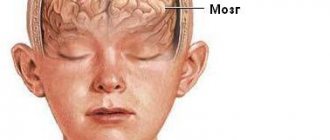
What brain diseases are visible on MRI?
A set of symptoms for which magnetic resonance imaging of the head is prescribed:
- Decreased breathing;
- Vomiting reflex;
- Loss of vision, speech;
- Unsteadiness of gait due to damage to the cerebellum;
- Twitching pupils;
- Strabismus;
- Lack of swallowing reflex;
- Difficulty in excreting feces;
- Periodic loss of consciousness;
- Frequent headaches.
The described clinical manifestations may be a consequence of brain contusion, traumatic brain injury, stroke, or severe head impacts.
The advent of MRI scanning made it possible to diagnose pathological changes in multiple sclerosis (destruction of the myelin sheaths of the nerves). After a bruise, headaches appear at a late stage, when pronounced microcirculation disorders occur. Contacts at the sites of destruction of nerve fibers cause a “short circuit” with subsequent neurological symptoms. Extensive disruption of nerve innervation is accompanied by vasoconstriction. Abnormal microcirculation provokes headaches, muscle disorders, visual and auditory disorders.
Concussion: classification, symptoms, diagnosis
The prevalence of disability and mortality after traumatic brain injuries (concussion, cerebral edema) in the Russian Federation exceeds fifty percent in people under 35 years of age.
According to statistics, approximately half of people die after a concussion due to untimely provision of qualified medical care. Concussions do not only occur with skull fractures. A strong external impact on the cranium can lead to a counter-impact, when the mass of the brain suddenly moves to the bones. Bleeding, hematoma, and swelling occur at the site of contact.
MRI of the head after a concussion: stages of edema formation
MRI of the head for injuries and bruises
Brain symptoms after bruises and injuries are varied. Manifestations depend on functional and organic changes in the brain matter. A mild concussion is rarely accompanied by loss of consciousness. More often, after a bruise of the skull, headaches, vomiting, and nausea appear. After a few weeks, the symptoms disappear on their own. The course of the pathology is monitored by neurologists.
Complications of a concussion are minor hemorrhages inside the subarachnoid space. Brain hyperintense lesions on MRI are limited. Enhanced spots after a concussion are combined with areas of normal or reduced signal. Dynamic observation allows you to visualize the normalization of the condition. The pathology persists when the vessel ruptures or the vascular wall dissects (aneurysm).
Complications after a concussion are clearly verified by MRI. A moderate bruise is characterized by fever, difficulty breathing, cardiac arrhythmia, twitching of the pupils, and loss of sensation in the limbs. Ocular nystagmus is accompanied by a movement to the right and left, which for a neurologist is a sign of damage to brain tissue.
Severe problems of intracerebral microcirculation occur when hemorrhage penetrates the brain parenchyma. With proper treatment, changes pass quickly.
Contrast MRI for severe concussion evaluates the condition of blood vessels, ventricles, and allows one to study the size of the pathological focus and location.
A severe contusion of the brain is accompanied by a blockage of the bronchial tubes due to damage to the corresponding center. Lack of oxygen supply, concomitant arterial ruptures, and aneurysms lead to muscle cramps and paresis of the limbs due to suppression of functional brain centers.
Severe brain contusion can lead to fractures and extensive hemorrhages. Intense brain lesions on MRI occur due to the accumulation of blood within the subarachnoid space. The heterogeneous density of magnetic resonance imaging is characterized by the appearance of dark and white areas in the images. Hyperintense spots do not have clear boundaries. Images indicate swelling and accumulation of blood clots.
A repeat MRI for a concussion is done after 40 days. By this time, the swelling disappears and the headaches disappear. A long course of the disease is expected with a skull fracture with increased intracranial pressure.
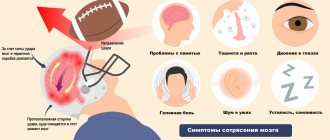
Symptoms of a concussion
Immediately after the injury, the patient experiences a disturbance of consciousness. The severity of this period can vary from stupor to short-term loss of consciousness. A mild concussion is manifested by stupor - deafness. The victim freezes due to inhibition of the transmission of neural impulses. The person feels confused, the muscles of the whole body are tense. With complete loss of consciousness, the patient does not respond to external stimuli. The unconscious state lasts from several seconds to several minutes.
Neurological symptoms of a concussion subsequently appear:
- there is a persistent feeling of nausea;
- vomiting - can be one-time or repeated after some time;
- dizziness, which intensifies when the patient’s position changes;
- headache – pressing, bursting or pulsating in the area of the bruise or over the entire surface of the head;
- noise, ringing in the ears;
- feeling of heat, increased sweating of the feet and palms.
Depending on which part of the brain is damaged, partial retrograde amnesia develops. The patient has no memory of what happened before or immediately after the injury.
When examining the patient, visual signs of a concussion are determined:
- the skin of the face and visible parts of the body turns pale and red alternately - this is due to dystonia of the blood vessels;
- passing asymmetry of the nasolabial folds, drooping of the corners of the mouth;
- involuntary eye movement;
- unsteadiness of gait.
Over the next few days, increased sensitivity to light and loud sounds persists. A person has problems sleeping. Signs of depression and increased anxiety appear.
A concussion in a child does not usually cause loss of consciousness. This is due to the peculiarities of the structure and functioning of the central nervous system. However, the clinical picture increases very quickly. Symptoms of a concussion in a child are intense:
- severe headache in the area of injury spreads throughout the head;
- breathing and heart rate increase significantly;
- the child's face turns red;
- a feeling of nausea appears immediately, vomiting - within the first hour after the impact.
The acute post-traumatic period lasts up to 10 days.
Concussion in infants manifests itself with symptoms of mild traumatic brain injury. The baby vomits during or immediately after feeding. The child's excitability increases, sleep and wakefulness patterns are disrupted, and crying appears when the position of the head changes.
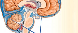
Venous and arterial aneurysms on MRI scans
Aneurysmal dilations of blood vessels have a special morphology - neck, body, dome. The cause of the disease is deposits of fatty complexes inside the vascular wall. The disease is called atherosclerosis and causes many complications in the brain and heart. Damage to the wall leads to disruption of permeability. Blood entering the interstitium causes dissection. Clots and formed elements accumulate between the individual layers. The movement of blood creates pressure on the thin wall. At a certain point, the vessel may not hold up. The appearance of a small rupture is accompanied by hemorrhage. Massive bleeding leads to death.
Contrast MRI for brain aneurysm is the best option for diagnosing the pathology. Tomograms reflect the full morphology - saccular, fusiform expansion of the arterial wall. Even a native MRI scan can reliably verify an aneurysm. Contrast improves visualization.
Tomograms show the size of the aneurysm. Formation over 10 mm is dangerous due to rupture.
Practical features of MRI of aneurysm after trauma:
- The spatial structure and relationships of individual parts are revealed by MR angiography. A bolus injection of contrast agent creates a clear image of the arteries and veins;
- Thrombosed and non-thrombosed aneurysmal dilatation is identified by taking multiple sections. The 3D modeling mode allows you to build a spatial projection of an object. The procedure helps determine the shape and size of the aneurysm;
- A combined study (CT and MRI) after head injuries allows you to diagnose thrombosed areas of the artery, measure the length of the neck, and verify the dimensions.
After severe trauma, angiography is performed to study the spatial relationships between various physiological and pathological formations. Magnetic resonance neuroimaging is performed to verify rupture, thrombosis, and aneurysmal dilatation of the artery. The study additionally reveals a whole list of pathologies.
Spasmodic contractions of arteries and veins of individual brain segments are diagnosed using a combination of methods - MR angiography, CT angiography, catheterization angiography. On the recommendation of a radiology doctor, additional examinations (MR spectroscopy, tractography) may be prescribed.
Magnetic resonance imaging of a cerebral cyst
A small brain cyst does not pose a danger if located outside the functional centers. Morphologically, the formation looks like a cavity with liquid internal contents. The bubble can remain inside the white or gray matter for life. If it forms after an injury and increases in size, dynamic control of the cavity is required.
Inflammatory cysts are formed by bacteria and parasites (echinococcus, alveococcus). After the infection is suppressed, the formations subside.
Autoimmune cysts gradually progress. Dynamic MRI helps monitor the condition of the bladder. With a growing formation, surgical treatment will be required. The absolute norm against the background of autoimmune damage to brain tissue requires repeated magnetic resonance imaging no later than 1 year.
The formation of antibodies to brain neurons and blood tests for infections are additional ways to verify pathological changes. After an MRI of the aneurysm, laboratory evaluation of cholesterol and blood clotting will be required.
Periodic rises in pressure are dangerous by rupture of cerebral arteries with the development of hemorrhagic or ischemic stroke. Classical MR angiography helps to obtain maximum information about the pathology.
Clinical signs of concussion
Determining the neurological symptoms of traumatic brain injury (TBI) allows timely verification of the nosology:
- Fear of light;
- Pupil dilation;
- Loss of coordination when walking;
- Memory disorder;
- Dizziness and headache;
- General weakness;
- Gradual descent into a comatose state.
The occurrence of any of the described signs after a traumatic brain injury requires immediate first aid:
- A person lies on the right or left side;
- To reduce swelling, apply a cold bandage to the forehead area;
- Remove or unbutton a shirt that is obstructing breathing;
- If necessary, perform cardiac massage and perform artificial pulmonary ventilation.
The procedures are performed at home until the ambulance arrives. Attentive attention to the person’s condition after a blow to the skull and early treatment prevents death.