What is angiography of the arteries of the lower extremities (LAN)
Angiography of the arteries of the lower extremities (LAN) is a minimally invasive study that is performed in a specially equipped cath lab. ANC is performed using ultra-low doses of X-ray and a special contrast agent, which is injected into the arteries of the lower extremities through the lumen of the catheter.
ANC allows you to determine the state of the blood supply to the legs and will help to develop the most optimal and effective further treatment tactics for the patient.
ANC will identify areas of narrowing of the leg arteries and/or areas of completely closed (occluded) arteries.
Why is an ANC necessary?
This study is prescribed if there is a suspicion of disruption of the lumen of the arteries supplying the legs with blood. An angiogram obtained after ANC will help decide what type of surgical treatment is necessary to restore the lumen of the arteries of the legs. There are two main types of treatment for leg artery disease: bypass surgery and peripheral artery stenting. Bypass surgery is a major surgical operation in which a shunt (bypass vessel) is sutured around a narrowed or closed section of an artery, which restores blood flow in the affected area of the vessel. Currently, either pieces of the patient's own superficial vein or artificial synthetic shunts are used.
Peripheral stenting is a type of minimally invasive operation, similar in technique to ANC. A special device (stent) is inserted into the site of narrowing of the artery and straightened, which allows thrombotic and atherosclerotic masses to be pressed against the artery wall and the lumen of the artery is restored.
A stent is a metal mesh that is expanded with an air balloon at the site of maximum narrowing. The stent is made of metal (usually stainless steel, nitinol or chromium-cobalt alloys) and within 6 months grows into the vessel wall (endothelialization.)
Endothelialization is the growth of the stent with connective tissue and, in fact, its ingrowth into the arterial wall. This is a normal and desirable process.
When is the study ordered?
CT angiography of the iliac arteries and lower extremities with bolus contrast and 3D reconstruction is prescribed for:
- suspected stenosis of the arteries of the lower extremities;
- progressive atherosclerosis;
- traumatic injuries to the area being examined;
- observation of acquired and congenital defects;
- postoperative monitoring of the condition of the iliac arteries and arteries of the lower extremities after bypass or stenting.
The obtained CT angiography data is processed by special programs with the ability to obtain three-dimensional images and multiplanar reconstructions. The method allows you to build a visual 3D model of blood vessels and assess the extent of the changes.
Possible complications during ANC
The risk of complications during ANC is very small, but the following complications may occur:
- Damage to an artery by a thin and flexible tube (catheter), with which an x-ray surgeon visualizes the arteries. In rare cases, emergency surgery is necessary.
- The development of an allergic reaction in a patient to a contrast agent containing iodine. It is imperative to inform your doctor if you are allergic to iodine, strawberries or seafood.
Progress of the procedure
To exclude allergic reactions, special tests are carried out in advance and antihistamines are prescribed.
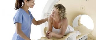
The patient is placed on a special angiography table, if necessary, secured with straps and placed inside the tomograph.
A special feature of the procedure when examining the heart and coronary arteries is that the pulse should be calm and not exceed 60-70 beats per minute. To do this, the patient takes a beta-blocker.
A contrast agent is necessarily administered, upon admission of which the patient may feel a feeling of heat or cold spreading through the veins. The skin at the puncture site is treated with antiseptic gel.
The study is performed after the introduction of contrast agents into the vascular bed. The procedure lasts 30-40 minutes. After completion, a sterile bandage is applied to the catheter insertion area. The patient's condition is monitored by a doctor. Bed rest is used to prevent the formation of a thrombotic process. In order for the contrast agent to be eliminated from the body faster, an enhanced drinking regime is used. In addition to water, patients can drink fruit drinks, dried fruit compotes and non-acidic juices.
If you need to undergo CT angiography in Sochi, we invite you to our medical center.
Preparing for the ANC
- As a rule, it is recommended to stop eating and drinking large amounts of liquids 6-8 hours before the test.
- You must tell your doctor about all medications/vitamins/herbs, etc. that you are taking. You may need to stop taking some or all of your medications until the study is done. We strongly recommend that you do not stop taking any medications yourself without the recommendation of your attending physician or x-ray surgeon.
- Tell your doctor if you have any allergies, including to iodine, seafood, latex or rubber, or to medications such as novocaine, lidocaine, heparin, nitroglycerin, etc.
- It is recommended to remove all jewelry and jewelry to avoid interfering with arterial imaging.
- On the eve of the examination or in the morning on the day of the examination, it is advisable to take a shower and change your underwear.
- It is necessary to shave the artery puncture site in the groin area or the wrist of the right hand (back side). Details can be obtained from your attending physician or x-ray surgeon.
- Take with you 1.0-1.5 liters of drinking water (with or without gas as you wish). Water is needed for hydration after the examination to quickly remove the contrast agent and minimize possible complications.
- You must have a CD or flash card with you in order to receive a record of the examination performed on you and the x-ray surgeon’s report in electronic form.
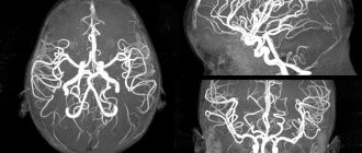
What does non-contrast MR arteriography of blood vessels show?
Magnetic resonance imaging of the brain using the vascular bed scanning mode allows us to identify:
- vascular aneurysms;
- vascular atherosclerosis;
- dissection of arterial walls;
- the presence or absence of stenoses and vascular occlusions;
- vascular malformations;
- vascular deformations affecting the passage of blood flow, including tortuosity of various degrees of severity;
- hypoplasia of brain segments, etc.
Vascular arteriography is an indispensable procedure for diagnosing brain diseases. Using magnetic resonance imaging, specialists can see minimal deviations from the norm, which is the key to providing timely medical care.
What happens during the study
The x-ray surgeon performs the examination only in a hospital setting in a special x-ray operating room. 30-40 minutes before the examination, the patient is given an intramuscular injection in the department. This is a premedication, which consists of a mild sedative and antiallergic drug. The patient will be slightly relaxed but alert and will be able to communicate with the x-ray surgeon throughout the procedure.
The study is performed on a special angiographic table in the supine position. Metal electrodes will be placed on the arms and legs, which will transmit information about each heartbeat and its frequency - to record an ECG. The area of the artery puncture will be treated with special and aseptic solutions. The patient is covered with a disposable, sterile surgical linen and local anesthesia is performed, during which a slight prick and numbness of the skin in the area of arterial puncture is felt. Then the x-ray surgeon will puncture the artery with a thin needle (less than 1.0 mm) and then through a thin and long tube (catheter) will introduce contrast into the lumen of the leg arteries being examined (see Figure).
This will allow you to see blocked or narrowed areas of the artery. When contrast is administered, the patient may feel heat in the groin for 10-15 seconds. On average, the study lasts no more than 30-40 minutes.
After completing the study, the x-ray surgeon will stop the bleeding at the site of arterial puncture within 10-15 minutes and apply a pressure, aseptic bandage, after which the patient is taken to the ward on a lying gurney.
Angiography
Angiography (Greek angeion vessel + graph to write, depict, synonym vasography) is an X-ray examination of blood vessels after the introduction of radiopaque substances into them. There are angiography of arteries (arteriography), veins (venography or phlebography), lymphatic vessels (lymphography). Depending on the purposes of the study, general or selective angiography is performed. With general angiography, all the main vessels of the studied area are contrasted, with selective angiography - individual vessels.
Angiography is carried out in specialized X-ray angiographic rooms, in which angiographic X-ray machines are installed, equipped with devices for fast serial large-format X-ray photography, high-speed fluorographic cameras, devices for X-ray filming and video recording, and special computer devices for recording and processing the resulting image. A method of computational angiography has been developed, in which X-ray imaging is carried out with a special device that allows computer processing of the image and obtaining additional information about the state of blood vessels and hemodynamics.
Angiography is used to diagnose developmental anomalies and diseases of the vascular system, as well as tumor, parasitic and other lesions of various organs. The method allows you to study the topographic and anatomical features of blood vessels, their functional state, blood flow speed, and circuitous circulation pathways.
Contraindications to angiography are: severe general condition of the patient, mental disorders, significant dysfunction of the heart, liver, kidneys, intolerance to radiocontrast drugs. Complications include allergic reactions to the administration of a radiocontrast agent, cardiac arrhythmias, bleeding from a vessel after puncture, hematomas, spasm and vascular thrombosis.
Cerebral angiography (angiography of the arteries of the brain) can detect, in particular, aneurysms, hematomas, tumors in the cranial cavity, stenosis and thrombosis of blood vessels. Angiography of the internal carotid artery (carotid angiography) is used in the diagnosis of pathological processes in the cerebral hemispheres.
Superselective cerebral angiography (catheterization of individual branches of the middle, posterior or anterior cerebral arteries) is usually used to identify vascular lesions and to perform endovascular interventions (for example, installing an occlusion balloon in the afferent vessel of the aneurysm to exclude it from the circulation).
Thoracic aortography (angiography of the thoracic aorta and its branches) is indicated for the recognition of thoracic aortic aneurysm, coarctation of the aorta and other anomalies of its development, as well as aortic valve insufficiency.
Angiocardiography (examination of the great vessels and cavities of the heart) is used to diagnose malformations of the great vessels, congenital and acquired heart defects, and to clarify the location of the defect, which allows choosing a more rational method of surgical intervention.
Angiopulmonography (angiography of the pulmonary trunk and its branches) is used in cases of suspected malformations and tumors of the lungs, thromboembolism of the pulmonary arteries. During angiopulmonography, if necessary, therapeutic procedures are performed, for example, a pulmonary artery thrombus is removed through a catheter or softened with enzymes.
Bronchial arteriography , in which an image of the arteries supplying the lungs is obtained, is indicated for pulmonary hemorrhages of unknown etiology and localization, enlarged lymph nodes of unknown origin, congenital heart defects (tetralogy of Fallot), lung malformations, and is carried out in the differential diagnosis of malignant and benign tumors and inflammatory processes in the lungs. In patients with pulmonary hemorrhage who are in serious condition, bronchial arteriography is performed in conditions of blood transfusion, anti-shock and other intensive care measures. In the process of bronchial arteriography, therapeutic measures are carried out, aimed, for example, at stopping pulmonary hemorrhage.
Abdominal aortography (angiography of the abdominal aorta and its branches) is used for lesions of parenchymal organs and retroperitoneal space, bleeding into the abdominal cavity or gastrointestinal tract. Abdominal aortography makes it possible to detect hypervascular kidney tumors; at the same time, metastases to the liver, another kidney, lymph nodes, and tumor growth into adjacent internal (visceral) organs and tissues can be detected. A radiocontrast agent is usually administered by catheterization of the aorta through the femoral or axillary artery. Abdominal aortography is used for therapeutic purposes for intravascular regional infusion of drugs, in particular in the treatment of peritonitis and pancreatitis.
Celiacography (angiography of the celiac trunk) is performed to clarify the diagnosis of tumors, injuries and other lesions of the liver and its vessels, spleen, pancreas, stomach, gall bladder and bile ducts, and greater omentum. Typically, catheterization of the femoral or axillary arteries is performed. It is important for assessing the results of surgical interventions on the abdominal organs. During celiacography, therapeutic measures can be carried out, for example, the administration of proteolytic enzyme inhibitors for acute pancreatitis.
Renal arteriography (angiography of the renal artery) is indicated in the diagnosis of various kidney lesions: trauma, tumors, hydronephrosis, urolithiasis. During renal arteriography, embolization of the renal artery can be performed, for example in case of inoperable kidney tumors. Renal arteriography is widely used in the complex therapy of renovascular hypertension with stenosis of the renal arteries.
Peripheral arteriography , in which images of the peripheral arteries of the upper or lower extremity are obtained, is used for acute and chronic occlusive lesions of the peripheral arteries, diseases and injuries of the extremities. The method is used for therapeutic purposes, in particular for long-term injections of drugs for thrombosis and arterial embolism.
What will need to be done in the ward
- You should drink plenty of fluids. This is necessary to compensate for the loss of water that you had during preparation for the study and to quickly remove the contrast agent from your body. For most people, this means drinking at least 6 glasses of water, juice or tea. Check the required volume with your attending physician or x-ray surgeon after the study.
- You must strictly adhere to bed rest for 6-8 hours after the examination and keep your leg straight on the side of the puncture site (when using the femoral approach). If using the radial carpal artery approach, you should remain in bed for 1.5 to 2.0 hours, but limit use of your right arm for the next 24 hours.
The puncture site may be slightly numb for a few days.
The next day the pressure bandage will be removed and you can return to your normal lifestyle. You will receive a conclusion about the study and will be able to discuss the results with your attending physician or x-ray surgeon.
Preparation for vascular angiography
MRI examination of the arteriovenous system with contrast is performed by appointment. You need to bring to the procedure:
- passport;
- Compulsory medical insurance policy, VHI policy;
- results of previous examinations;
- referral from the attending physician.
You should arrive 10-15 minutes before the scan starts.
Before MRI angiography of cerebral vessels, it is recommended to rest well and avoid stressful situations. It is advisable to monitor your blood pressure several days before the procedure.
Alcohol and smoking should be avoided 2-3 days before MRI with MR angiography. Avoid taking tonic substances, including strong tea, coffee, and energy drinks.
Before MRI angiography of veins and arteries, you need to warn your doctor about possible contraindications, diseases, and use of medications.
In case of suspected pregnancy, the woman needs to undergo additional examination. To clarify the condition, an analysis is performed to determine the level of the hCG hormone, and an ultrasound is performed. MRI angiography of blood vessels is a contrast procedure, which is a contraindication for diagnostic purposes.
During lactation, women are advised to prepare milk or formula to feed the baby. After administration of the contrast solution, the breasts must be expressed for 6-12 hours.
Before MRI angiography of veins and arteries, the patient removes jewelry and metal accessories. You can change into a comfortable home set and pajamas.
What you should pay close attention to
There may be a slight hemorrhage (subcutaneous hematoma) at the puncture site. If bleeding starts from the puncture site, immediately call the attending physician or the doctor on duty.
Also, be sure to inform your attending or duty doctor about the following cases:
- If your leg feels numb or tingling, or if your foot turns blue or cold.
- If the area around the puncture area appears blue.
- If the puncture area swells or fluid is released from it.
What can be done to improve your condition if damage to the arteries of the legs is detected.
The most important steps are:
- Quit smoking. Try not to be a passive smoker.
- Get physically active. Try to follow the level of physical activity recommended by your doctor. Ideally (unless contraindicated), walk, ride a bike, or do other types of cardio exercise 5 times a week for at least 30 minutes.
- Monitor your blood pressure. Consult your doctor to help you achieve your blood pressure goals.
- Try to reduce your blood cholesterol levels. Eat a healthy diet (high in fiber and low in saturated fat and trans fat) and take cholesterol-lowering medications if necessary.
- If you have diabetes, monitor your blood sugar levels. And aim to achieve and maintain an HbA1c level of less than 7%. HbA1c (hemoglobin A1c) is a blood test that shows the average blood sugar level over the previous 2-3 months.
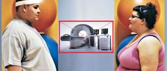
- If you are obese, then set a goal to lose 5-10 kg. If you need to lose more, it is recommended to lose about 1 kg per week until you reach the desired weight.