Raeography of the head (REG)
Posted at 23:55h in Services by doctor
Rheoencephalography of cerebral vessels is a simple but effective diagnostic method. As a result of this procedure, pathological processes such as circulatory disorders, as well as other deviations from the normal functioning of this important organ, are identified.
The method is popular with patients and doctors. This is explained not so much by the affordability of the survey, but by its high information content and the ability to quickly obtain accurate results.
A big advantage over other methods of examining cerebral vessels is its minimal invasiveness, which becomes a factor favoring the use of this diagnostic even for pediatric patients.
Features of the event
REG is performed using a rheographic attachment and a recording device. The child sits comfortably. The doctor applies a special gel to his head and places electrodes on rubber bands. The location of the application is determined by the assigned tasks. During the examination, you may need to perform simple commands: turn, tilt your head. Functional tests provide the most accurate data.
The indicators are recorded by a computer, and the results are displayed graphically on a special tape, similar to an ECG.
The procedure takes no more than half an hour and does not cause discomfort or pain to the little patient. Parents are allowed to be near the child, since it is necessary to maintain a motionless body position.
General information about the method
Rheoencephalography (REG) allows you to identify circulatory disorders in the brain even in the early stages of pathology and thereby prevent the possibility of developing complications that pose a danger to the health and life of patients.
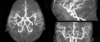
Its invaluable advantage over MRI and CT is the ability to be examined without waiting in line, which in other places takes about six months. Without detracting from the effectiveness of magnetic resonance and computed tomography, it should be noted that timely treatment is the key to victory over the disease, and in some cases, the ability to save the patient’s life.
What kind of procedure is this, who needs it, how to prepare for the examination - these are the questions that will be discussed in the article.
For what purpose is it carried out?
In addition to procedures related to the need to study pathological changes in the arteries and vessels of the brain, it is advisable to conduct REG for preventive purposes.
Types of REG
Among the main types of REG, under certain conditions, the following are prescribed:
- Rheopulmonography (RPG).
Prescribed for bronchopulmonary pathologies. The essence of the method is to record the electrical resistance of the lung tissue and makes it possible to clarify the expected extent of resection. - Rheoencephalography (REG).
A technique that allows you to determine the degree of damage or abnormalities in the central nervous system, vascular system and cerebral blood supply. - Rheovasography (RVG).
One of the practical ways to diagnose blood circulation in the patient’s arms and legs. - Rheocardiography (RKG).
The study is aimed at studying the work of the heart and allows us to determine the degree of its stroke volume. - Integral rheography (IR).
A procedure that makes it possible to accurately measure the baseline impedance of the patient's entire body. - Polyrheocardiography (PRCG).
Diagnostics aimed at studying blood flow in the pulmonary and systemic circles to determine hypertension, hypotension or heart failure in the earliest stages. - Rheoophthalmography (ROG).
A technique for diagnosing and studying blood circulation in the tissues of the eye, with its reaction to the action of an electric discharge. - Rheohepatography (RGG).
A graphical method for studying blood circulation in the liver, based on recording frequency oscillations of the tissues of this organ in response to a high-frequency electrical discharge. - Rheorenography (RRG).
A special non-invasive method for studying blood flow in the kidneys, which makes it possible to identify or prevent renal failure and other pathologies associated with the functioning of this organ. - Reuterography (RUG).
A non-invasive way to study blood circulation in the uterus and pelvic organs. Allows you to detect vascular disorders in the early stages of pregnancy and other gynecological diseases. - Rheoprostatography (RPrG).
An effective way to study blood flow in the prostate gland, allowing timely detection of various diseases in the early stages and making a decision on the choice of quality treatment.
Operating principle of the device
The essence of rheoencephalography is that with the help of a special device - a rheograph - an electric current of low frequency is passed through the brain, as a result of which the resistance of brain tissue is visualized on the monitor. In this way, disorders in arteries, veins and small vessels are detected .
The presence of six channels in the device makes it possible to simultaneously examine several areas of the brain.
In the projection of the studied areas, metal electrodes are installed using an elastic rubber band, which transmit the image to the monitor.
The essence and principle of the study
What is REG? If we decipher the abbreviation and translate it into Greek, it turns out that rheoencephalography is “reo” - “flow”, “encephalo” - “brain”, “graphia” - “I write, I depict”. All together forms a record of the blood supply to the brain. For such an examination of the head, a weak electric high-frequency current is used, the passage of which changes the value of tissue resistance.
Blood, like almost all liquids, is an electrolyte, and when blood vessels are filled with it, electrical resistance values decrease. The obtained values are recorded on a tape by a specially designed device (rheograph), which results in a curve called an encephalogram. Afterwards, based on the data, the rate of change in resistance is assessed, which indicates the rate of blood flow, vessel filling and other indicators.
When is an REG prescribed?
- patient complaints of dizziness;
- deterioration of condition with changes in atmospheric pressure;
- osteochondrosis;
- noise in ears;
- debilitating headaches;
- suspicion of ischemic disease;
- memory losses;
- weakened vision;
- hearing loss;
- atherosclerosis;
- hypertensive crisis;
- dystonia;
- hypertension of the cerebral arteries.
For all pathologies associated with a violation of the condition of blood vessels - their blood supply, changes in blood flow speed and viscosity, it is necessary to conduct an REG.
Decoding
When analyzing the rheogram, the data obtained as a result of the examination, as well as age, health status and chronic diseases are taken into account. The graph that appears on the screen or paper tape when examining a teenager will be different from the graph of an adult. In the first one, it looks like a series of uneven waves with clearly defined periodic peaks. In an adult, it looks like a straight line with evenly spaced vertices. The graph shows anacrotes, catacrotes and incisuras.
Anacrota is a wave that shoots sharply upward with a slightly rounded top. Catacrota is a wave that goes down smoothly. An incisura is a notch between a downward wave and a tooth that anticipates the next wave.
Decryption is carried out only by a specialist. During the analysis, the regularity and type of waves, features of the rounding of the top, the dicrotic tooth located after the descent of the wave, and the position of the notch are assessed. Based on the results of the analysis, the doctor determines the signs of pathologies:
- With the hypotonic type, the waves have sharp, high peaks, the tooth or additional waves are displaced, the incisura and amplitude are increased.
- The hypertensive type is characterized by prolonged anacrosis with displaced waves, additional waves are possible. The amplitude is reduced and uneven. The teeth have different shapes.
- Cerebral atherosclerosis is indicated by flat tops, soft vibrations, and the absence of additional waves on the catacrota. In severe pathology, the waves have a dome shape.
- Spasm of the walls is characterized by round peaks.
- With stagnation of venous blood and difficulties in its outflow, increased catacrota and a large number of small waves between the main ones are observed.
- Angiodystonia is manifested by floating teeth and waves on the catacrota.
Only a functional diagnostics doctor is involved in decoding.
A rheoencephalogram is a graphic representation of vibrations corresponding to a certain blood supply to the brain.
The rheogram distinguishes the main elements:
- Anacrota is the ascending part of the wave. Displays blood flow to the organ being examined. The steeper the rise, the higher the tone and less elasticity of the arteries.
- Top of the wave. Corresponds to the period of time during which blood inflow is equal to outflow. Changes with disturbances in the venous bed.
- Catacrota is the downward part of the wave. Displays the condition of veins and arteries, after incisura - only veins.
- Incisura, also known as cutting, is a short-term flattening of the curve before the rise. The moment corresponds to the transition from systole to diastole. Characterizes the elasticity of blood vessels.
- Diastolic wave is a wave following an incisura, with a smaller amplitude than an anacrotic wave. Shows total vascular resistance.
Previously, the encephalogram was deciphered using tables that show all the values of the components of the graph. Now these calculations are carried out by a special computer program.
When analyzing the obtained REG data, the patient’s age must be taken into account, because the parameters for determining the norm for each age are different.
In conclusion, the doctor does not indicate the probable diagnosis, but the type of blood flow disorder:
- Normotonic – adequate vascular resistance and normal venous outflow.
- Dystonic – constant vasodilation, stagnation of venous blood.
- Angiodystonic – low elasticity of blood vessels associated with pathology of the walls of arteries and veins.
- Hypertensive – persistent increase in peripheral vascular resistance. Violation of venous circulation. Characteristic is the lack of rhythm in the appearance of waves. Most often it indicates the presence of sclerotic changes in the vessel wall.
It should be understood that determining the presence of disorders is not even half of the diagnosis. This is only a determination of the functional state of the vascular bed, and to make a full diagnosis, additional, more informative diagnostic methods are required (ECG, ultrasound of the neck vessels, ECHO of the heart, ultrasound, CT or MRI of the brain). But it is possible to suspect the presence of diseases with a high degree of probability.
Characteristic signs of some of the most common variants of pathological conditions in neurology that are visible on a rheoencephalogram:
- Migraine – higher waves on one side.
- Vegetovascular dystonia (VSD) – the top of the wave is rounded, low amplitude.
- Hypertension - the diastolic wave becomes more pronounced and closer to the top. The amplitude of the waves decreases.
- Atherosclerosis - the apex is smoothed out, the waves gradually take on the shape of an arch, and the amplitude decreases.
- Liqueur hypertension - disappearance of incisura, the curve is expressed in the shape of a dome.
What the study shows
- Based on rheoencephalography of the vessels of the head, specialists receive significant information about the condition of the object of examination. Among them is the possibility of studying vascular tone, their elasticity, blood circulation speed and blood inflow/outflow.
- The use of rheoencephalography makes it possible not only to identify abnormalities in the vessels of the brain, but also to monitor blood flow after complex operations or severe injuries.
- With the help of REG, various pathologies are detected, and the severity of the pathological process is established.
In this case, the high speed of obtaining results is of no small importance.
What problems are being identified?
- presence of traumatic brain injuries;
- localization of hematomas formed as a result of head trauma;
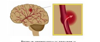
- pre-stroke condition;
- damage to blood vessels by atherosclerotic plaques (atherosclerosis);
- thrombus formation in the vessels of the brain;
- predisposition to increased blood pressure;
- diseases associated with circulatory disorders.
The procedure facilitates the task of making an accurate diagnosis, on the basis of which the doctor prescribes an adequate course of treatment. With the help of it, he subsequently monitors the effectiveness of therapy.
Due to the complete safety of such an examination for the patient’s health, it can be performed repeatedly.
One of the most significant advantages of encephalography is the ability to distinguish between pre-stroke indicators, which have certain differences for men and women.
Other features of the method
Specialists obtain even more information by conducting functional tests.
The simplest and most accessible of them is with nitroglycerin. This substance helps reduce vascular tone. This test is used to differentiate organic and functional disorders.
Diagnostics
During the procedure, the doctor may ask the patient to change his body position, turn his head, stand up and lie down. The fact is that changing the position of the head allows the specialist to draw conclusions about the nature of the disorders - to understand whether they are functional (situational) or organic, associated with certain diseases.
Most often, rheoencephalography (REG) of cerebral vessels is performed to diagnose atherosclerosis of cerebral vessels, its prevalence and stage. In case of traumatic brain injury, the study provides important information about the size of the injured areas, the presence of intracranial hematomas, their depth and the severity of the condition.
JSC "Medicine" (academician Roitberg's clinic) in Moscow is equipped with modern high-precision electrocardiographs that allow diagnostics. Data processing is carried out using computer programs, and therefore decryption requires a minimum of time. Rheoencephalography allows you to obtain high-precision results that reflect a comprehensive picture of the dynamics of the development of diseases affecting the brain. The procedure takes place in comfortable conditions. For patients with limited mobility, rheoencephalography can be performed at home. Optimal price, professionalism of doctors and individual approach are the main advantages of JSC Meditsina.
Go through rheography at JSC "Medicine" (clinic of Academician Roitberg) with comfort!
How to decipher the results
When assessing the examination results, the patient’s age must be taken into account. This is explained by the fact that the walls of blood vessels lose their elasticity over the years, become more fragile, and react differently to various stimuli.
An REG shows graphical wave fluctuations. The following indicators are taken into account:
The specialist reads the diagnostic results, taking into account the regularity of the waves, the appearance and rounding of the apex, as well as the location of the tooth and incisura.
The rate of oscillations of the wave depicted on the screen in adults differs from the manifestations of acceptable indicators in a child.
Rheoencephalographic study makes it possible to classify the condition of blood vessels according to three types of their behavior:
- Dystonic. Characterized by frequent manifestations of changes in vascular tone. Hypotonia with difficulty in venous outflow of blood and low pulse filling is more often observed.
- Angiodystonic. Its symptoms are similar to those of the previous type. The difference is that the cause of the tone disorder is a defect in the vessel wall.
- Hypertensive type according to REG. Significantly different from the species described above. Vascular tone is significantly increased. Venous outflow is impaired.
These types of behavior are not independent pathologies. They are only signs of other diseases and make it possible to identify them in the early stages of development.
You should not attempt to decipher the examination results yourself. It is better to leave this to qualified doctors who will do it professionally and establish an accurate diagnosis.
Description of the examination result
Studying the blood supply to the head using REG is a simple procedure, but deciphering its results requires special education and skills. Therefore, only a specialist can make a diagnosis and correlate changes in the rheogram with possible pathologies of the vascular system. In the process of analyzing the data obtained, the diagnostician studies deviations in the amplitude of the waves from the norm, the reaction of the REG wave to functional tests, and makes a conclusion about the normality or pathology of the vessels supplying the brain with blood. REG waves have several types. Common types are:
- hypertensive type - characterized by persistent hypertonicity of the vascular wall of the vessels delivering blood to the brain and impaired outflow of venous blood from the brain structures;
- dystonic type - a violation of vascular tone, which is characterized by regular changes - for example, hypertonicity may predominate, combined with low pulse pressure, venous stagnation may be observed;
- angiodystonic is close to the previous version, the characteristic difference is the presence of a defect in the vascular wall, which leads to a decrease in elasticity and disruption of the blood supply to individual brain structures.
The type of rheoencephalogram is not a diagnosis. The curve graph shows only the changes that the blood flow undergoes. And how it differs from the norm. Based on deciphering these changes and data from other diagnostic procedures, the doctor can determine the cause of suffering and make a diagnosis.
The result of the study shows deviations from the norm in the blood supply to the brain, that is, pathological conditions of the blood vessels
How is the procedure performed?
The described diagnostic method is completely painless and safe. During the procedure, there is no impact on the patient’s skin, and no various instruments are used.
During the procedure, the patient is placed on a couch or offered to sit on a chair. To obtain more accurate information, the patient is asked to tilt his head forward, turn it to the right or to the left.
The procedure lasts 10-15 minutes. The results of the study are displayed immediately on the monitor screen and are assessed by a neurologist.
Recommendations
To avoid distorting the results, you should consider some simple tips:
- Before installing the electrodes, some areas of the head are treated with alcohol. It is advisable not to stress and take it calmly.
- Eyes should be kept closed during the procedure.
- You need to completely relax. Anxiety can cause a sharp constriction of blood vessels. This will affect the wave oscillation performance.
- It is advisable to rest for 15-20 minutes before the procedure.
- The day before the scheduled examination, you should not take medications that can affect the speed of blood flow.
- The session should not be interfered with by any objects, so you need to remove chains, earrings, hairpins and let your hair down.
If you are examining a small child, you should tell him in advance everything about the upcoming procedure. You can pick him up and sit on a chair with him. Then he will not be afraid and nervous.
Characteristic
The doctor writes a mysterious abbreviation in the direction and, without going into too much detail, sends home the patient who forgot to ask what it is - REG.
REG, rheoencephalography, is a non-invasive type of study of cerebral vessels, which is based on measuring the electrical resistance of tissues at the moment a weak high-frequency impulse passes through them. Blood is a conductor of electric current and, filling the vessels, reduces their resistance, as can be seen on the rheoencephalogram.
Clinical rheography is used to show cerebral blood supply. Another type of study, integral, is aimed directly at assessing vascular resistance.
The information obtained from REG allows you to see:
- tone of the walls of blood vessels;
- time of change in their resistance;
- elasticity;
- condition of arteries and veins;
- blood volume;
- viscosity;
- speed of pulse wave propagation.
The rheoencephalogram shows signs of impaired blood flow, but this is not enough to reliably say about the presence of a particular pathology and, moreover, to differentiate between diseases. In this regard, recently there has been a lot of debate about the advisability of conducting REG and replacing it with more informative research methods.
However, it is too early to write off rheoencephalography. It is indispensable in cases where MRI and CT are contraindicated: during pregnancy, lactation, the presence of pacemakers or other implants, obesity, claustrophobia, contrast agent intolerance, and the need to avoid radiation. Sometimes, to clarify the diagnosis, this procedure is complemented by echoencephalography.
REG is often confused with EEG. To carry out these studies, electrodes are placed on the head. The main difference is in the purpose: electroencephalography examines brain activity, and REG examines the state of blood vessels.
Advantages of REG
The rheoencephalographic examination method is absolutely safe and can be performed on patients of all ages, even small children. It is painless, does not require surgical intervention, and does not pose a risk to the patient’s health. The procedure takes a few minutes, and the result can be obtained almost immediately after its completion. REG provides accurate information about the condition of the arteries and veins of the brain.
An important factor is the availability of REG. The survey not only has a low price. Equipment for its implementation is usually available in all clinics and paramedic centers.
About contraindications
Due to the absolute absence of harm to the body, rheoencephalography has virtually no contraindications or side effects.
Such examination is contraindicated for newborn children . This is explained by the small amplitude of the reflected waves, the large size of the anacrote and the complete absence of incisura. Such readings do not provide an accurate picture of the condition of the vessels of the head.
Rheoencephalography is an effective and affordable method for examining cerebral vessels. Its widespread use is due to the presence of the device in every hospital and, of course, the absence of side effects and contraindications for use.