Angiography is a method for examining blood vessels. CT angiography helps to obtain a three-dimensional picture of the vessels. The examination does not require special patient preparation or hospitalization. Angiography is used to diagnose vascular diseases and monitor changes in the area of surgical interventions. The method is based on the use of an X-ray contrast agent. An angiographic image makes it possible to identify tumors, developmental anomalies, and vascular pathologies (stenosis or aneurysms). The procedure is used by various specialists: oncologists, surgeons, phlebologists, pulmonologists, angio-surgeons. In addition to computed tomography, angiography is used in fluorescence studies in ophthalmology, in studies using magnetic tomography and others.
Progress of the procedure
To exclude allergic reactions, special tests are carried out in advance and antihistamines are prescribed.
The patient is placed on a special angiography table, if necessary, secured with straps and placed inside the tomograph.
A special feature of the procedure when examining the heart and coronary arteries is that the pulse should be calm and not exceed 60-70 beats per minute. To do this, the patient takes a beta-blocker.
A contrast agent is necessarily administered, upon admission of which the patient may feel a feeling of heat or cold spreading through the veins. The skin at the puncture site is treated with antiseptic gel.
The study is performed after the introduction of contrast agents into the vascular bed. The procedure lasts 30-40 minutes. After completion, a sterile bandage is applied to the catheter insertion area. The patient's condition is monitored by a doctor. Bed rest is used to prevent the formation of a thrombotic process. In order for the contrast agent to be eliminated from the body faster, an enhanced drinking regime is used. In addition to water, patients can drink fruit drinks, dried fruit compotes and non-acidic juices.
If you need to undergo CT angiography in Sochi, we invite you to our medical center.
Contraindications for CT angiography of lower extremity vessels
Computed tomography of the veins and arteries of the legs is performed with a contrast agent, which improves the visualization of the area under study. The main contraindication may be an allergy to iodine, which is part of the contrast agent. In addition, contraindications are also represented by pregnancy and lactation; renal failure).
A CT scan is not recommended if the patient has severe pain, as well as involuntary movements (hyperkinesis), because the patient will not be able to remain motionless for a long time. The doctor himself clarifies what contraindications the patient has at a preliminary appointment.
When is CT angiography needed?
For the circulatory system to function normally, it is important to have healthy blood vessels. Ekaterina Aleksandrovna Nekhaeva, a radiologist at the Expert Clinic Kursk, told us how CT angiography helps check their condition and the indications for such a study.
— Among the methods of studying blood vessels, CT angiography occupies a special place. Ekaterina Alexandrovna, what is this? What is the point of this research?
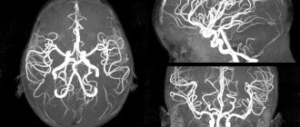
— This is a modern and one of the most informative methods for diagnosing vascular pathology, based on the use of X-ray radiation. It is distinguished from ordinary x-rays by much greater information content. Using a special program, the doctor can create a three-dimensional model of the vessels of the area under study.
CT angiography is always performed with contrast, that is, with intravenous administration of a contrast agent. This makes blood vessels more visible on images compared to surrounding tissue. This makes it possible to accurately assess their lumen, location, syntopy (relationship with surrounding anatomical structures) and other parameters. These parameters determine the availability of vessels for the surgeon, for example, when planning interventions. We try to reflect all this information in the protocol in as much detail as possible (especially if the referring doctor asks us specific questions).
— Vessels of which parts of the body and human organs can be examined using CT angiography?
— CT angiography is widely used to evaluate the vessels of the head, neck, large vessels of the abdominal cavity and retroperitoneal space, vessels of the iliac regions, lower extremities, coronary arteries, as well as pulmonary arteries and veins.
Read material on the topic:
Who can benefit from CT coronary angiography?
— When might a patient need CT angiography?
— Basically, the indications for the study are determined by the referring doctor. These are usually vascular surgeons, because CT angiography is often performed immediately before the proposed surgery so that the doctor can take into account the location and size of the vessels when performing the operation.
The most common pathologies and other reasons for which patients come (or are admitted) to us are:
- obliterating atherosclerosis of the vessels of the lower extremities (damage to the large arteries of the legs, leading to narrowing of the arteries and poor circulation);
- atherosclerotic lesions of the arteries of the head and neck, coronary arteries, etc.;
- vascular development abnormalities;
- aneurysm;
- aortic rupture;
- arterial thrombosis;
- cardiac ischemia;
- extravasal blood vessel compression syndrome (i.e. when the vessel is compressed by something from the outside);
- vascular damage;
- examination after surgery, etc.
Read materials on the topic:
Find and neutralize. How to protect against blood clots? Coronary heart disease: diagnosis and treatment
— What does CT angiography reveal?
— It helps to detect stenotic changes (areas of narrowing) or aneurysmal areas (areas of expansion) of blood vessels, the presence and prevalence of atherosclerotic changes (with determination of the exact size and structure of plaques), compression of blood vessels by a tumor, etc.
CT angiography also makes it possible to identify abnormalities in the development of blood vessels, the presence of various variants of the origin and relative position of vessels, which are also often clinically significant.
— Ekaterina Aleksandrovna, what are the advantages of CT angiography when compared, for example, with MRI and ultrasound? And can it completely replace these methods?
— CT angiography is definitely more informative than the listed methods, since it allows us to visualize the vascular wall itself, while on MRI and ultrasound we see only the signal of blood flow through the vessels. Also, only thanks to such CT diagnostics we can see atherosclerotic plaques, assess their size, structure, and shape.
With CT angiography of cerebral vessels, intracranial (intracranial) vessels can be seen much better than with ultrasound, since the capabilities of ultrasound are limited due to the screening of ultrasound by the bones of the skull.
CT angiography can completely replace MR angiography, since the latter is inferior to it in sensitivity and specificity. On the other hand, when performing MRI, there is no radiation exposure inherent in computed tomography. The only advantage of ultrasound is the ability to assess the speed of blood flow in dynamics, as well as the absence of contraindications typical for X-ray studies and intravenous contrast.
In any case, the optimal choice of one or another research method, depending on the questions facing the radiologist, always remains with the referring clinician.
Read more about vascular ultrasound in our article: Doppler, duplex, triplex... What is vascular ultrasound?
— Is preparation necessary before CT angiography?
— In general, no special preparation is needed. But if the patient underwent an X-ray examination of the stomach and (or) intestines with barium sulfate a few days before the computed tomography scan, then you should wait 3-4 days until the barium is removed from the body. Also, if the patient uses metformin (a drug for the treatment of type 2 diabetes), then, after agreement with the attending physician, it is necessary to stop taking the medication two days before the examination.
— Tell us in more detail how CT angiography is performed
— The person is placed on the retractable table of the computed tomograph. First, a catheter is installed in his cubital vein, through which a radiopaque contrast agent is injected using a special device (injector). First, a “native” (preliminary non-contrast) scan is performed, and then a scan with the introduction of contrast. Some areas may require holding your breath for an average of 10 seconds. During the entire session, the specialist remains in the next room and communicates with the patient via speakerphone. The duration of the procedure is approximately 20 – 30 minutes.
— Can everyone do this research? Or are there contraindications for CT angiography?
- Yes, they are. This:
- impaired renal function;
- thyrotoxicosis (increased levels of thyroid hormones) and thyrotoxic crisis;
- allergy to iodine-containing contrast agents;
- pregnancy.
— Is a referral required to undergo CT angiography?
— Yes, a referral from the attending physician is necessary in the vast majority of cases.
— Ekaterina Aleksandrovna, is this research carried out for children?
- Yes. To perform computed tomography for children, special scanning protocols are used, the use of which implies the maximum possible reduction in radiation exposure to the child’s body. However, CT angiography is performed for children according to strict indications and with a mandatory referral from the attending physician.
If you need a CT angiography, you can sign up for this test here ATTENTION: the service is not available in all cities
Interviewed by Marina Volovik
The editors recommend:
How will MRI of cerebral vessels help a patient? When is ultrasound of cerebral vessels prescribed? Take care of your vessels! How not to miss vascular diseases of the brain?
For reference:
Nekhaeva Ekaterina Aleksandrovna
In 2013 she graduated from Kursk State Medical University.
In 2015 – residency in general surgery.
Completed professional retraining in radiology.
Currently, he is a radiologist at the Expert Clinic Kursk. Receives at the address: st. Karl Liebknecht, no. 7.
Computed tomography angiography
This technique is fast. To carry it out, there is no need to invade the patient’s body. The patient is injected with a contrast agent containing iodine into a vein. Using a computed tomograph, the necessary images are obtained.
CT angiography makes it possible not only to visualize the bed of the vessel, but also to give an idea and reconstruction of the blood supply at any time, to obtain a picture of the lumen of the vessel “in its natural form,” and to accurately assess the degree of its narrowing. Difficulties can arise when calcium is deposited in an atherosclerotic vessel, since it is impenetrable to X-rays.
The number of images in one projection per second is the main principle of classification of computed tomographs. Today, 16-, 32-, 64-, 256- and 320-slice tomographs are used. The more pictures you take, the clearer the image. In addition, increasing the number of images minimizes the negative impact of the patient’s breathing on the image and reduces the amount of contrast agent in the body.
With all the advantages of CT angiography, there are contraindications and limitations:
- Patient intolerance to iodine preparations;
- Serious renal impairment;
- Dangerous for children and pregnant women;
- Within 24 hours after the examination, a nursing mother should not feed her baby breast milk;
- It is difficult, and sometimes impossible, to perform the study if the patient is hypermobile or overweight;
- It is impossible to carry out therapeutic actions during the study.
Preparation for aortography
In preparation for angiography, the patient must donate blood for a general analysis, a biochemical analysis to exclude kidney and liver pathologies, and a coagulogram (blood test for clotting).
It is necessary to notify your doctor about allergic reactions, especially to iodine-containing drugs.
Aortography is best performed on an empty stomach to avoid nausea and vomiting. Fluid intake is not limited. If endovascular intervention is planned, you may be given antithrombotic drugs before the angiography.
Immediately before the procedure, you must sign an informed consent for the procedure.
Before the examination, the patient must go to the toilet, remove all metal objects (since they are reflected on x-rays and can distort the picture) and change into special clothes
During angiography, the patient is in a supine position and fixed to the table, since movements during the examination can distort the results.
X-ray angiography
Angiography is classified depending on the organ in which the vessels are contrasted.
For example, angiopulmonography is used to visualize the vascular network of the lungs; angiocardiography is used to visualize the atria and ventricles with large vessels.
The study requires access to a vessel that is located in close proximity to the tumor and feeds it. A catheter is inserted into the artery and, under X-ray monitoring, this catheter is brought to the vessel of interest. The contrast agent contained in the catheter allows images to be taken.
The resulting images help to see the pathological characteristics of the organ and its functions, which affect the function of the blood supply.
Stomach biopsy
X-ray allows you to contrast arteries, veins, and can also be used to obtain images of the lymph nodes of blood vessels. This study is called vasography. Study of arteries - arteriography, study of veins and lymph nodes, respectively - venography (phlebography) and lymphography.
The condition of the arterial bed in the area where the tumor is located is important for deciding the issue of surgical intervention. It is this condition that indicates the boundaries of tumor growth, as well as its growth into the artery.
The pressure that allows blood to flow through the arteries is high, and during surgical manipulations, a large volume of blood will be lost from a vessel damaged by a cancerous tumor. The speed at which blood flows through the veins is small, the pressure is insignificant, which means that it is not difficult to compress a large vein damaged by cancer without a large loss of blood.
The information obtained using angiography allows you to avoid all sorts of surprises. In addition, taking into account the results of the study, the doctor can immediately carry out a therapeutic measure, for example, installing a stent in a narrowed vessel or removing a blood clot.