Ultrasound diagnostics is one of the safest and most effective ways to detect various diseases of internal organs. This technique is used in various fields of medicine, including assessment of the condition and possible pathologies of blood vessels. In this case, Doppler ultrasound or ultrasound is performed, which is used to examine the neck and head. Early diagnosis allows you to choose gentle and effective treatment methods and prevent the development of complications. First you need to understand what an ultrasound scan of the vessels of the head and neck shows, why this method is effective in diagnosing many diseases.
The Doppler ultrasound effect is based on the ability of ultrasound waves to be reflected from blood cells. As a result, electronic impulses appear that appear on the device screen in the form of images of veins and arteries with blood flow.
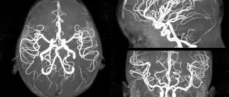
This ultrasound examination allows you to assess the condition of the large arteries that supply the brain, carotid and vertebral arteries, determine the intensity of blood flow, and also identify traces of obstruction.
How is Dopplerography performed?
This examination is carried out in the presence of certain symptoms. Preliminary preparation is necessary so that the results of the ultrasound examination give an accurate picture of the patient’s condition.
There are two types of procedure:
- Transcranial. The sensor is installed on the skull bones in those places where they are thinnest. The procedure gives an accurate picture of the vessels located directly in the brain.
- Extracranial. The technique is used to examine large vessels passing through the neck (carotid arteries, jugular veins, etc.). The sensor is located at each site separately.
What ultrasound methods exist?
1. Doppler ultrasound is standardly included in ultrasound examination of the heart (Echocardiography, echocardiography, echocardiography with Doppler are synonyms). Allows you to assess the condition of the heart valves and blood flow in the cavities and large vessels.
2. Doppler ultrasound of the branches of the aortic arch (synonyms: Doppler ultrasound of the neck vessels, Doppler ultrasound of the brachiocephalic vessels) - study of the main arterial and venous vessels involved in the blood supply to the neck, head and brain. This is one of the most frequently performed studies and allows us to identify impaired patency of the carotid and vertebral arteries due to atherosclerosis, vascular abnormalities and spinal pathology. As an additional method, the so-called TCD (transcranial Doppler sonography) can be performed, which allows you to directly assess the blood supply to the brain.
3. Doppler ultrasound of the arteries of the upper or lower extremities . It is carried out in cases of impaired patency of the peripheral arterial bed (atherosclerosis, obliterating endarteritis, diabetes mellitus).
4. Doppler ultrasound of the veins of the upper or lower extremities . It is carried out in case of blood clots in peripheral veins, as well as in case of varicose veins to assess the consistency of venous valves and determine the patency of the veins.
Indications for the procedure
Doppler sonography is indicated in the following cases:
- Problems with coordination and other symptoms indicating a malnutrition of the brain (the patient is dizzy, has tinnitus, etc.).
- Unstable heart rhythm.
- Hypertonic disease.
- Transient ischemic attacks.
- IOP.
- Previous stroke or heart attack.
- There are suspicions of atherosclerosis.
- Diabetes.
- Fatigue quickly for no apparent reason.
- Cervical osteochondrosis.
This procedure is also indicated if heart surgery is to be performed, if there are pulsating formations in the neck, or if thrombosis is suspected. In children, it is performed in case of serious postural problems and delayed development of the child.
When is it necessary to do an ultrasound scan of the lower extremities?
Ultrasound Dopplerography of blood vessels is considered one of the most informative and universal diagnostic methods, while it is painless and safe. It is prescribed as part of a comprehensive examination if the patient complains of:
- feeling of heaviness in the legs;
- cramps in the calf muscles;
- feeling of numbness, burning in the legs;
- sudden pain when walking or during physical activity;
- redness or pallor, dryness, itching of the skin;
- obesity;
- swelling of the legs;
- the appearance of spider veins or a protruding section of a vein.
In addition, Doppler ultrasound is almost always prescribed to assess the effectiveness of therapy if a vascular disease - thrombosis, varicose veins, angiopathy or others - has already been diagnosed and treatment has been prescribed.
For preventive purposes, ultrasound scanning of the lower extremities is recommended for elderly people, patients after a stroke or heart attack, with diabetes mellitus, and long-term smokers.
A temporary contraindication to the analysis may be the presence of wounds, abrasions and other defects in the area of the body being examined, due to which the sensor will not fit tightly to the skin.
Are there any contraindications?
Ultrasound waves are absolutely safe for humans, so there are no special contraindications to this type of ultrasound. The only exception is a violation of the integrity of the skin. The procedure involves contact of sensors with the head or neck, so the examination is postponed until the wound heals.
However, there are a number of special cases that make duplex examination of blood vessels difficult:
- cardiac arrhythmia, which changes the speed of blood circulation through healthy arteries and veins;
- the area under study is hidden under bone tissue;
- slow blood flow in the patient.
These are not contraindications, but in this case changes in the location of the sensors are required. It will be necessary to navigate non-standard conditions, which requires professionalism on the part of the doctor.
Price for ultrasound examination of the veins of the lower extremities
Doppler ultrasound is a primary diagnostic method and is prescribed either for prevention or to study an existing diagnosis. Hence the low price of ultrasound examination of leg veins in Moscow with excellent information content of the examination. Affordable cost, safety and painlessness, absence of contraindications at any age, accuracy - all this makes Doppler ultrasound a popular procedure.
Comprehensive examination of deep and superficial veins with an accurate diagnosis of the disease
2000 rub.
Expert consultation with a phlebologist
1500 rub.
| Fill out an application on the website, we will contact you as soon as possible and answer all your questions. | Registration online |
| Fill out an application on the website, we will contact you shortly. Or call us by phone | Registration online |
Find out the price of ultrasound examination of the arteries and veins of the lower extremities at the First Phlebological Center, as well as make an appointment with a doctor conveniently online. Fill out the short form on our website, and a Center specialist will contact you shortly.
Features of ultrasound examination
As a rule, Doppler sonography is planned in advance, but it can be performed urgently. The procedure itself is painless, so patients tolerate it calmly.
The examination is carried out according to the following scheme:
- The patient lies on his back. The head should be relaxed (a flat pillow is placed under it).
- You need to remain calm, breathe evenly and, if possible, not move.
- A special gel is applied to the area under study to prevent the penetration of air between the skin and the sensor.
- The movement of the sensors is controlled by a specialist. As a result, an image of the vessels appears on the monitor screen.
The study lasts no more than 1 hour. During the study, the doctor may ask the patient to do some action: turn his head, squeeze a vein in a certain place, breathe less often, etc.
Dopplerography of extracranial vessels begins with examination of the right lower segment of the carotid artery. The sensor is then moved up the neck. Next, the color mode is activated, which makes it possible to identify a number of pathologies: deformations of the walls of blood vessels, impaired blood flow in certain areas, etc.
How is the ultrasound procedure performed?
Doppler ultrasound is performed on an outpatient basis. The patient stands or lies on the couch lying down, on his back or on his stomach. The doctor applies a conductor gel to the areas being examined, then moves the sensor over the skin, and the image of the vessels is displayed on the screen of the diagnostic device. During the procedure, the patient may be asked to tense his muscles and change his body position. The duration of the study is from 20 to 40 minutes.
Ultrasound scanning of the vessels of the lower extremities is performed in several modes:
- black and white B-mode is designed to measure the diameter of the vessel, the thickness of the vascular walls, and assess the condition of the venous valves;
- color mode: at the site of blood flow disturbance, the color of the vessel is changed;
- spectral mode demonstrates the main characteristics of the blood flow of a vessel in spectrogram format.
What can she show?
Duplex scanning allows you to determine areas of narrowing of the lumen of blood vessels, the direction of blood flow, its speed, as well as the condition of the vascular walls. This study can show the location of cholesterol plaques and the presence of blood clots, which is very important when diagnosing atherosclerosis.
A decrease in the elasticity of the walls of the arteries, as well as their thickening, indicate that the patient has hypertension (high blood pressure). If a change in blood flow is diagnosed, this indicates the presence of obstacles. For example, an aneurysm is a sac-like protrusion of a vessel.
Indications for Dopplerography of head vessels:
- chronic and acute cerebral circulatory disorders or suspicion of them;
- the patient belongs to groups at increased risk of atherosclerosis and other pathological vascular conditions;
- absence of pulse and blood pressure in the arteries of the arms against the background of intense deterioration of visual acuity;
- abnormal changes in the cervical spine due to diseases or congenital anomalies, which can lead to compression of the vertebral artery;
- suffered traumatic brain injuries, as a result of which the vessels were damaged;
- vascular damage due to toxic effects or surgery.
What pathologies can be diagnosed?
Doppler ultrasound has certain limitations in diagnosing diseases. For example, she will not be able to study small veins - for this she needs to perform angiography. But there are a number of pathologies that can be identified:
- Atherosclerosis. By examining the head and neck using ultrasound, it is most often possible to diagnose atherosclerotic disorders. This condition is indicated by echogenicity, the presence of plaques, and thickening of the vascular walls. Moreover, if the lumen of the artery narrows by more than 20%, then the patient is diagnosed with “stenosis.”
- Thrombosis. Thrombosis is characterized by blocking of the arterial lumen by a blood clot. On Doppler ultrasound, inflammation of the vessel wall with a homogeneous formation inside is clearly visible.
- Compression of blood vessels. The examination allows you to see areas where blood flow is impaired due to the impact of swollen tissue on the channels. This allows you to diagnose cervical osteochondrosis and identify problems with blood circulation after injuries.
In addition, these ultrasound examinations can detect a number of other diseases, including vasculitis, post-traumatic dissections of the arterial walls and other pathologies.
Doppler ultrasound is a safe diagnostic method that can be performed in any case if there is a medical need for it. Timely implementation of the procedure allows you to identify the disease at an early stage of its development.
Contraindications to Dopplerography of head vessels
It is no coincidence that Doppler sonography of the head and neck is considered safe and universal. It has no absolute contraindications, although it can sometimes be difficult if:
- the vessel being diagnosed is located under the bone or a thick layer of subcutaneous fat;
- the patient suffers from diseases characterized by changes in blood flow even in healthy vessels.
If the skin of the area being examined is damaged or there is a rash or ulcers on it, the procedure is not performed.
Ultrasound Dopplerography (USDG)
Home / Diagnostic department / Ultrasound diagnostic room / Doppler ultrasound (USD)
|
Ultrasound Dopplerography of blood vessels is used to detect disturbances in blood flow in vessels.
The ultrasound examination is carried out on the vessels of the head, neck, vessels of the lower and upper extremities, and kidneys. The examination is informative for both venous and arterial circulatory systems.
Recently, new ultrasound techniques using the Doppler effect have found widespread use. Based on the Doppler effect, a new complex section of ultrasound angiography has emerged, which makes it possible to determine the state of the lumen wall of blood vessels and record blood flow parameters.
Dopplerography makes it possible to identify early lesions: arterial stenoses, determine their significance, characterize the condition of the vascular walls, tortuosity, and quantify arterial and venous blood flow.
Modern ultrasound diagnostics is unthinkable without Dopplerometry, since it allows us to identify predisposing factors for the development of circulatory disorders. Using Doppler ultrasound, you can study the nature of blood flow in the vessels and its disturbances caused by various factors.
The results obtained using Doppler ultrasound make it possible to successfully treat heart and vascular diseases, neurological diseases and many others.
It is recommended to perform Doppler ultrasound in the following cases:
- hypertension
- headaches
- repeated cases of loss of consciousness
- varicose veins
- seizures
- chilliness of hands and feet
What is the difference between ultrasound, duplex scanning and triplex scanning?
- Ultrasound Dopplerography ( ) Dopplerography or Doppler is an inexpensive study of blood vessels that allows you to study only one function - the patency of the vessel, based on the graph of the blood flow. It is performed blindly, the sensor is placed on the approximate projection points of the vessel. If a violation of the patency of a vessel is detected, it is impossible to clarify the cause of this violation. No vessel visualization.
- Duplex scanning of blood vessels (ultrasonic duplex scanning). The vessel is visible on the screen - you can evaluate not only its patency, but also the reasons for the obstruction of patency : tortuosity of the course, thickening of the walls (stenosis), the presence of blood clots, plaques, developmental anomalies, installed stents, postoperative junctions of vessels, etc., and also evaluate speed and direction of blood flow (as ultrasound Doppler), i.e. simultaneously perform two functions (duplex): - study of vascular anatomy; — assessment of blood flow (velocity). This is a more expensive study.
- Triplex scanning This is the same as duplex scanning, but a color image is added - a more accurate diagnosis of vascular patency and the degree of stenosis is performed. Three functions are performed simultaneously (triplex): - study of vascular anatomy; — blood flow assessment; — accurate assessment of vascular patency in color mode.
Contraindications to Dopplerography : There are no absolute contraindications to Dopplerography. Relative (temporary) contraindications for Doppler ultrasound may be the patient’s severe general condition or other reasons due to which he cannot lie down. This technique is completely harmless to the person being examined and is highly informative in the early stages of the development of pathological processes, allowing a correct diagnosis to be made in a timely manner and treatment to begin in a timely manner. Vascular examination is a painless diagnostic method that has no side effects, radiation exposure or contraindications. For maximum diagnostic efficiency, it is desirable that the decision to conduct the study is made after consultation with a neurologist.
Doppler ultrasound requires highly qualified specialists performing Doppler ultrasound . At the Branch of the FBLPU "LRC "Moscow Region" of the Federal Tax Service of Russia, diagnostics using Doppler are carried out by experienced doctors who have undergone special training and are certified, doctors of the highest category.
Prices for services
Photo gallery
Branch phone: 8 (495) 020-65-71
What can be seen during the procedure
If we consider the procedure for ultrasound scanning of the brain from a technical point of view, it is quite simple and easily tolerated by patients. However, to carry it out, a medical center or clinic must be equipped with a special device. At the same time, only a specialist who has certain skills in this area of diagnostics can decipher the results of an ultrasound scan. Therefore, the choice of the clinic where the examination will be carried out must be approached responsibly.
During the procedure, ultrasound examination of the brain, the specialist sees a picture on the monitor screen of the device, displaying the state of the diagnosed area of the vascular system in cross-section. Thanks to the ability to rotate the image in several projections, the doctor receives information regarding the speed of blood circulation, the diameter of blood vessels, and the level of patency. During the diagnosis, aneurysms, narrowing or blockage of brain vessels, as well as the thickness of the vessel wall are visible. In addition, the structure and direction of arteries and veins, the presence of atherosclerotic plaques or blood clots are visualized.
Doppler ultrasound scanning of the vascular system is quite common due to its high information content and speed of obtaining diagnostic results.
What is ultrasound
Doppler ultrasound is an ultrasound examination based on the Doppler effect, which consists in the fact that the length and frequency of the sound wave changes depending on the speed of the object.
When scanning blood vessels, waves are reflected from red blood cells flowing through blood vessels. This information is read by equipment (a sensor that the doctor passes over the area under study) and displayed on the monitor. The following types of ultrasound examination are distinguished:
- B-mode, or duplex scanning, is the simplest, inexpensive and quite informative for preliminary diagnosis. In this study, veins and arteries are visible only in black and white, which allows the doctor to evaluate the main parameters: the diameter and curvature of the blood vessel, the thickness of its wall, the presence or absence of plaques and blood clots in it.
- Color or triplex scanning allows you to obtain an image on the monitor screen in which different sections of blood vessels, depending on the presence or absence of pathology, are painted in different colors. In color mode, the doctor evaluates, first of all, the uniformity of filling a vein or artery with blood, which makes it possible to identify blood flow anomalies.
- In addition to duplex and triplex scanning, in some cases it is necessary to study the hemodynamics of blood flow. In this case, the spectral mode of ultrasound with Doppler is used, through which a spectrogram is formed on the screen. Such an examination is indispensable during pregnancy, when it is necessary to determine whether the placenta is sufficiently supplied with blood.
How is the examination carried out?
When examining the head and neck, ultrasound examination is performed in a sitting position.
All other types of examination are performed lying down. In some cases, when examining the veins of the lower extremities, the patient will be asked to stand or place the leg in a certain position. A gel is applied to the area under study to facilitate better penetration of ultrasound into the vessels. Next, a sensor is passed over this place several times in different directions, which transmits data to the monitor.
Based on the data obtained, a conclusion is formed. Based on it, a neurologist, phlebologist, cardiologist or other specialized specialist makes a diagnosis and prescribes treatment