Introduction
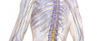
The pathways of the nervous system and the complex reflex arcs consisting of them are the most important and complex section of neurology. It is important because it affirms the cellular nature of the nervous system (neural doctrine) and shows the ordered nature of the arrangement and connections of neurons (in the form of reflex arcs), which underlies its regulatory function.
There is a significant difference from the method of descriptive anatomy. The latter makes it possible to demonstrate the shape, size and localization of a particular formation of the nervous system, as well as its belonging to gray or white matter, but does not reveal at all the structural organization of the nervous system and the mechanisms of its functioning.
This “separation” of structure from function, which is dangerous for the worldview, eliminates the systematic approach to the nervous system in the form of the study of reflex arcs. Here the emphasis is placed precisely on the presence of connections between neurons, on their interaction, leading to the functioning of both the nervous system itself and the entire organism. However, at the same time, the number of mental operations among students increases (synthesis is added to analysis), which increases the complexity of mastering the material and its subjective complexity. Nevertheless, only the study of the nervous system as a set of reflex arcs allows us to understand its organization and functional significance. Finally, only knowledge of neural connections and interactions allows for topical diagnosis of damage to the nervous system, i.e. take a meaningful approach to the diagnosis and treatment of nervous and many other diseases and injuries.
How do the concepts of “conducting path” and “reflex arc” relate to each other? Here it should be clearly understood that any conductive path is part of one or another reflex arc. Since there are two main links in the reflex arc: afferent and efferent, the pathways are classified into afferent and efferent. Taking into account the hierarchical principle of construction of the central nervous system (the presence of higher and lower nerve centers subordinate to them) and the possibility of closing reflex arcs at the level of higher nerve centers, it is clear that both afferent and efferent pathways should be localized in both the peripheral and central parts nervous system. Since the closure of somatic reflex arcs (the connection of afferent and efferent links through interneurons) always occurs in the central nervous system, the latter also contain an associative link and corresponding associative pathways, localized only within the central nervous system.
Afferent nerve pathways conduct impulses from the receptor to the nerve center and are sensitive. Afferent nerve pathways ending in the projection centers of the cerebral cortex are classified as pathways of conscious sensitivity. The same afferent pathways that end in the subcortical sensory nerve centers are classified as pathways of unconscious sensitivity.
Efferent nerve pathways conduct impulses from nerve centers to the working organ. Since we are talking here only about the somatic nervous system, the working organ is the skeletal muscle, therefore the efferent nerve pathways are called motor. Depending on which nerve centers the efferent pathways are connected to, the latter are responsible for performing both conscious and unconscious movements.
Any conducting pathway (afferent, associative or efferent), depending on the level of closure and complexity of the reflex arc, can be single-neuron or multi-neuron (several neurons connected in series in a chain). If we consider a multineuron pathway as a chain, then within its boundaries we can distinguish links represented by the corresponding neurons. Compactly located neuron bodies form nerve centers (nodal, nuclear or screen type), and axons collected in bundles form nerve tracts. Thus, a multineural pathway consists of nerve centers and tracts. In this case, the nerve centers and tracts of the same pathway are localized in certain but different parts of the nervous system. Each tract within the central nervous system conducts nerve impulses usually in one direction and in most cases - of the same functional content. It should be clearly understood how the tracts within the central nervous system differ from the bundles of fibers that form the cranial or spinal nerves. Nerves contain both afferent and efferent fibers, and different afferent fibers can carry different sensory impulses.
In the future, material will be presented that concerns primarily the somatic part of the nervous system.
MAIN CONDUCTING PATHWAY OF VOLUNTARY MOVEMENTS
4.3.1. Motor cortex
The main pathway providing voluntary movements is the path along which nerve impulses from the motor zone of the cerebral cortex pass to the striated muscles. The motor zone of the cerebral cortex is mainly the cortex of the precentral gyrus (areas 4 and 6, according to Brodmann) and its territories adjacent to this zone. The cortex here consists of 6 cell layers. Layer V contains large pyramidal cells, described in 1874 by V.A. Betz (1834-1894). They are recognized as the bodies of central (upper) motor neurons, the axons of which have monosynaptic connections with peripheral (lower) motor neurons.
4.3.2. Corticonuclear and corticospinal connections
Nerve impulses from central motor neurons (fields 4 and 6), motor cells located in the adjacent zones of the frontal (field and parietal lobes (fields 5 and 7), as well as in the cingulate gyrus (fields 23c and 24c), move in a centrifugal direction along their axons that take part in the formation of the corona radiata, and then the knee and the anterior two-thirds of the posterior leg of the internal capsule (Fig. 4.7).
The knee of the internal capsule is made up of axons of central motor neuron cells that carry nerve impulses to the motor nuclei of the cranial nerves located in the tegmentum of the brain stem. This part of the axons of central motor neurons forms the corticonuclear (corticonuclear) pathway. Its fibers are directed to the motor nuclei of the cranial nerves, consisting of the bodies of peripheral motor neurons. Approaching these nuclei, part of the fibers of the cortical-nuclear tract passes to the opposite side, i.e. a partial supranuclear decussation of cortical-nuclear fibers is formed. The only exceptions to this rule are the fibers going to the lower part of the nucleus of the facial and to the nucleus of the hypoglossal cranial nerves, since they make an almost complete decussation above the level of the corresponding nuclei. As a result, the lower part of the nucleus of the facial nerve and the nucleus of the hypoglossal cranial nerves receive nerve impulses, as is commonly believed, only from the opposite hemisphere of the brain, while the rest of the peripheral motor neurons located in the nuclei of the cranial nerves receive nerve impulses from both the opposite cerebral hemisphere (by crossed paths) and from the homonymous hemisphere of the brain (along uncrossed paths).
The axons of central motor neurons, constituting the anterior 2/3 of the posterior limb of the internal capsule, pass as part of the base of the brain stem, and along the way, most of them (approximately 97-98%) end at the nerve cells of the reticular formation of the brain stem and in nuclear formations related to to the extrapyramidal system.
Approaching these formations, the axons of the cortical cells branch; as a result, the majority of efferent nerve impulses coming from the cortical cells excite not one, but several intermediate cells encountered along the way. A process of divergence of efferent motor impulses occurs and, as a result, after the involvement of intermediate neurons in the motor pathway, the number of fibers through which motor impulses continue to move increases significantly. Efferent motor pathways that have and have not passed through intermediate synoptic apparatuses (these are axons of Betz cells, the number of which is 25-30 thousand on each side), subsequently constitute pyramids located on the ventral surface of the medulla oblongata (it is estimated that each of the pyramids is approximately 1 million nerve fibers). Having passed the pyramids, these fibers, named in this regard in 1885 by P. Flexig pyramidal, at the border of the medulla oblongata and the spinal cord undergo partial decussation (decussatio pyramidum). The pyramidal fibers that have passed to the opposite side (approximately 80%) enter the lateral cord of the spinal cord (funiculus lateralis) and form the lateral pyramidal tract in it, and the motor fibers remaining on the same side (20%) are part of the anterior cord ( funiculus anterior) and constitute the anterior pyramidal tract in it. The nerve fibers that make up the pyramidal tracts end by approaching the peripheral motor neurons located in the anterior horns of the spinal cord segments. All these fibers participate in the formation of corticospinal (corticospinal) pathways. Among the nerve fibers that form them, only 2-2.5% are axons of Betz cells. They are monosynaptic, have the thickest myelin sheath
ku and provide the highest speed of movement of nerve impulses along the corticospinal pathways.
Thus, the pyramidal tract is currently considered as a heterogeneous system, consisting of several subsystems with different types of fibers, starting from different parts of the cerebral cortex. The vast majority of them are interrupted in certain structures of the subcortical formations and the brain stem. In this regard, the definition of the corticospinal system as monosynaptic and “pyramidal” does not correspond to reality, but it is traditional, accepted in classical neurological literature, so in the future we will not avoid the term “pyramidal tract”, but its understanding should correspond to modern concepts.
In the spinal cord, the pyramidal tracts descend down, and at the level of each segment, part of their constituent nerve fibers ends at the cells of the anterior horns, which are peripheral (lower) motor neurons.
The fibers of the lateral pathway approach each peripheral motor neuron, while the fibers of the anterior pyramidal tract end only at the peripheral motor neurons, which provide innervation on the same side to the muscles of the neck and trunk and, in particular, the respiratory muscles, helping to maintain breathing in the event of development of patient with hemiplegia. In this regard, peripheral mononeurons related to the innervation of the muscles of the neck and trunk receive nerve impulses from both sides, which creates conditions for maintaining vital functions in patients with central hamiparesis.
Summarizing the above, it can be noted that in the anterior horns of the spinal cord segments, where the bodies of peripheral motor neurons are located, the axons of neurons coming from nerve cells located in numerous supraspinal motor centers end. Afferent structures also end here, bringing impulses from receptors of various types of sensitivity located within the corresponding segment of the body.
Efferent and afferent formations directly or indirectly (through interneurons) approach peripheral motor neurons, in which the summation of excitatory and inhibitory postsynaptic potentials (EPSP and IPSP) coming to them occurs. As a result of the synthesis of impulses entering the peripheral neuron, a bioelectric charge is formed. It determines the nature of the nerve impulse formed in this neuron and directed to the muscle fibers associated with it.
A large number of neurotransmitter systems are involved in the implementation and regulation of voluntary motor acts. Clarification of the nature of mediators in different synaptic zones of the active movement system is under study. However, it is already known that the key excitatory neurotransmission in central motor neurons is carried out mainly by glutamate and aspartate, while GABA and taurine help reduce the activity of motor reactions. The neurotransmitter of the peripheral motor neuron, which ensures the transmission of nerve impulses at neuromuscular synapses, is acetylcholine.
The so-called pyramidal tract is not a single, homogeneous system of fibers; it consists of several subsystems with different connections and functions. Direct corticonuclear and corticospinal projections, i.e. fibers that provide monosynaitic connections between the pyramidal cells of the cerebral cortex and peripheral motor neurons, mainly with those that provide innervation to the distal limbs, represent the youngest system in phylogenetic terms, existing only in primates. It is currently known that in the conduction pathway, usually called the lateral corticospinal, or pyramidal, the axons of Betz's giant pyramidal cells, located in the cortex of the anterior central gyrus, and having monosynaptic connections with peripheral motor neurons, make up only 2- 2.5%.
Rice. 4.7. Pyramid system (diagram). a - pyramidal tract: 1 - cerebral cortex; 2 - internal capsule; 3 - cerebral peduncle; 4 - bridge; 5 - intersection of pyramids; 6 - lateral corticospinal (pyramidal) tract, 7 - spinal cord; 8 - anterior corticospinal tract; 9 - sensory motor fibers of the peripheral nerve; III, VI, VII, IX, X, XI, XII - corresponding cranial nerves, b - convexital surface of the cerebral hemisphere: motor zone of the cortex (fields 4 and 6); topographic projection of parts of the opposite half of the body: 1 - legs; 2 - torso; 3 - hands; 4 — brushes; 5 - faces; c — horizontal section through the internal capsule, location of the main conducting pathways in it. 1 - visual and auditory radiance; 2 - temporopontine and cervical-occipital-pontine fibers; 3 - sensitive thalamocortical fibers; 4 - pyramidal tracts that conduct impulses to motor neurons of the lumbosacral enlargement of the spinal cord; 5 - pyramidal fibers, conducting impulses to the motor neurons of the thoracic spinal cord; 6 - pyramidal tracts that conduct impulses to the motor neurons of the cervical enlargement; 7 - cortical-nuclear pathways; 8 - frontal-pontine tract; 9 - corticothalamic tract; 10 — anterior leg of the internal capsule; 11 — knee of the internal capsule; 12 - posterior leg of the internal capsule; d - anterior surface of the brain stem: 1 - intersection of the pyramidal tracts.
4.3.3. Peripheral (lower) motor neurons
In 1904, Sherrington coined the term “terminal motor pathway.” In it he included peripheral motor neurons and their axons, ending in branches that deliver the total nerve impulse to muscle fibers through neuromuscular synapses, the mediator of which is acetylcholine-N.
The cells of the anterior horns of the spinal cord are represented by two types of motor neurons: I) motor neurons, the axons of which have a thick myelin sheath (A-alpha fibers), and 2) motor neurons, the axons of which have a thin myelin sheath (A-gamma fibers). Nerve cells, by analogy with the designation of nerve fibers, are called alpha and gamma motor neurons. The axons of gamma motor neurons are directed into the muscles, where they penetrate the receptor apparatus - muscle spindles containing intrafusal (located inside the spindle) fibers. In cases of muscle stretching, for example, when inducing a tendon reflex, a change in the length and intrafusal muscle fibers occurs: the configuration of the muscle spindles changes, and at the same time, irritation of the spiral-shaped receptor structures located on them occurs. The nerve impulses arising in these receptor structures are sent along the afferent fibers of the peripheral nerve structures to the pseudounipolar cells of the spinal ganglia. Further, along the axons of these cells, impulses enter the spinal cord and directly through collaterals or through interneurons of the same segment of the spinal cord provide excitation of large motor cells (alpha motor neurons) of the anterior horns. Nerve impulses along the thick myelin sheathed axons of alpha motor neurons are directed to the extrafusal fibers, which make up the bulk of the previously stretched striated muscles. The resulting excitation of these muscle fibers causes tension or contraction of the muscles, which ensures staying in a fixed position or causes a certain motor act.
Some of the dendrites of the nerve cells of the spinal ganglia originate in muscle tendons, or more precisely, in the receptor apparatus located in these tendons - the Golgi tendon organs. These receptors have a high threshold of excitation, which occurs only when the muscle tendons are overstretched, threatening traumatic damage (rupture, separation). In such cases, impulses are sent from the excited Golgi tendon organs to the spinal cord, ensuring a decrease in the activity of alpha motor neurons and a decrease in tension (tone) of the corresponding muscles.
In addition, a decrease in muscle tone when alpha motor neurons are overexcited is facilitated by impulses that, through collaterals extending from their axons, reach the Renshaw cells located in the anterior horn of the spinal cord. Having undergone excitation, these cells send inhibitory biopotentials to alpha motor neurons, thereby reducing the degree of their tension.
Thus, the peripheral motor neuron, along with the influences transmitted to it along the efferent pathways from the cortical and extrapyramidal structures of the brain, is influenced by impulses arising in the receptor apparatus at the level of the same spinal segment or in the myotome of the same name as this segment. All this greatly contributes to the regulation of muscle tone, its optimization in accordance with changing requirements, depending on the nature of the motor acts performed.
So, the corticomuscular pathway includes central and peripheral motor neurons. Each peripheral neuron receives impulses along several, sometimes many, nerve fibers coming from the cerebral cortex, from its numerous extrapyramidal structures, as well as from the tissues of the body metamer of the same name. Some of them are excitatory, some are inhibitory bioelectric potentials. All of them are processed and summed up in the peripheral motor neuron. As a result, bioelectric potentials are sent from the body of the peripheral motor neuron to the muscle, which are the result of the synthesis of the properties of the impulse acting on it. The peripheral motor neuron provides the innervation of the associated striated (from 5 to 1000) muscle fibers and the trophic processes occurring in them.
A group of muscle fibers innervated by one peripheral motor neuron and its axon constitutes a motor unit, while the nerve fibers (axon of the motor neuron) going to the muscle providing fine movements innervate 5-12 muscle fibers, while large muscles performing gross movements receive impulses along nerve fibers, each of which innervates from 100 to 1000 of their muscle fibers. The concept of a motor unit as a functional unit of the neuromotor apparatus was introduced by the English neurologist Ch. Sherrington (Sherrington Ch., 1857-1952). An average-sized muscle has several hundred motor units. A group of peripheral motor neurons innervating one muscle makes up the motor pool.
Damage to peripheral neurons leads to peripheral paralysis, which is characterized by complete immobility of the corresponding part of the body. Violation of the function of central motor neurons causes central paralysis, in which voluntary movements are absent, while reflex movements and synkinesis are excessive. In case of damage to part of the central motor neurons, pyramidal tracts or motor units involved in the innervation of the muscles of a certain part of the body, paresis occurs in it, which can be central or peripheral, depending on the damage to the elements of which neurons (central or peripheral) this paresis is caused .
Graphic basis for depicting conduction paths and reflex arcs
The diagram of pathways or reflex arcs requires a symbolic representation of the sections of the nervous system and the chain of neurons located in these sections.
Let's start with a picture of neurons. The first in the chain of neurons of the afferent pathway is the ganglion nerve cell, the body of which is always located in the peripheral part of the nervous system, in most cases in the sensitive nerve ganglion. In shape, a ganglion cell looks either like a bipolar neuron or (more often) like a pseudounipolar neuron (Fig. 1).
Rice. 1. Schematic representation of neurons and synaptic connections. Bipolar neuron (a); pseudounipolar neuron (b): 1 - receptor nerve ending, 2 - peripheral process (dendrite), 3 - neuron body, 4 - central process (axon), 5 - ending in contact with the second neuron in the chain, 6 - direction of impulse. Interneuron contact (c): 1 - first neuron, 2 - synapse, 3 - second neuron, 4 - direction of impulse. Motor neuron (d): 1 - neuron body, 2 - central process (axon), 3 - effector nerve ending, 4 - direction of impulse
The bodies of communication and motor neurons are located only in the central nervous system: communication - in centers of either the screen or nuclear type; motor - only in the motor nuclei (see Fig. 1).
For diagrams of most pathways and reflex arcs, a graphic representation of the parts of the nervous system is used (Fig. 2).