Location and functional significance of the crown
Parietal region (vertex)
The fontanel is separated from the brain by three membranes. The small spaces between them are filled with cerebrospinal fluid. As the cranial bones fuse, the area of the fontanel tightens, and it disappears by 1.5 years. If this does not happen at a later date, the reason for this may be disturbances in the general development of the child.
The parietal part of the head performs several functions. First, the fontanelle helps the baby to be born. The sutures and soft crown make the bones of the skull flexible and plastic, so they can fit into each other. This helps the baby's head change its shape, which is extremely important when passing through the mother's birth canal.
The fontanel serves as a natural shock absorber. It softens the impact of a head injury. It also takes over some of the functions of the thermoregulation system. If a child’s temperature rises, the brain naturally cools through the thin covering of the fontanel.
A soft crown is one of the health indicators for a pediatrician. Based on its size and the nature of the pulsation, an experienced doctor can assess the level of development of a small organism and even make a preliminary diagnosis.
The crown can be felt even in an adult; in most people it is slightly thickened. This is where hair growth begins for almost all people.
Why does a person have two or three crowns on his head: reasons, medical opinion
These protrusions on the skull do not in any way affect the health and psychological development of a person. Medical specialists did not notice any supernatural features or abnormalities or health threats. Doctors believe that the cause of their appearance is only parental genes and nothing more.
According to incomplete studies, medical professionals have not confirmed the fact that people with two or three vertices have more developed brain activity than those with one. Other factors influence this. Several crowns on the back of the head are a manifestation of the special structure of the skull. This factor was influenced by the genetic component.
Doctors' opinions on the presence of two crowns on the head
Anatomy of the parietal region
The parietal region covers the area of the parietal bones. Its boundaries are clearly visible only on the skeleton. They can be represented by drawing projection lines:
- in front – coronal suture (connecting the frontal and parietal bones);
- behind – lambdoid suture (connecting the parietal bones with the occipital bone);
- from the sides - temporal lines.
The parietal region contains the following layers:
- leather;
- subcutaneous tissue;
- tendon helmet;
- subgaleal tissue;
- periosteum;
- subperiosteal tissue;
- parietal bones;
- meninges.
Location of the parietal bone
The skin has a strong connection with the underlying tendon helmet, which is why it is inactive. Subcutaneous tissue contains a large number of sebaceous and sweat glands. It contains the arteries and nerves of the parietal zone. The tendon helmet has the appearance of a monolithic tendon stretch, connecting both bellies of the occipitofrontal muscle. Beneath it lies a layer of loose fatty tissue, separating it from the periosteum. This structure makes it possible to scalp the skin along with the tendon helmet in case of certain skull injuries.
The periosteum is connected to the bones of the skull through subperiosteal tissue, but along the line of the sutures of the skull it is firmly fused with them. The parietal bones are light but strong. In newborns, a section of connective tissue (the aforementioned fontanel) remains between them, which ossifies by 1-1.5 years. On the inside, the parietal bones lack periosteum, are adjacent to the dura mater of the brain, and have depressions from arteries, pachyonic granulations, and brain convolutions.
The dura mater of the brain has a process called the falx cerebri. It corresponds to the midline of the cranial vault and separates the cerebral hemispheres. All along its length. In the upper part there is the superior sagittal sinus, which belongs to the venous system of the brain.
What does science say about two crowns?
Doctors explain that the presence of several “peaks” does not in any way affect a person’s health or his character. Doctors say that this is an individual characteristic of a person. It does not pose a threat to life or health.
But even the luminaries of science tend to endow such a person with special talents. Doctors say that the presence of several crowns indicates that the two hemispheres of the brain are developing equally. This means that his abilities are manifested in different areas, he will achieve a lot in life. But it is difficult for such a person to concentrate on one thing and develop only one talent. He is “scattered” and may not achieve anything at all.
A version has also been put forward that several “tops” on the head are a sign of possible autism. But this hypothesis has not found scientific confirmation. Now she is rarely mentioned.
Innervation and blood supply
Blood supply to the parietal region of the skull
Vessels and nerves penetrate the parietal region from the back of the head, forehead and temporal zones, from where they are directed radially (i.e. from bottom to top). Here, the hollow organs are connected to each other by anastomoses, which form collateral networks that pass to the opposite side.
The arterial network of the parietal region consists of branches of the supraorbital artery (from the internal carotid artery system), which connects with the branches of the superficial temporal, posterior auricular and occipital arteries (from the external carotid artery system).
The venous system is well developed and is located in three levels: extracranial veins of soft tissues; diploetic veins located in the spongy substance of bones; venous sinusitis of the dura mater. All of them communicate with each other by emissary veins. The largest of them is the parietal sinus, which opens into the superior sagittal sinus.
Lymphatic vessels of the parietal region flow into the preauricular and inferior auricular lymph nodes. Sensory nerves come from the frontal, temporal, and occipital zones, accompanying the arteries and veins.
Causes of headaches in the crown area
Cervical osteochondrosis can cause pain in the parietal part of the head.
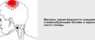
Headache in the crown area can occur in both adults and children. Its character can be different: it is accompanied by a feeling of squeezing, bursting from the inside, tingling. Sometimes the pain is pulsating, or penetrating into the ears or optic nerve. The causes of this phenomenon are of different origins and are accompanied by specific symptoms.
Dystonia of cerebral vessels is provoked by high blood pressure. The headache increases gradually. It’s as if the head is being pulled together by a “hoop.” Cephalgia has a descending nature, affecting the parietal, frontal, and occipital parts. “Floaters before the eyes” and a feeling of fullness in the ears often appear.
The cranial cavity contains cerebrospinal fluid (CSF), which maintains optimal intracranial pressure. Its volume is small and stable. An imbalance leads to intracranial hypertension. An increase in ICP manifests itself as pressure in the crown of the head, which increases with physical activity and a change in position. The headache is usually mild but long lasting. The situation is aggravated by dizziness, nausea, and visual impairment.
Cervical osteochondrosis, spondylosis, herniated disc are the most common causes of cephalalgia in the crown area. Pain is felt along the back of the neck, on the back of the head, and on the top of the head. The attacks are strong, pulsating, radiating to the lower jaw, shoulders, and neck. They are provoked by compression of the nerve roots and impaired blood flow.
Parietal cephalohematoma in a newborn
The consequences of a TBI can appear within a month and result in chronic cephalgia. Exacerbations are possible after viral infections, stress, and excessive physical exertion. The pain is dull, aching, and occurs along with attacks of nausea and vomiting, sleep disturbance, and pressure changes.
Formations in the crown area, tumors and cysts, increasing in size, compress the brain tissue. The pain is paroxysmal, from oppressive to unbearable. Temporary disturbances of vision, speech, fainting, and symptoms of epilepsy are observed. This pathology is a real threat to human life. Urgent diagnosis is required.
Infectious diseases that affect the protective membranes of the brain manifest themselves with high fever, spasms of the neck muscles, and severe headache in the crown of the head. Secondary signs are manifestations of general intoxication.
Rigidity of the neck muscles and shoulder girdle occurs as a result of prolonged stay in a static position, or due to myositis (inflammation of the muscle). In the first case, headache attacks are aching with a feeling of twisting, muscle tension. With myositis, acute local cephalgia is observed at the site of inflammation with limited movement.
Most common reasons
Among the many factors mentioned above, there are causes of pain in the top of the head that are particularly common and have the most specific manifestations.
Occupational pain or tension pain
Both children and adults are susceptible to this type of headache, which most often affects the crown of the head. A dull pain presses on the head from above, compresses it and absorbs it entirely. There is a feeling as if an invisible helmet, helmet or spacesuit is being worn on top. The nature of occupational pain lies in the fact that a person remains in an uncomfortable position for a long time, without moving, in the presence of poor lighting, or in the workplace not meeting ergonomic standards and requirements.
Solution:
- Change working conditions - adjust the height of the chair, adjust the lighting, position the computer at the correct distance from the eyes.
- After every hour of work, get up from the table and do a little exercise, stretching your neck and shoulders.
- Regularly ventilate the room, maintain normal humidity and temperature.
- Perform a light self-massage of the head.
During the week, a person spends most of his time at work. To avoid headaches in the workplace, you need to properly organize your work-rest schedule and create suitable working conditions.
Neuroses and psychoemotional disorders
Over half of the complaints about pain that occurs where the crown of the head is located in a person is associated with neurotic diseases and disorders. If a squeezing or constricting headache occurs, often accompanied by dizziness and even numbness of the limbs, then this is a serious reason to consult a specialist.
Take care of your nerves. They cause most diseases
Approximately 60% of people diagnosed with neurasthenia or hysteria suffer from pain in the upper part of the head. Painful sensations are constant or periodic, they can intensify or weaken, but they always accompany the patient and do not leave him for a long time.
A person suffering from neurosis experiences emotional instability and is prone to panic attacks, and the fear is always very specific. Someone is afraid of getting a certain disease, someone is worried that they might lose a loved one, etc.
Unfortunately, a person with a neurotic disorder is constantly in a vicious circle of “fear-pain”. Fear increases pain, and pain causes new fears. Most often, people who experience prolonged stress or psycho-emotional stress suffer from neuroses.
Solution: seek help from a psychotherapist or psychiatrist and undergo a course of medication.
Post-traumatic conditions
People who have suffered even minor traumatic brain injuries complain that the top of their heads hurts. At the same time, there is a decrease in memory, performance, concentration and an increase in psycho-emotional exhaustion. The nature of such pain is dual in nature: physiological and psychosocial.
Physiological manifestations of pain are determined by the following factors:
- disturbance of the movement of cerebrospinal fluid;
- compression of the nerve roots;
- excessive pressure on the membranes of the brain.
The psychosocial component consists of increased suspiciousness of the victim, fear of complications after injury, and mistrust of doctors.
Editorial: Causes and treatment of focal epilepsy
Solution: treatment from specialized specialists and consultation with a psychologist.
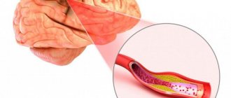
Vascular pain
Often the parietal area of the head suffers from pain that occurs with hypertension, hypotension, and vegetative-vascular dystonia. Decreased or increased vascular tone prevents them from performing their functions of maintaining optimal blood pressure. Either compression of the nerve cells by the vascular walls or vasospasm occurs.
- Equalizing blood pressure with medications prescribed by a doctor.
- Dieting.
- If the pain takes you by surprise, then people with high blood pressure need to lie down in bed and put a high pillow under their head, and people with low blood pressure need to put it under their feet.
Pain in the vertex associated with neck pathologies
Osteochondrosis, cervical migraine, pinched nerves and blood vessels, instability of the cervical vertebrae can lead to pain in the upper part of the head.
- Examination by a neurologist and surgeon.
- Massage and manual therapy sessions.
- Physiotherapy.
- Physiotherapy.
Treatment methods
Pain in the crown of the head may be a symptom of high blood pressure.
Self-administration of medications will help eliminate the symptoms, but not the cause of cephalalgia. Discomfort in the crown area is dealt with by combining medications and non-drug therapy.
In accordance with the problem, the following is prescribed:
- If blood pressure changes, use antihypertensive drugs and analeptics, moderate physical activity, sufficient sleep, and changes in diet.
- For high intracranial pressure - nootropics, correctors of cerebrovascular accidents, diuretics, avoidance of bending work, eye strain.
- For osteochondrosis - analgesics and antispasmodics, manual therapy, massage, gymnastics.
- For infectious diseases - antibacterial and antiviral drugs.
- For muscle spasms - antispasmodics, local anesthetics, sedatives, acupressure, walks in the fresh air.
- For neoplasms, specific treatment is required, often surgery.
Usually, pain in the parietal region is caused by serious causes. Early diagnosis of diseases allows for timely and effective treatment.
What does one crown on the head mean for women, men, children: folk signs, esoteric opinion
According to doctors' studies, people with one crown do not differ in anything special from individuals with two or three. But according to esotericists, there are differences (this has already been discussed above). Individuals with one crown are endowed with a personal rhythm, unlike men and women with two crowns. That is why he is able to control his feelings and is more organized.
What is the difference between people with one and two crowns on their heads?
Pathological features of the parietal region
Benign tumors of the parietal region are removed surgically.
A feature of penetrating injuries to the parietal region (not counting the danger of brain damage) is the risk of damage to the sagittal sinus, which is accompanied by severe bleeding and requires urgent surgery. Depressed (closed) fractures of the parietal bones also cannot be avoided without surgical interventions, both in the presence and absence of neurological symptoms of brain compression.
In the parietal region, dermoid cysts, lipomas, atheromas, acute purulent-necrotic inflammations and other formations can be observed. These phenomena in this zone have limited development, which is due to the density of the tissues adjacent to the skull.
Surgical approaches are used through the parietal region during some neurosurgical operations. They are usually used to expose the superior surface of the parietal lobe of the brain, enter the longitudinal fissure of the cerebrum, and perform surgical interventions for damage to the superior sagittal sinus.