Nerve impulses - the alphabet of the brain - are of an electrochemical nature
Magazine reader L. Gorbunova (village of Tsybino, Moscow region) writes to us: “I am interested in the mechanism of signal transmission through nerve cells.”
1963 Nobel Prize laureates (from left to right): A. Hodgkin, E. Huxley, D. Eccles.
Scientists' ideas about the mechanism of nerve impulse transmission have recently undergone significant changes. Until recently, Bernstein's views dominated science.
‹
›
The human brain is, without a doubt, the highest achievement of nature. A kilogram of nervous tissue contains the quintessence of the whole person, starting from the regulation of vital functions - the work of the heart, lungs, digestive tract, liver - and ending with his spiritual world. Here are our thinking abilities, our entire perception of the world, memory, reason, our self-awareness, our “I”. Knowing the mechanisms of how the brain works is knowing yourself.
The goal is great and tempting, but the object of research is incredibly complex. Just kidding, this kilogram of tissue represents a complex system of communication between tens of billions of nerve cells.
However, the first significant step towards understanding how the brain works has already been taken. It may be one of the easiest, but it is extremely important for everything that follows.
I mean the study of the mechanism of transmission of nerve impulses - signals running along the nerves, as if through wires. It is these signals that are the alphabet of the brain, with the help of which the senses send information-dispatches about events in the outside world to the central nervous system. The brain encodes its orders to the muscles and various internal organs with nerve impulses. Finally, individual nerve cells and nerve centers speak the language of these signals.
Nerve cells - the main element of the brain - are varied in size and shape, but in principle they have a single structure. Each nerve cell consists of three parts: a body, a long nerve fiber - an axon (its length in humans ranges from several millimeters to a meter) and several short branched processes - dendrites. Nerve cells are isolated from each other by membranes. But the cells still interact with each other. This happens at the junction of cells; this junction is called a “synapse”. At a synapse, the axon of one nerve cell and the body or dendrite of another cell meet. Moreover, it is interesting that excitation can be transmitted only in one direction: from the axon to the body or dendrite, but in no case back. A synapse is like a kenotron: it transmits signals in only one direction.
In the problem of studying the mechanism of a nerve impulse and its propagation, two main questions can be distinguished: the nature of the conduction of a nerve impulse or excitation within one cell - along a fiber, and the mechanism of transmission of a nerve impulse from cell to cell - through synapses.
What is the nature of the signals transmitted from cell to cell along nerve fibers?
People have been interested in this problem for a long time; Descartes assumed that the propagation of the signal was associated with the transfusion of fluid through the nerves, as if through tubes. Newton thought it was a purely mechanical process. When the electromagnetic theory appeared, scientists decided that a nerve impulse is analogous to the movement of current through a conductor at a speed close to the speed of propagation of electromagnetic oscillations. Finally, with the development of biochemistry, a point of view emerged that the movement of a nerve impulse is the propagation along a nerve fiber of a special biochemical reaction.
Yet none of these ideas came to fruition.
Currently, the nature of the nerve impulse has been revealed: it is a surprisingly subtle electrochemical process, which is based on the movement of ions through the cell membrane.
The work of three scientists made a major contribution to the discovery of this nature: Alan Hodgkin, professor of biophysics at the University of Cambridge; Andrew Huxley, Professor of Physiology, University of London, and John Eccles, Professor of Physiology, University of Canberra, Australia. They were awarded the Nobel Prize in Medicine for 1963.
The famous German physiologist Bernstein was the first to suggest the electrochemical nature of the nerve impulse at the beginning of this century.
By the early twentieth century, quite a lot was known about nervous excitation. Scientists already knew that a nerve fiber can be excited by electric current, and the excitation always occurs under the cathode - under the minus. It was known that the excited area of the nerve is charged negatively in relation to the non-excited area. It was found that the nerve impulse at each point lasts only 0.001-0.002 seconds, that the magnitude of excitation does not depend on the strength of the irritation, just as the volume of the bell in our apartment does not depend on how hard we press the button. Finally, scientists have established that the carriers of electric current in living tissues are ions; Moreover, inside the cell the main electrolyte is potassium salts, and in the tissue fluid - sodium salts. Inside most cells, the concentration of potassium ions is 30-50 times higher than in the blood and in the intercellular fluid that washes the cells.
And based on all this data, Bernstein suggested that the membrane of nerve and muscle cells is a special semi-permeable membrane. It is permeable only to K+ ions; for all other ions, including negatively charged anions inside the cell, the path is closed. It is clear that potassium, according to the laws of diffusion, will tend to leave the cell, an excess of anions appears in the cell, and a potential difference will appear on both sides of the membrane: outside - plus (excess cations), inside - minus (excess of anions). This potential difference is called the resting potential. Thus, at rest, in an unexcited state, the inside of the cell is always negatively charged compared to the outer solution.
Bernstein suggested that at the moment of excitation of the nerve fiber, structural changes occur in the surface membrane, its pores seem to increase, and it becomes permeable to all ions. In this case, naturally, the potential difference disappears. This causes a nerve signal.
Bernstein's membrane theory quickly gained recognition and existed for over 40 years, until the middle of our century.
But already at the end of the 30s, Bernstein's theory encountered insurmountable contradictions. It was dealt a major blow in 1939 by the subtle experiments of Hodgkin and Huxley. These scientists were the first to measure the absolute values of the membrane potential of a nerve fiber at rest and during excitation. It turned out that upon excitation, the membrane potential did not simply decrease to zero, but crossed zero by several tens of millivolts. That is, the inner part of the fiber changed from negative to positive.
But it is not enough to overthrow a theory, we must replace it with another: science does not tolerate a vacuum. And Hodgkin, Huxley, Katz in 1949-1953 proposed a new theory. It is called sodium.
Here the reader has the right to be surprised: until now there has been no talk about sodium. That's the whole point. Scientists have established with the help of labeled atoms that not only potassium ions and anions are involved in the transmission of nerve impulses, but also sodium and chlorine ions.
There are enough sodium and chlorine ions in the body; everyone knows that blood tastes salty. Moreover, there is 5-10 times more sodium in the intercellular fluid than inside the nerve fiber.
What could this mean? Scientists have suggested that upon excitation, at the first moment, the permeability of the membrane only to sodium sharply increases. The permeability becomes tens of times greater than for potassium ions. And since there is 5-10 times more sodium outside than inside, it will tend to enter the nerve fiber. And then the inside of the fiber will become positive.
And after some time - after excitation - equilibrium is restored: the membrane begins to allow potassium ions to pass through. And they go outside. Thus, they compensate for the positive charge that was introduced into the fiber by sodium ions.
It was not at all easy to come to such ideas. And here's why: the diameter of the sodium ion in solution is one and a half times larger than the diameter of potassium and chlorine ions. And it is completely unclear how a larger ion passes where a smaller one cannot pass.
It was necessary to radically change the view on the mechanism of ion transition through membranes. It is clear that reasoning about pores in the membrane alone is not sufficient here. And then the idea was put forward that ions could cross the membrane in a completely different way, with the help of secret allies for the time being - special organic carrier molecules hidden in the membrane itself. With the help of such a molecule, ions can cross the membrane anywhere, not just through the pores. Moreover, these taxi molecules distinguish their passengers well; they do not confuse sodium ions with potassium ions.
Then the general picture of the propagation of a nerve impulse will look like this. At rest, carrier molecules, negatively charged, are pressed to the outer boundary of the membrane by the membrane potential. Therefore, the permeability for sodium is very small: 10-20 times less than for potassium ions. Potassium can cross the membrane through pores. As the excitation wave approaches, the pressure of the electric field on the carrier molecules decreases; they throw off their electrostatic “shackles” and begin to transfer sodium ions into the cell. This further reduces the membrane potential. There is a kind of chain process of recharging the membrane. And this process continuously spreads along the nerve fiber.
Interestingly, nerve fibers spend only about 15 minutes a day on their main job—conducting nerve impulses. However, the fibers are ready for this at any second: all elements of the nerve fiber work without interruption - 24 hours a day. Nerve fibers in this sense are similar to interceptor aircraft, whose motors are continuously running for instant departure, but the departure itself can only take place once every few months.
We have now become acquainted with the first half of the mysterious act of passing a nerve impulse along one fiber. How is excitation transmitted from cell to cell, through junctions - synapses? This question was explored in the brilliant experiments of the third Nobel laureate, John Eccles.
Excitation cannot directly transfer from the nerve endings of one cell to the body or dendrites of another cell. Almost all of the current flows through the synaptic cleft into the outer fluid, and a tiny fraction of it enters the neighboring cell through the synapse, unable to cause excitation. Thus, in the region of synapses, the electrical continuity in the propagation of the nerve impulse is disrupted. Here, at the junction of two cells, a completely different mechanism comes into force.
When excitation approaches the end of the cell, the site of the synapse, physiologically active substances - mediators, or intermediaries - are released into the intercellular fluid. They become a link in the transfer of information from cell to cell. The mediator chemically interacts with the second nerve cell, changes the ionic permeability of its membrane - as if punches a hole into which many ions rush, including sodium ions.
So, thanks to the work of Hodgkin, Huxley and Eccles, the most important states of a nerve cell - excitation and inhibition - can be described in terms of ionic processes, in terms of structural and chemical rearrangements of surface membranes. Based on these works, it is already possible to make assumptions about the possible mechanisms of short-term and long-term memory, and about the plastic properties of nervous tissue. However, this is a conversation about mechanisms within one or more cells. This is just the ABC of the brain. Apparently, the next stage, perhaps much more difficult, is the discovery of the laws by which the coordinating activity of thousands of nerve cells is built, the recognition of the language that the nerve centers speak among themselves.
In our knowledge of how the brain works, we are now at the level of a child who has learned the letters of the alphabet, but does not know how to connect them into words. However, the time is not far when scientists, using the code - elementary biochemical acts occurring in a nerve cell, will read the most fascinating dialogue between the nerve centers of the brain.
Detailed description of illustrations
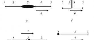
Scientists' ideas about the mechanism of nerve impulse transmission have recently undergone significant changes. Until recently, Bernstein's views dominated science. In his opinion, in a state of rest (1) the nerve fiber is charged positively on the outside and negatively on the inside. This was explained by the fact that only positively charged potassium ions (K+) can pass through the pores in the fiber wall; Large negatively charged anions (A–) are forced to remain inside and create an excess of negative charges. Excitation (3) according to Bernstein is reduced to the disappearance of the potential difference, which is caused by the fact that the pore size increases, anions come out and equalize the ionic balance: the number of positive ions becomes equal to the number of negative ones. The work of 1963 Nobel Prize winners A. Hodgkin, E. Huxley and D. Eccles changed our previous ideas. It has been proven that positive sodium ions (Na+), negative chlorine ions (Cl–) and negatively charged carrier molecules are also involved in nervous excitation. The resting state (3) is formed in principle in the same way as previously thought: an excess of positive ions is outside the nerve fiber, an excess of negative ions is inside. However, it has been established that during excitation (4) there is not an equalization of charges, but a recharging: an excess of negative ions is formed outside, and an excess of positive ions inside. This is explained by the fact that when excited, carrier molecules begin to transport positive sodium ions through the wall. Thus, the nerve impulse (5) is a recharge of the electrical double layer moving along the fiber. And from cell to cell, excitation is transmitted by a kind of chemical “battering ram” (6) - an acetylcholine molecule, which helps ions break through the wall of the neighboring nerve fiber.
CENTRAL NERVOUS SYSTEM
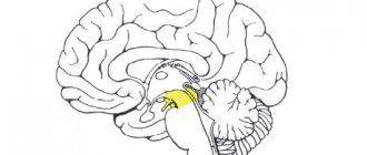
The central nervous system consists of the brain and spinal cord and their protective membranes. The outermost is the dura mater, under it is the arachnoid (arachnoid), and then the pia mater, fused with the surface of the brain. Between the pia mater and the arachnoid membrane is the subarachnoid space, which contains cerebrospinal fluid, in which both the brain and spinal cord literally float. The action of the buoyant force of the fluid leads to the fact that, for example, the adult brain, which has an average mass of 1500 g, actually weighs 50–100 g inside the skull. The meninges and cerebrospinal fluid also play the role of shock absorbers, softening all kinds of shocks and shocks that tests the body and which could lead to damage to the nervous system.
The central nervous system is made up of gray and white matter. Gray matter is composed of cell bodies, dendrites, and unmyelinated axons, organized into complexes that include countless synapses and serve as information processing centers for many functions of the nervous system. White matter consists of myelinated and unmyelinated axons that act as conductors transmitting impulses from one center to another. The gray and white matter also contains glial cells.
CNS neurons form many circuits that perform two main functions: they provide reflex activity, as well as complex information processing in higher brain centers. These higher centers, such as the visual cortex (visual cortex), receive incoming information, process it, and transmit a response signal along the axons.
The result of the activity of the nervous system is one or another activity, which is based on the contraction or relaxation of muscles or the secretion or cessation of secretion of glands. It is with the work of muscles and glands that any way of our self-expression is connected.
Incoming sensory information is processed through a sequence of centers connected by long axons that form specific pathways, for example pain, visual, auditory. Sensory (ascending) pathways go in an ascending direction to the centers of the brain. Motor (descending) tracts connect the brain with motor neurons of the cranial and spinal nerves.
The pathways are usually organized in such a way that information (for example, pain or tactile) from the right side of the body enters the left side of the brain and vice versa. This rule also applies to the descending motor pathways: the right half of the brain controls the movements of the left half of the body, and the left half controls the movements of the right. There are, however, a few exceptions to this general rule.
AUTONOMIC NERVOUS SYSTEM
The autonomic, or autonomic, nervous system regulates the activity of involuntary muscles, the heart muscle, and various glands. Its structures are located both in the central nervous system and in the peripheral nervous system. The activity of the autonomic nervous system is aimed at maintaining homeostasis, i.e. a relatively stable state of the body's internal environment, such as a constant body temperature or blood pressure that meets the body's needs.
Signals from the central nervous system enter the working (effector) organs through pairs of sequentially connected neurons. The bodies of neurons of the first level are located in the CNS, and their axons end in the autonomic ganglia, which lie outside the CNS, and here they form synapses with the bodies of neurons of the second level, the axons of which are in direct contact with the effector organs. The first neurons are called preganglionic, the second - postganglionic.
In the part of the autonomic nervous system called the sympathetic nervous system, the cell bodies of preganglionic neurons are located in the gray matter of the thoracic (thoracic) and lumbar (lumbar) spinal cord. Therefore, the sympathetic system is also called the thoracolumbar system. The axons of its preganglionic neurons terminate and form synapses with postganglionic neurons in ganglia located in a chain along the spine. Axons of postganglionic neurons contact effector organs. The endings of postganglionic fibers secrete norepinephrine (a substance close to adrenaline) as a neurotransmitter, and therefore the sympathetic system is also defined as adrenergic.
The sympathetic system is complemented by the parasympathetic nervous system. The bodies of its preganglinar neurons are located in the brainstem (intracranial, i.e. inside the skull) and the sacral (sacral) part of the spinal cord. Therefore, the parasympathetic system is also called the craniosacral system. The axons of preganglionic parasympathetic neurons terminate and form synapses with postganglionic neurons in ganglia located near the working organs. The endings of postganglionic parasympathetic fibers release the neurotransmitter acetylcholine, on the basis of which the parasympathetic system is also called cholinergic.
As a rule, the sympathetic system stimulates those processes that are aimed at mobilizing the body's forces in extreme situations or under stress. The parasympathetic system contributes to the accumulation or restoration of the body's energy resources.
The reactions of the sympathetic system are accompanied by the consumption of energy resources, an increase in the frequency and strength of heart contractions, an increase in blood pressure and blood sugar, as well as an increase in blood flow to the skeletal muscles by reducing its flow to the internal organs and skin. All of these changes are characteristic of the “fear, flight or fight” response. The parasympathetic system, on the contrary, reduces the frequency and strength of heart contractions, lowers blood pressure, and stimulates the digestive system.
The sympathetic and parasympathetic systems act in a coordinated manner and cannot be viewed as antagonistic. They jointly support the functioning of internal organs and tissues at a level corresponding to the intensity of stress and the emotional state of a person. Both systems function continuously, but their activity levels fluctuate depending on the situation. See also HEART; EMOTION.
DISEASES OF THE NERVOUS SYSTEM
Damages to the nervous system occur due to organic diseases or injuries of the brain and spinal cord, meninges, and peripheral nerves. Diagnosis and treatment of diseases and injuries of the nervous system are the subject of a special branch of medicine – neurology. Psychiatry and clinical psychology deal primarily with mental disorders. The scope of these medical disciplines often overlap. See selected diseases of the nervous system: ALZHEIMER'S DISEASE; STROKE; MENINGITIS; NEURITIS; PARALYSIS; PARKINSON'S DISEASE; POLIO; MULTIPLE SCLEROSIS; TETANUS; CEREBRAL PALSY; CHOREA; ENCEPHALITIS; EPILEPSY. See also COMPARATIVE ANATOMY; HUMAN ANATOMY.
PERIPHERAL NERVOUS SYSTEM
The PNS provides two-way communication between the central parts of the nervous system and the organs and systems of the body. Anatomically, the PNS is represented by the cranial (cranial) and spinal nerves, as well as the relatively autonomous enteric nervous system, located in the intestinal wall.
All cranial nerves (12 pairs) are divided into motor, sensory or mixed. Motor nerves begin in the motor nuclei of the trunk, formed by the bodies of the motor neurons themselves, and sensory nerves are formed from the fibers of those neurons whose bodies lie in ganglia outside the brain.
31 pairs of spinal nerves depart from the spinal cord: 8 pairs of cervical, 12 thoracic, 5 lumbar, 5 sacral and 1 coccygeal.
They are designated according to the position of the vertebrae adjacent to the intervertebral foramina from which these nerves emerge. Each spinal nerve has an anterior and a posterior root, which fuse to form the nerve itself. The posterior root contains sensory fibers; it is closely connected with the spinal ganglion (dorsal root ganglion), consisting of the cell bodies of neurons, the axons of which form these fibers. The anterior root consists of motor fibers formed by neurons whose cell bodies lie in the spinal cord. Table: Cranial nerves
| CRANIAL NERVES | |||
| Number | Name | Functional characteristics | Innervated structures |
| I | Olfactory | Special sensory (olfaction) | Olfactory epithelium of the nasal cavity |
| II | Visual | Special sensory (vision) | Rods and cones of the retina |
| III | Oculomotor | Motor | Most extrinsic muscles of the eye Smooth muscles of the iris and lens |
| IV | Block | Motor | Superior oblique muscle of the eye |
| V | Trigeminal | General sensory Motor | Facial skin, mucous membrane of the nose and mouth Chewing muscles |
| VI | Abductor | Motor | External rectus oculi muscle |
| VII | Facial | Motor Visceromotor Special sensory | Facial muscles Salivary glands Taste buds of the tongue |
| VIII | vestibulocochlear | Special sensory Vestibular (balance) Auditory (hearing) | Semicircular canals and spots (receptor areas) of the labyrinth Auditory organ in the cochlea (inner ear) |
| IX | Glossopharyngeal | Motor Visceromotor Viscerosensory | Muscles of the back of the pharynx Salivary glands Taste and general sensitivity receptors in the back of the mouth |
| X | Wandering | Motor Visceromotor Viscerosensory General sensory | Muscles of the larynx and pharynx Heart muscle, smooth muscle, glands of the lungs, bronchi, stomach and intestines, including digestive glands Receptors of large blood vessels, lungs, esophagus, stomach and intestines External ear |
| XI | Additional | Motor | Sternocleidomastoid and trapezius muscles |
| XII | Sublingual | Motor | Muscles of the tongue |
| The definitions “visceromotor” and “viscerosensory” indicate the connection of the corresponding nerve with the internal (visceral) organs. | |||