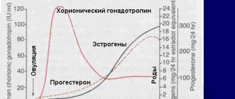
In this article we will tell you:
- Symptoms of anisocoria
- Classifications of anisocoria
- Physiological and pathological anisocoria
- Congenital and acquired anisocoria
- Diagnosis and treatment of anisocoria
Anisocoria is present in 20% of the world's population, however, in most cases it is a physiological disease. A person with this diagnosis has pupils of different diameters
. It looks unusual, but physiological anisocoria does not pose a health threat.
It’s another matter when the condition is accompanied by unpleasant symptoms: photophobia, double vision, pain, decreased visual acuity. In this case, consultation with an ophthalmologist and selection of effective therapy is indicated. Pupillary anisocoria may be an important diagnostic criterion indicating ocular or nervous system involvement. The disease can occur at any age, most often in young females. If a child suffers from the pathology, there is a high risk of developing various refractive errors.
About physiological and pathological anisocoria, signs, causes of anisocoria in adults and treatment methods - in our article.
So,
anisocoria
is an ophthalmological symptom in which a person’s pupils are asymmetrical (that is, they have different diameters)
. In this case, one eye functions normally, and the other reacts to light in a pathological way, not expanding or contracting as in a normal, healthy state.
Symptoms of anisocoria
Anisocoria can be asymptomatic, but can bring many unpleasant minutes, causing:
- dizziness, headaches;
- decreased visual acuity, appearance of spots before the eyes, diplopia (double vision);
- nausea leading to vomiting;
- motor dysfunctions: paresis and partial paralysis, hand tremors;
- impaired coordination of movements.
In addition, anisocoria leads to:
- increased eye fatigue, especially during exercise;
- drooping, drooping of the upper eyelid – so-called ptosis;
- corneal edema, pain;
- deterioration of eyeball mobility;
- protrusion of the eyeball forward. This phenomenon is called “proptosis”.
Reasons for the development of pathology
Many factors cause different pupil sizes. The causes in adults are most often associated with external factors. The most common causes of anisocoria are:
- Traumatic injuries to the eyeball, which also lead to an abnormal shape of the pupil.
- Infectious diseases affecting the brain: meningitis, meningoencephalitis, syphilitic brain damage.
- Regular drug use, which has a detrimental effect on the parasympathetic nervous system.
- Pathological conditions affecting nerve and visual fibers.
- Diseases of the visual apparatus of an inflammatory nature, as well as those associated with constant fluctuations in intraocular pressure: iridocyclitis, glaucoma, keratitis.
- Malignant brain tumors, accompanied by the development of hematoma.
- Cerebrovascular accidents: stroke.
- Genetic predisposition.
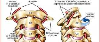
- Pathologies of the cervical area that provoke compression of the cervical plexus: plexitis, osteochondrosis.
The disease can also be a consequence of enlarged lymph nodes in the neck, thrombosis of the carotid artery.
Classification of the disease
The pathological condition can be congenital or acquired. There is also an ocular or non-ocular form, which depends on the cause of the development of the pathology.
According to the degree of spread of the disease, dilation of the pupil is distinguished as unilateral, when only one eye is affected, and bilateral, when the disease affects both eyes. Quite a few cases of bilateral anisocoria have been recorded. Doctors say that such patients account for one in a hundred patients.
Symptoms of anisocoria
Dilation of the pupil of one eye, the causes of which are described above, most often does not bother the patient until a certain point, until the disease begins to manifest itself.
- The first sign is the inability of one pupil to react to light, which most often appears in a dark room.
- One pupil is larger than the other and the difference is up to 1 mm.
- A common symptom is an irregularly shaped pupil.
- Blurred vision.
- Signs of ptosis (drooping of the upper eyelid).
- Increased sensitivity of the eyes to light.
- Headaches, nausea.
- The affected pupil does not respond not only to mechanical damage and bright light, but also to drugs that are instilled to achieve mydriasis.
Quite often, anisocoria and heterochromia (different eye colors) are concomitant pathologies.
Diagnostic methods
First of all, the ophthalmologist must determine which eye is affected, as well as whether the pupil is wide or narrow, the cause of the pathology, and differences in size. During the collection of anamnesis, the specialist learns from the patient about the deterioration in the quality of vision and the presence of double vision and pain in the eyes. The following methods are required in the diagnosis.
- Examination of the eyeballs in a special slit lamp.
- General and biochemical blood tests from veins and capillaries.
- Magnetic resonance and computed tomography of the skull.
- X-ray examination of the skull and cervical spine to detect compression of the nerve roots.
- Repeated measurement of blood pressure at certain intervals.
- Spinal tap to examine spinal cord fluid.
If the ophthalmologist suspects that the pathological condition is caused by abnormalities of the blood vessels, additional angiography and ultrasound examination are prescribed.
Classifications of anisocoria
There are several classifications of this condition.
Physiological and pathological anisocoria
With physiological anisocoria, the difference in pupil size is observed more often at rest, and the diameter of the affected pupil does not differ from the healthy one by more than 1 mm and does not depend on the level of illumination.
Physiological anisocoria is characterized by the disappearance of symptoms when using special drops that dilate the pupil.
Pathological anisocoria signals a malfunction in the body: ophthalmological or neurological diseases. The pupil can contract and dilate depending on the brightness of the light.
Congenital and acquired anisocoria
Congenital anisocoria is a consequence of genetic diseases, disorders in the embryonic development of the nervous or muscular apparatus of the eyes. Often accompanied by strabismus and may disappear with age. If the condition does not go away, it usually does not affect the quality of vision in adulthood.
Causes of acquired anisocoria:
- neurology;
- disturbances in the functioning of the nervous system;
- migraine (impaired pupil symmetry occurs as a result of swelling of brain tissue);
- vascular diseases of the brain, strokes, cerebral infarctions (in this case, anisocoria is one of the symptoms and is usually accompanied by increased blood pressure, vomiting, nausea, headache, impaired motor function, loss of coordination);
- injuries of the iris, ligaments of the eye apparatus, brain;
- foreign body getting into the eye;
- oncological diseases of the brain;
- eye surgery (for example, cataract removal);
- inflammatory eye lesions (iritis, iridocyclitis);
- brain infections (meningitis, encephalitis, etc. - with these diseases the functionality of the nuclei of the optic nerves suffers);
- taking certain medications;
- drug use;
- tuberculosis of the upper lungs;
- glaucoma;
- paralysis and paresis of the oculomotor nerve of various origins.
In addition, the cause of different pupil sizes in an adult can be:
- Horner's syndrome. This symptom complex is characterized by lesions of the sympathetic nervous system. In addition to ophthalmological ones, it also causes vascular tone disorders and sweating.
- Argyle Robertson syndrome. The cause of the phenomenon in which the pupils stop responding to changes in lighting is often infection of the organ of vision due to neurosyphilis and neuropathy of diabetic origin.
- Holmes-Adie syndrome. With Adi syndrome, abnormal mydriasis (dilation) of the pupil is observed, accompanied by impaired sweating, twitching of the limb, and farsightedness. Occurs due to viral or bacterial inflammation of postganglionic fibers.
- Parinaud's syndrome. The cause of the disease is damage to the posterior parts of the midbrain due to tumors, injuries, and multiple sclerosis.
How to treat anisocoria
Physiological anisocoria does not affect vision or eye health. Therefore, it does not require treatment.
With pathological anisocoria, the cause of the appearance of different pupils is first identified. Then they carry out treatment.
For example, for a brain infection, treatment is carried out in a specialized hospital. A course of antibiotics and antiviral drugs is prescribed.
Head tumors and aneurysms of the head vessels require surgical treatment.
For glaucoma, treatment is aimed at normalizing eye pressure and preventing the development of glaucoma attacks.
In case of inflammatory eye diseases, a course of treatment with antibiotics is carried out.
For eye tumors, surgical treatment is indicated.
Diagnosis and treatment of anisocoria
An ophthalmologist will be able to make a diagnosis of “anisocoria” based on examination, studying the reaction of the pupils in the dark, in the light, studying the speed of their reaction, and symmetry under different conditions.
In addition, a biomicroscopic examination, diascleral transillumination using a diaphanoscope, and, if necessary, angiography, ultrasound, MRI and CT will be performed. The doctor can also use mydriatics - special drops that can cause artificial dilation of the pupil.
There are also special tests - tropicamide and phenylephrine and pilocarpine - to help make the diagnosis.
Physiological anisocoria, which does not cause any problems to the patient, does not require treatment. Only a cosmetic defect can cause inconvenience.
For anisocoria associated with diseases of the body, treatment consists of eliminating the underlying disease.
Is treatment required?
It is worth understanding that anisocoria is only a sign. Therefore, first of all, it is necessary to find out the reason for its appearance. So, if the difference in pupil size exceeds 2 mm, I would recommend contacting a neurologist, and also making an appointment with an osteopath. Anisocoria, which does not occur on its own, but has specific and understandable causes (you can read about them above), can only be eliminated through a thorough and thorough multifaceted approach, which involves mandatory treatment, including from a specialist working using special techniques with muscles and bones.
Anisocoria - causes, symptoms and treatment. After injury and without
Anisocoria is characterized by inequality among students. This seemingly harmless disease can cause many diseases. This is why, when detected, it should always be carefully diagnosed and treated. What are the causes and symptoms of this disease? The treatment of anisocoria depends on the answers to these questions.
The width of the pupil can be different for everyone, and there is no need to worry when both pupils are almost the same diameter and the difference in length does not exceed 0.6 mm. These types of changes do not necessarily mean defeat. They affect about 20 percent. healthy population. But when the difference between students is greater, the reasons for such inequality should be quickly diagnosed.
Anisocoria in children and adults: features
A congenital disorder in the functioning of the pupil can be noticed in infants, but this may be a physiological phenomenon that takes place over several years.
Dysfunction of the pupil of one eye can develop as a result of birth trauma or genetic predisposition. If the child’s parents discover that his pupils are unevenly located or of different sizes, then you should consult a doctor and check for concomitant diseases, such as drooping upper eyelid, strabismus, or restriction in the movement of the eyeball.
In children older than one year, the symptom of different pupils may appear as a result of a brain tumor.
In such cases, there is a decrease in the diameter of the pupil in a dark room, although the child’s image clarity does not suffer, he sees well what is far or close. The abnormality of the pupil is manifested by visual impairment, the appearance of fear of light. This should alert the child’s parents, and a visit to the doctor is mandatory.
The causes and diseases that provoke pupils of different sizes can manifest themselves both in young people and in adults after fifty years.
Prescription of medications occurs after a complete examination and identification of the cause of the deviation. The main efforts are directed at treating the underlying disease, a sign of which is a narrowing or dilatation of the pupil in one eye.
Among the prescribed drugs: corticosteroids to relieve inflammation, antibacterial agents that actively affect pathogenic microorganisms.
Anisocoria caused by eye trauma is treated with medications that relax the muscles of the iris. These include drops Irifrin, Atropine. The ophthalmic drug Cyclomed and Midriacil, which belongs to the group of anticholinergics, is used to dilate the pupil.
Among folk remedies, inflammation of the membranes of the eye is relieved by liquid aloe extract used for lotions. An infusion of a mixture of carrots and dry stinging nettle, taken in the amount of two tablespoons, is prepared from half a liter of boiling water. After two hours, drink the drink; such daily treatment will strengthen your eyesight.
Correctly selected treatment will help you get rid of the disease. In some cases, surgical intervention may be necessary.
Causes of anisocoria
The most dangerous causes of the development of anisocoria are considered to be vascular catastrophes of the brain caused by a sharp disruption of blood circulation. This category includes hemorrhagic and ischemic strokes.
When an attack develops, in addition to visual asymmetry, a person experiences headache, nausea, vomiting, tachycardia, and increased blood pressure. The patient loses the ability to move, sensitivity in his limbs decreases, speech and coordination of movements are impaired. At the same time, sweating increases and the skin turns red.
Also, the main causes of the acquired form of the disease include the following:
- traumatic brain injuries that lead to damage to the visual centers in the brain. In this case, the neurons of the retina suffer. As a result, the affected pupil dilates. A person with this diagnosis may experience strabismus;
- traumatic injuries that lead to disruption of the structure of the iris and ligaments. In such a situation, intraocular pressure often increases. This category of factors also includes ophthalmological operations - for example, cataract removal;
- paralysis of the oculomotor nerve - it is responsible for the movement of the eyeball and raises the eyelid;
- various pathologies of the organ of vision - for example, Adi syndrome leads to the fact that the pupil does not respond to light. In this case, glaucoma provokes visual asymmetry associated with damage to the optic nerve;
- tumor formations in the brain. They can be benign or malignant;
- neurological disorders - these include migraine, aneurysm, diabetic neuropathy;
- pathologies of the upper lungs - tuberculosis may be the cause of the problems;
- the use of medications that affect pupil size. This category includes Adrenaline, Atropine, Pilocarpine;
- taking drugs;
- infectious pathologies that are accompanied by pathogens entering the central nervous system. Pathology may accompany tick-borne encephalitis, meningitis, and neurosyphilis.
Reasons for development
Based on their origin, the following forms of pathology are distinguished:
- Congenital and acquired.
- Ocular and extraocular.
The congenital form of narrowing or dilation of the pupil is often associated with an abnormal structure of the muscular or nervous apparatus of the eye. Often accompanied by strabismus or limited mobility of the eyeball. It is diagnosed in an infant from the first days of birth, but often goes away by 5-7 years.
Congenital anisocoria can be physiological, i.e., not directly indicating a pathology of the eye structure. In this case, the difference between the sizes of the pupils does not exceed 1 mm, does not affect visual acuity, and during diagnosis no significant damage is detected. According to statistics, a congenital physiological manifestation occurs in every fifth inhabitant of the Earth.
The acquired form develops for a number of reasons:
- Neurological pathologies affecting the conductivity of the visual nerve pathways.
- Ophthalmological diseases.
- Injuries.
- Exposure to various substances.
Neurological pathologies
Of the neurological diseases that lead to different reactions of the pupils to light, meningitis and tick-borne encephalitis are most often diagnosed. In more rare cases, meningoencephalitis or neurosyphilis (extremely rare). The occurrence of a large difference in the diameter of the pupils is associated with damage to the muscular and nervous apparatus of the eye, deterioration of innervation and a decrease in the activity of areas of the brain responsible for the visual organs.
Ophthalmic diseases
Among ophthalmological diseases, the most common cause of the development of anomalies and inconsistency of pupil reactions are infectious or non-infectious iritis and anterior uveitis (iridocyclitis) - inflammation of the iris or choroid. This leads to disruption of the normal functioning of the motor muscles of the eye, their spasm and pathological contraction and dilation of the pupil.
Glaucoma often leads to miosis of one of the pupils, because when the hole contracts, the outflow of fluid from the anterior chamber of the eye improves, which leads to a decrease in intraocular pressure. It is also worth noting the influence of tumors and neoplasms in the area of the visual center, directly on the conducting fibers or in the eye itself. The tumor can compress nerve fibers, leading to:
- Miosis, if sympathetic innervation is insufficient.
- Mydriasis, if parasympathetic innervation is insufficient.
Injuries
Trauma is one of the most common causes of anisocoria. Both damage to the eye itself and traumatic brain injury can lead to a mismatch in the reaction of the pupils. When the eye is injured, primary infectious (traumatic) uveitis or iritis may develop. This will increase intraocular pressure, which will cause the pupil in the affected eye to constrict.
With a traumatic brain injury, the nervous system of the eye or the visual centers located in the cerebral cortex can be damaged. In the first case, there is concomitant esotropia (inward squint) or exotropia (outward squint). When the visual analyzer is damaged, a pronounced dilation of the pupil is observed in the cerebral cortex on the affected side. The same thing can happen with a stroke.
Exposure to substances
Some medicinal substances, including those that are no longer used for medical purposes in the modern world, can cause uneven dilation or constriction of the pupils due to psychotropic effects. The following have a mydriatic effect:
- scopolamine;
- atropine;
- homatropine;
- tropicamide
Other substances that are often used in ophthalmological practice to constrict the pupils after studies and the use of mydriatic drugs have a pronounced pupil-constricting effect:
- pilocarpine;
- physostigmine.
Of the narcotic drugs, cocaine and amphetamine have the greatest activity on unilateral constriction and dilation of the pupils.
Signs of illness in children
However, if a child is diagnosed with congenital anisocoria, especially when it progresses or is combined with neurological symptoms, then it is necessary to contact doctors - an ophthalmologist and a neurologist, who will conduct a detailed examination and be able to confirm or exclude possible pathological processes.
It is especially important to immediately undergo an examination if, together with a change in the size of the pupils, the following phenomena are observed:
- headache;
- decreased visual acuity;
- nausea and vomiting;
- the appearance of blurred images or double vision;
- symptoms of fever;
- photophobia.
The neurological causes that cause such a symptom can manifest themselves in different ways. Increased anisocoria in bright light indicates that the sympathetic innervation of the eye predominates, this is accompanied by mydriasis (dilation of the pupil), this is due to damage to the oculomotor nerve.
Additional symptoms for this disorder are limited eye mobility, double vision, and divergent strabismus. In this case, a larger pupil is abnormal.
Damage to sympathetic innervation manifests itself in increased anisocoria in a dark room. This often occurs when brain stem structures are damaged and may be accompanied by drooping eyelids. At the same time, accommodation and convergence remain normal. An abnormal reaction occurs in the pupil, which is smaller in diameter - it does not dilate in the dark.
Viewed by: 6,144 people.
Anisocoria is a pathology that involves detecting a difference between the parameters of a person’s pupils. Sometimes the deviation in size is accompanied by deformation of one or both pupils. In most cases, with anisocoria, one pupil can narrow and dilate, that is, respond correctly to lighting, while the other has a fixed size.
It is believed that a difference between pupil sizes of up to 1 mm is not a deviation from the norm. A more significant difference is a sign of various pathologies.
Symptom Definition
The pupil is the black area in the center of the iris. Depending on the lighting, it can change its size (from one to six millimeters).
Manifestation of anisocoria
Many factors can affect pupil size. For example, heredity. If one of the family members had anisocoria, then it is possible that it will be inherited. In this case, the pathology does not cause harm and no treatment is required. When exposed to light, the pupils contract, and if the muscles work incorrectly, external signs of anisocoria appear.
If there is any pathology, then anisocoria can be supplemented by such manifestations as:
- Limited movement of the eye or both eyes.
- Ptosis (drooping of the upper eyelid).
- High temperature, febrile condition.
- Headaches, nausea, vomiting.
- Deterioration of visual acuity.
- Double vision of objects.
Anisocoria has three types. It can be physiological, congenital and pathological.
Congenital anisocoria occurs due to the presence of defects in the visual apparatus, developmental disorders or damage to the nervous system.
Pathological anisocoria is associated with various eye diseases, such as glaucoma, uveitis, tumors, as well as general diseases, such as brain tumors, migraines, syphilis and so on.
Symptoms of such changes can be noticed visually. When a child has different pupils, most likely this is a disease from the field of ophthalmology or neurology. The name of this pathology is anisocoria.
As soon as parents notice this deviation from the norm, they should be examined immediately. Anisocoria usually impairs vision and also makes images blurry. You can check for the presence of the disease in the following way: if you turn on the light, you can see that one pupil reacts to it, while the other remains the same. The child usually reacts to many symptoms and tells his loved ones about it.
Symptoms can include darkening or double vision, and in particularly severe cases, sometimes vomiting or nausea. However, different pupils are not always a serious problem; it depends on the difference in their diameter. Under normal conditions, the size should not differ much; if it is no more than 1 mm, this is not considered a pathology.
The physiological form is detected when the pupil difference is 0.5-1 mm. Such a deviation is not considered significant and is an individual characteristic of the child. In this case, no diseases are diagnosed and is considered a fairly common phenomenon, since a fifth of the entire population has this form.
The normal width of each pupil in standard lighting should be from two to four millimeters, in twilight lighting - from four to eight. In this case, the difference between the widths should be no more than half a millimeter. Synchronous changes in pupil sizes are ensured by the autonomic nervous system. At the same time, the parasympathetic narrows them, and the sympathetic expands.
Pathology is currently quite common. According to statistics, about twenty percent of the world's population may have anisocoria. At the same time, it appears more in the dark.
Reasons and features of different pupil sizes
There are several environmental factors and a number of diseases that can affect the diameter of the hole in the iris.
In adults
Among the reasons that influence the development of anisocoria, there are a number of factors:
- Impact of medications. In this case, giving up medications and looking for alternative options will help get rid of the deviation;
- Optic nerve damage;
- Traumatic brain injury causing hemorrhage;
- Drug use (cocaine, tropicamide, etc.).
In children
Pupils of different sizes in a child can be caused by several reasons, which can be divided into three groups.
Physiological
According to statistical data, anisocoria due to physiological abnormalities is diagnosed in every fifth child and goes away without additional therapy by the age of seven. Such asymmetry may appear for the following reasons:
- Use of psychostimulants;
- Stressful situation;
- Emotional outburst or fear;
- Poor lighting in the room where the child spends most of his time.
It is quite simple to understand that pupils have different sizes not due to destructive processes. Shine a flashlight into your child's eyes. If the holes in the iris react identically to changes in light levels, then everything is in order with the baby’s health.
| If one of the pupils does not “respond” to bright light, you should visit a doctor. Perhaps the child has a malfunction in the functioning of internal organs or systems. |
Pathological causes
If anisocoria develops due to a disease, the functionality of one of the pupils is impaired. It “freezes” in a certain position (expanded or narrowed) and does not react in any way to lighting or hormonal release.
Reasons for different pupil diameters:
- Incomplete development of the main organ of the central nervous system. As a result, the functionality of the nerve endings responsible for the movement of the eye muscles and contraction of the pupil is disrupted;
- Traumatic brain injury. If asymmetry occurs after injury, by the nature of the disease it is possible to recognize which part of the brain experiences maximum pressure from the formed hematoma;
- Infectious pathologies in which inflammation is concentrated in the membranes or tissues of the main organ of the central nervous system;
- Damage to the muscle responsible for reducing the diameter of the pupil;
- Neoplasms inside the skull. As they increase in size, they put pressure on the optic nerve. The functioning of the pathways responsible for transmitting the signal to the visual apparatus about the narrowing or dilation of the pupil is also disrupted;
- Anomalies of the autonomic nervous system. Including nerve endings that regulate the ability of the pupil to change diameter.
Genetic causes
If a baby is born with pupils that differ in diameter, or if one of your close relatives suffered from similar asymmetry, there is no reason to panic. In this case, anisocoria is hereditary.
In infants
Different pupil sizes may be due to genetic predisposition. In this case, parents do not need to worry about the health of the baby. He will not have mental or mental retardation. If the difference does not exceed one millimeter, mom and dad can sleep peacefully.
However, if the asymmetry is greater than the specified parameters, there remains a risk that the baby develops pathology:
- Inflammation of the membranes of the brain;
- Congenital dystrophy of the ocular muscles;
- Damage to the cervical vertebrae during childbirth;
- Violation of the structure of the iris;
- Bleeding in the brain.
Causes
The reasons for asymmetrical pupil diameter in a child can be different. This includes physiology, which is quite natural under certain circumstances, and pathology, and a genetic feature that the baby can inherit from one of the relatives.
Physiological
Such completely natural causes of imbalance are usually observed in every fifth child. At the same time, in many children the problem goes away on its own closer to 6-7 years. The dilation of the pupil can be affected by the use of certain medications, for example, psychostimulants, severe stress, strong emotions, fear experienced by the child, as well as insufficient or unstable lighting where the child spends most of his time.
In most cases, there is a symmetrical decrease or increase in pupils relative to the norm, but this does not always happen. And then they talk about physiological anisocoria. It is quite simple to distinguish it from pathology - just shine a flashlight into the child’s eyes. If both pupils react to changes in light, then there is most likely no pathology. In the absence of one pupil, changes in the intensity of artificial lighting indicate pathological anisocoria.
Pathologies
For pathological reasons, one pupil is not just visually larger than the other, the functionality of the pupils changes. The healthy one continues to respond adequately to light tests, to changes in lighting, to the release of hormones (including fear, stress), while the second one is fixed in an abnormal or narrowed position.
Less commonly, the cause lies in underdevelopment of the brain and dysfunction of the nerves that move the extraocular muscles and the sphincter of the pupil.
An acquired problem in children may be a consequence of a birth injury, especially if the cervical vertebrae were injured. Such anisocoria is diagnosed already in a newborn, as is genetic asymmetry of the pupils.
Different sized pupils may be a sign of traumatic brain injury. If a symptom first appears precisely after a fall or a blow to the head, then it is considered one of the main ones in the diagnosis of traumatic changes in the brain. Thus, by the nature of anisocoria, one can determine which part of the brain is subjected to the strongest pressure during a cerebral hematoma or brain contusion.
Other reasons
Other causes:
- Taking narcotic drugs. At the same time, parents will be able to notice other oddities in the behavior of their child (usually adolescence).
- Tumor. Some tumors, including malignant ones, if they are located inside the skull, may well put pressure on the visual centers during growth, and also interfere with the normal functioning of the nerve pathways through which the brain sends a signal to the visual organs to narrow or dilate the pupil depending on the surrounding environment. conditions.
- Infectious diseases. Anisocoria can become one of the symptoms of an infectious disease, in which the inflammatory process begins in the membranes or tissues of the brain - with meningitis or encephalitis.
- Eye injuries. Blunt trauma to the pupillary sphincter usually leads to anisocoria.
- Diseases of the nervous system. Asymmetry in pupil diameters can be caused by pathology of the autonomic nervous system, in particular, the cranial nerves, the third pair of which is responsible for the ability of the pupil to contract.
Diseases that cause anisocoria:
- Horner's syndrome - in addition to the reduction of one pupil, there is a retraction of the eyeball and ptosis of the upper eyelid (drooping eyelid);
- glaucoma - in addition to constriction of the pupil, severe headaches caused by increased;
- the Argyll-Robinson phenomenon is a syphilitic lesion of the nervous system, in which photosensitivity decreases;
- Parinaud's syndrome - in addition to pupil asymmetry, multiple neurological symptoms associated with damage to the midbrain are observed.