The brain is one of the most complex structures studied in physiology. It consists of several parts, each of which is unique and no less difficult for science. The brainstem, which is a part of the brain, seems to be its most interesting component, because responsible for the functioning of many systems. In recent years, scientists have been able to study it in detail and give precise characteristics. Knowledge of the structure and functions of the brain stem will allow you not only to increase your erudition, but also to avoid some diseases associated with the head.
Stem section
The first living beings to appear on Earth had only a medulla oblongata. It was he who provided them with all the necessary instincts that helped them survive. But this is not enough, because... they needed to constantly develop their reflexes and thinking. After some time, new organisms began to be born with larger brains. Such changes occurred shortly before the appearance of man, with whom the formation of the cerebellum occurred. The remaining parts of the brain began to form only hundreds of years later.
The brain stem, which appeared during evolution, was responsible for ensuring respiratory function and blood supply to all necessary parts of the body. As it developed, it began to consist of a huge number of different centers, which began to form a very complex system. Now this section is a necessary part of the brain, without which life is impossible.
It is located between the large opening of the head in the occipital region and the slope of the inner part of the skull. The trunk extends the spinal cord, connecting it with the main one located inside the head. Its length is about 7 cm, and it includes several separate parts that are very important for the body.
Brain stem structure
The brainstem contains a large number of different components, which are combined into three different brain regions:
- The middle one is formed from the left and right legs, as well as the quadrigemina, a section of the organ that guarantees communication with the cerebellum and the pons. The third and fourth pairs of nerve cords emerge from it.
- The pons is a thickened part of the stem organ, from which the 5th, 6th, 7th and 8th pairs of nerve ganglia emerge. This part of the trunk is connected to the base, tegmentum, ventricle and quadrigeminal tract of the main human brain system.
- Oblong is the part of the trunk that resembles an onion, which is separated from the pons by a transverse groove. This section of the trunk produces 9, 10, 11 and 12 pairs of nerve cords. At the same time, it also contains 7 pair nuclei.
The brain stem is characterized by structural features - it contains two types of neurons: dendrites and axons.
They, in turn, are components of the reticulum. The reticular formation is associated with the structure of the central nervous system. This connection is provided by two types of nerve conductors: afferent and efferent. Afferent conductors work along the fibrous system of the trigeminal nerves and spinorecticular tracts, by conducting pain and temperature impulses. The movement begins from the sensory and other parts of the cerebral cortex, along the corticoreticular pathway to the nuclei, which in turn send signals to the cerebellum. Efferent conductors project to the spinal cord along the reticulospinal tract, to the upper part of the brain along the ascending tract in the pons and medulla oblongata. Efferent conductors are also projected into the cerebellum, starting their path in the paramedial, lateral and reticular nuclei.
Anatomical features
The brain is a complex organ that acts as the center of the human nervous system. Scientists estimate that it may contain more than 20 billion different neurons that transmit signals to other parts of the body. The brain stem includes several sections, each of which is responsible for specific functions. There are 5 of them in total:
- Oblong;
- Intermediate;
- Rear;
- Average;
- Finite.
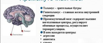
Anatomy also involves the identification of several equally important parts: the cerebral cortex, the cerebellar cortex, the vermis with nuclei, the pons, the thalamus, the hypothalamus, the pituitary gland, the basal ganglia.
The structure itself presents the following picture:
- The medulla oblongata acts as a continuation of the spinal cord, emerging from the vertebral region. It contains two types of substance: white and gray. The function of the first is to conduct information between body systems. The second is the nerve nuclei, which mature by age 7.
- Valoriev Bridge. It is the next section emerging from the medulla oblongata, located in the middle part of the trunk, formed by the base, quadrigeminal, components of the cranial ventricles and tegmentum. Consists of longitudinal and transverse fibers. The former are built from neural clusters, presented in the form of nuclei, from which the latter pass. The latter include the upper and lower layers, through which pyramidal paths are laid.
- Cerebellum. It consists of small hemispheres that are covered with white and gray matter. Reaches its maximum size by the age of 15.
- Midbrain. Attached to the cerebellum by two peculiar legs, it includes 2 visual and 2 auditory sections in the form of separate tubercles through which nerve fibers pass.
- Hemisphere cortex. Between the hemispheres is the corpus callosum, which provides communication between all parts. All thought processes take place in the cortex.
The structure of the brain stem includes another significant section. It is called the reticular formation, which includes dendrites and axons that form the reticulum, which is a special network. The main function of this area is to manage information transmitted from the brain to other parts of the body. There are 2 types of information conduction: afferent, which directs data to the formation, and efferent, which performs the opposite effect.
The brain is well protected. Three shells are responsible for this: soft, hard, arachnoid. Additional protection is provided by the surface of the skull.
Understanding the Brain Stem
The healthy human brain is a master regulator, consisting of 20-25 billion neurons.
They help the central nervous system correctly form complex electrical impulses that control the functioning of the human body. One of the most important parts of a GM is its barrel. It contains cranial formations, which are a bundle of nerve fibers. They, in turn, are surrounded by connective tissue, with respiratory, vasomotor and other centers that ensure the proper functioning of this part of the body.
The human brain stem is made up of different nuclei, each of which helps perform different functions and ensures the proper functioning of other organs through electrical impulses.
The main components of the brain stem are white and gray matter, which are concentrated in the nuclei:
- motor;
- parasympathetic;
- sensitive;
- salivary;
- vestibular;
- cochlear
Each of these nuclei provides the functionality of the brain stem. With the help of willpower, they work correctly, in the way the person himself desires. This organ ensures proper functioning of the entire brain system, saturation with oxygen and the removal of harmful substances from it, since blood vessels of various kinds pass through it. But the main feature of this organ is the large concentration of nuclei and nerve cords, with the help of which a person can sense, feel, hear, smell and see the world. Special attention should be paid to the cores - which made the functionality of the trunk so wide.
Cranial nerve nuclei
One of the most important components of the brain stem is the cranial nerve nuclei, which extend from its base. They are located between the rear and oblong parts, with a small number of them located on the bridge. The nuclei consist of nerve endings that have a direct effect on the trunk. They are presented in the form of branches that penetrate through its most important parts.
Each core has its own purpose. The following nerves emerge from this zone:
- Olfactory;
- Visual;
- Oculomotor;
- Facial;
- vestibulocochlear;
- Block;
- Discharge;
- Trigeminal;
- Glossopharyngeal;
- Sublingual;
- Additional;
- Wandering.
Their full functioning is very important for the human body. Dysfunction of any nerve can have serious consequences that impair quality of life and even lead to death.
Structure and functions
The brain stem consists of sections - medulla oblongata, middle and pons. Sometimes the intermediate section is also referred to as the trunk. Brain structures primarily consist of white matter, within which are grouped areas of gray matter - the basal ganglia, from which the cranial nerves originate. The medulla oblongata is a continuation of the spinal column.
The structure of the brain stem includes centers that control respiratory and cardiovascular activity. The centers regulate unconditioned autonomic reflexes and spontaneous motor activity, for example, maintaining reflexive postures. One of the tasks of the centers is to conduct impulses coming from sensory systems (visual, auditory).
The brainstem includes a reticular structure studied in anatomy called the reticular formation. This is a network consisting of many neurons that form complex connections. The reticular formation runs along the entire section of the trunk. Neurons of the reticular formation provide inhibition or excitation of the peripheral parts of the nervous system, which determines the control of reflex activity.
Neurons in the reticular formation regulate the tone of skeletal muscles, transmitting inhibitory or excitatory impulses to muscle tissue. Another task of the formation within the brainstem is the integration and interaction of the influences of the sympathetic and parasympathetic parts of the nervous system. The main functions of the neurons that form the basis of the reticular formation:
- Somato-motor regulation (maintaining a certain tone of skeletal muscles).
- Somato-sensitive regulation (modification of external stimuli coming from the organs of vision, hearing, olfactory and vestibular systems).
- Viscero-motor regulation (the nature of the activity of the cardiovascular and respiratory systems, the activity of smooth muscles lining the walls of blood vessels and internal organs).
- Neuroendocrine transduction (transfer of active substances - hormones, neurotransmitters).
- Regulation of the physiological state of the body (sleep state, awakening, staying conscious).
Axons extend from the neurons forming the reticular formation in the trunk; one branch enters the spinal cord, the other rises to the intermediate section and higher, to the cortical layer of the hemispheres. As a result, two-way neuronal connections are formed. Neuron dendrites are connected to sensory receptors.
Thanks to the continuous activity of sensory receptors, the neurons of the formation are constantly in a state of excitation. The continuous two-way transmission of nerve impulses causes the excitability of parts of the nervous system. The intermediate portion of the brain, which is often considered the terminal part of the brainstem, consists of the thalamus and hypothalamus, connected to and closely interacting with the pituitary gland.
The multilevel and complex structure of the brainstem determines the variety of functions assigned to this part of the brain. The trunk is a section that provides connection between the cortex of the brain and the spinal cord. The main function of the trunk is to conduct impulses from the cortical parts of the brain to the peripheral parts of the nervous system and back.
Medulla
The oblong section enters the trunk and is a continuation of the dorsal section, which determines its leading role in conducting impulses from the central zones of the brain to peripheral structures. Anatomically it combines the characteristics of the spinal and cerebral substances. This is the caudal (extreme, tail) section of the trunk. The oblong region is formed from white and gray medulla. The nuclei are formed by gray matter. White nerve fibers connect the structures of the spinal cord and brain.
The bottom of the 4th ventricle is represented by the surfaces of the oblongata and pons. In the anterior part of the oblongata there are medullary pyramids (nerve fibers collected in bundles and forming a pyramidal tract), separated by a median fissure. In anatomy, medullary pyramids are structures of the core of the trunk, consisting of motor neurons, forming the corticospinal and corticobulbar tracts, which connect the cortical parts of the brain and skeletal muscles, as well as the muscles of the face and tongue.
The fibers of the corticospinal (pyramidal) tract intersect at the junction of the 2 parts of the brain - the medulla oblongata and the spinal cord, forming a structure known as the pyramidal decussation. On the side of each pyramid there is an olive - a rounded area of gray matter, which is the intermediate section of the vestibular system. Functions of olives:
- Regulation of muscle tone at the reflex level.
- Reproduction of reflex movements under the influence of vestibular loads, for example, due to loss of balance.
- Maintaining relationships with the cerebellum, red nucleus, cortical structures, and spinal cord.
When a person finds himself in an extreme situation, reflex reactions arise at the level of the olive, from which the corresponding impulses enter the spinal cord and peripheral parts of the nervous system. The medial lemniscus is formed by fibers of sensory pathways that terminate in the lateral (lateral) nucleus of the thalamus.
The medial lemniscus is a segment of the conductive tract (bulbo-thalamic tract), which provides proprioceptive sensitivity (muscular-articular sense, sensation of the position of the body in space). The structure of the medial lemniscus is formed from axons belonging to the cuneate nucleus. The bulbo-thalamic pathway carries impulses that provide the function of touch in the direction of the cerebellum and the parietal lobule located above.
The superior parietal lobule synthesizes complex types of sensitivity, including two-dimensional spatial, localization (in certain areas of the body), discriminatory (exclusive). For example, to determine local sensitivity, the doctor lightly presses his finger on a certain area of the subject’s body. The patient, with his eyes closed, must determine where the doctor pressed and indicate the exact location.
The test for determining discriminatory sensitivity involves the use of a special compass, the legs of which are spread at a distance from 2 mm to several tens of millimeters. The doctor touches an area of the subject’s body with both ends of the compass at once. The minimum distance between the legs is determined experimentally when the subject perceives the touch as separate (separate perception of the touch of each leg).
For example, the norm for the fingertips is 3 mm, for the tongue area 1-2 mm, in the torso area this distance can reach several centimeters. Pseudo-unipolar neurons forming the bulbothalamic tract originate in the spinal ganglia. Peripheral processes of pseudo-unipolar neurons are components of spinal nerves that approach all parts of the musculoskeletal system where they end in receptors.
Pons
The pons is a part of the trunk within the boundaries of the brain, which is responsible for motor and sensory function, ensures integration, which is associated with the harmonious interaction of all departments. In addition, it is in the area of the bridge that the nuclei of the cranial nerves (V-VIII pairs) are located. All descending and ascending tracts run through the Varoliev pons, connecting the pons with the cerebellum, the cortex of the cerebral hemispheres, and the spinal cord.
Midbrain
The midbrain responds to changes in environmental conditions, regulates pain sensitivity, as well as sleep and wakefulness. The brain stem is responsible for regulating body temperature, salivation, gastric juice secretion and swallowing, which makes it important in the development of eating behavior.
Functions
All parts of the brain stem are equally necessary. They provide people with the opportunity to smell, hear sound, understand speech, think about any serious things. If not for them, humanity could have remained in the Stone Age forever.
The functions of the brain stem are reduced to the distribution of information between the brain and the central nervous system. They are provided by nuclei and nerve endings. In this case, the trunk is a physiological connecting step between the spinal cord and the brain. If it is damaged, then signals from the brain will not be able to reach the end point, which will completely eliminate the normal functioning of the human body.
There are several groups of functions that are characteristic of the brain stem. Among them:
- Motor. This includes all actions associated with the muscles of the eyes and eyelids. The function is also responsible for the reflexes of the eyeballs and controls the chewing muscles.
- Sensitive. Ensures the functioning of taste buds, as well as all reflexes that affect the digestive system. Helps transmit signals for swallowing and many other actions, including even vomiting. Additionally responsible for sneezing.
- Parasympathetic. Affects movement and dilation of the pupils, controls the ciliary muscles. Managed by cores, ensuring the execution of a block function.
- Upper salivary. It affects the salivary glands, ensuring timely and necessary formation of saliva.
- Vestibular. Responsible for the functioning of the vestibular apparatus, which helps control body balance and stay on your feet.
- Swallowing. Ensures the swallowing reflex works. Complements the work of the sensitive function.
- Auditory. Transmits information to the cerebellum, is responsible for hearing, as well as recognizing heard sounds.
- Sensory. Gives sensitivity to the skin on the face, analyzes taste and sound, and recognizes vestibular stimuli.
The brain stem has the most important functions. It gives every person the opportunity to hear, feel, see, move, think. All of them are necessary for a full life.
If you distribute individual functions across parts of the brain stem, you get the following:
| Brainstem region | Functions |
| Midbrain | · Functioning of visual and auditory organs; · Management of relevant bodies; · Orientation in space. |
| Medulla | · Reflexes associated with coughing, vomiting, sneezing; · Breath control; · Cardiovascular system management; · Functioning of the digestive tract. |
| Pons | · Providing blood supply to the brain; · Fast transmission of signals between the brain and the central nervous system. |
| Cerebellum | · Coordination of movements, balance; · Muscle tissue tone. |
| Diencephalon | · Function of the thyroid gland; · Control of the adrenal glands. |
The importance of such functions makes us take the condition of the brain stem more seriously. He is no exception and may be susceptible to various life-threatening diseases.
If there are disturbances in one section of the trunk, failures may occur in others, because they are all closely interconnected.
Brain stem functions
The significance of the brain stem functions realized by the nuclei of the cranial nerves:
- motor nucleus, responsible for the work of the muscles of the eye and eyelid, as well as all eye reflexes;
- parasympathetic nucleus or trochlear nerve, responsible for the pupil and ciliary muscle;
- the motor nucleus is located in the pons and monitors the masticatory muscles;
- sensitive nuclei receive impulses from the facial organs, participate in the reflexes of swallowing and sneezing;
- the sensitive nucleus of the solitary tract is responsible for the functioning of the taste receptors of the tongue;
- superior salivary nucleus;
- the vestibular nuclei are involved in maintaining the balance of the body;
- cochlear nuclei - auditory receptors;
- double nucleus - swallowing reflex;
- The nucleus of the solitary tract is sensitive - digestive reflexes.
Thanks to the functions of the brain stem, a person can think, touch, understand speech and sounds, see, and make movements. The brain performs all the vital functions and processes that the human body performs. A person controls his muscles using willpower; the muscles receive impulses from the central nervous system and react with long and short contractions.
It is worth noting that the left hemisphere is responsible for the right half of the body, and the right hemisphere is responsible for the left half.
Anatomical and physiological education is responsible for the perception and analysis of information from the environment. The brain stem is connected to the spinal cord. Its main task is to transmit all instructions from the central nervous system of the brain to all human organs. If a stroke occurs unexpectedly, the thalamic part of the brain, the pons, and the cerebellum are affected.
This part of the brain is also responsible for the reflexes of the facial muscles, eyes, and the important swallowing reflex. As a result of a stroke, severe hemorrhage occurs, as a result of which a hematoma appears, blocking the path of oxygen to the brain, which, as a consequence, leads to atrophy of all internal vital organs of the human body.
Diseases
Like any other organ, the brain can fail. The same applies to its trunk. Most problems become the consequences of injuries or other diseases, and sometimes simply manifestations of age. There are several diseases:
- Stroke;
- Tumor;
- Cysts;
- Chordomas;
- Ischemia;
- Malformation;
- Aneurysms;
- Epidermoids;
- Meningiomas.
Most of them appear extremely rarely. The bulk of reported cases of brainstem lesions are stroke and various tumors. They are the most dangerous and require the highest quality and fastest treatment. But why do they arise?
Causes
A particular disease can develop for many reasons. Those most at risk are those who already have serious brain diseases, lead an unhealthy lifestyle, or suffer from regular stress. But even healthy people can get problems with the brain stem. Violations occur for the following reasons:
- Diseases associated with blood vessels, as well as their damage;
- Traumatic brain injuries;
- Poor circulation;
- Nervous breakdowns, severe stressful situations;
- Extreme sports, as well as extreme sports in everyday life;
- Eating junk food or untreated water;
- Alcohol abuse, smoking;
- Congenital diseases associated with the brain stem.
When any diseases appear, they must be treated immediately. Lack of necessary medical intervention can lead to severe irreversible consequences or death.
Stroke
The most common disease affecting the brain stem is stroke. It is always associated with disturbances in the functioning of blood vessels. As the body ages or certain diseases occur, their walls become thinner and more inelastic, and may become covered with plaques or become completely clogged. Then a stroke occurs, which can lead to death.
There are two types of stroke: ischemic and hemorrhagic. The first is an infarction of the brain stem and is considered extremely dangerous due to blockage of blood vessels and subsequent oxygen starvation of nerve cells. The second manifests itself in the form of hemorrhage in the brain tissue. There is a risk of death in both cases.
Mechanism of action
In most cases, a hemorrhagic stroke occurs like this: first, a blockage of a vessel occurs, and then, under increased pressure, it bursts. When thinning, the vessels can immediately burst or become damaged without the formation of blood clots or any plaques. Immediately after the rupture, severe bleeding occurs in the brain, after which a hematoma appears, limiting the access of oxygen to the neurons. This becomes a failure, the consequence of which is a disruption of the functioning of all body systems.
With an ischemic stroke, severe damage to brain tissue also occurs, which greatly complicates the survival of the patient. After damage, the tissue gradually begins to die. Therefore, it is important to provide medical assistance to the victim as quickly as possible.
Causes
You can prevent a stroke if you try to exclude from your life all the moments that lead to this dangerous phenomenon. Doctors have been able to identify several main factors that increase the risk of cerebral infarction. Among them:
- Diabetes;
- Rheumatism;
- Hypertension;
- Atherosclerosis.
Everyone who is affected by at least one point needs to be as attentive to their health as possible and consult a doctor at the first alarming sensation.
Symptoms
A stroke is always sudden. A person can feel great throughout the day, and at one point hemorrhage occurs. Occasionally, shortly before a stroke, discomfort or pain may appear in the head. Symptoms indicating a cerebral hemorrhage are as follows:
- Dizziness;
- Increased sweating;
- Pale skin color;
- High body temperature;
- Pressure interruptions;
- Cardiopalmus;
- Breathing problems;
- Muscle paralysis.
The brain stem may be significantly damaged, making full recovery impossible. In this case, the development of severe complications associated with other diseases or characteristics of the body cannot be ruled out.
Treatment
Providing prompt assistance is the most important condition for preserving the life of the patient. But even she does not give any guarantees. Approximately 60% of victims die in the first days after a major stroke. In some cases, a person may die within two weeks. Only 20% of stroke survivors survive. If help is provided in the first hour after an attack, there is a chance of successful therapy. However, all consequences can be treated with great difficulty.
Transfer to a hospital is a prerequisite. It will not be possible to treat the victim at home; refusal to hospitalize will lead to death. Treatment involves constant monitoring by doctors and taking medications aimed at:
- Elimination of the formation of blood clots in blood vessels;
- Thinning of blood and existing blood clots;
- Reduced pressure;
- Normalization of cholesterol levels.
Additionally, physiotherapy is prescribed. In severe cases, emergency surgery may be performed. It is required to stop hemorrhage when conventional means do not have the desired effect.
Rehabilitation after successful treatment can take several years. Its duration depends on many factors and is individual in each individual case.
Tumor
The second most common tumor is the brain stem tumor. Some of them can be very dangerous, but most do not require any medical intervention. There are several types of tumors:
- Primary. Appear when brain tissue is damaged.
- Secondary. They are a consequence of other diseases.
- Deforming. They negatively affect the shape of the brain stem, deforming it. They may be located on the stem or some other parts.
- Diffuse. They merge with the brain matter, which creates serious difficulties in treatment. Cases of successful therapy are very rare.
- Parastem. They grow to the trunk, causing deformation.
- Diamond-shaped. Appear in the occipital part of the skull.
- Cerebellar. The cerebellum is affected along with the trunk.
- Exophytic. They form on the cerebellum, then reach the trunk.
Neoplasms develop gradually, increasing in size. Sometimes their growth may slow down or stop completely, eliminating the need for treatment. The reasons for their appearance are various injuries and complications after serious illnesses.
Symptoms
Identifying neoplasms that affect the brain stem is not so easy. If they are small in size, they may not cause any symptoms at all, which creates certain difficulties in diagnosis. By the time a tumor is discovered, as a rule, it has already grown to a large size.
Symptoms that may indicate the growth of a tumor are as follows:
- Headache;
- Dizziness;
- Coordination problems;
- Problems with vision or hearing;
- Disorientation in space;
- Tremor of the hands or head;
- Unstable mood.
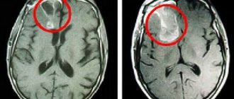
If such symptoms occur, you should consult a doctor. The patient will be scheduled for an MRI, which will determine the presence of a tumor.
Treatment
The prognosis always depends on what kind of tumor the patient has. Its growth rate, size and exact location are of great importance. Benign neoplasms are easily removed surgically, for which an incision is made through which the tumor itself is excised. Malignant tumors cannot be removed using this method, so you will have to give preference to radiation therapy or other methods.
Methods for treating tumors:
- Surgical removal. Excision of the tumor using physical pressure with a knife; it requires making an incision. Suitable only for benign tumors.
- Radiation therapy. X-ray exposure to the tumor through all other structures of the head. Effectively slows down the growth of tumors.
- Stereotactic. A combination of several types of exposure is used, including radiation. It is characterized by the absence of any painful sensations for the patient.
If necessary, doctors can combine several treatment methods at once. This will increase the chances of successful tumor removal.
Drug treatment for tumor development is almost impossible. Cytostatics are the only drugs that can produce the desired effect. They are classified as chemotherapy drugs.
conclusions
The brainstem is the most important part of the brain, as well as the entire body. The general condition of a person depends on his health. The slightest damage can cause serious consequences: loss of hearing or vision, inability to taste food, or maintain balance. The most dangerous is damage to the respiratory center, which leads to respiratory arrest. Prevention of brain stem diseases consists of maintaining a healthy lifestyle, avoiding head injuries and timely elimination of factors that can trigger the pathological process.