What is Arnold-Chiari Malformation?
Arnold-Chiari malformation is a condition in which a part of the brain at the back of the skull, called the cerebellum, protrudes through the foramen magnum into the spinal canal (normally through this opening the spinal cord exits the skull).
This protrusion of part of the cerebellum creates a disruption in the outflow of cerebrospinal fluid (CSF) from the cranial cavity, which can cause increased intracranial pressure with the development of hydrocephalus and symptoms of varying severity. In most cases, this problem is congenital.
In rare cases, malformation may develop at a later age, in which case it is referred to as acquired or secondary malformation.
There are several types of Arnold-Chiari malformation, but the most common is type I, also called primary Arnold-Chiari malformation type I. Although it is congenital, the first symptoms of the disease may appear in infancy or early childhood, but they are most often detected in adolescence or early adulthood.
Get an MRI of the brain in St. Petersburg
Treatment in adults
The main treatment method is surgery. Conservative therapy can only be considered as an additional method. But in most cases, the disease is accompanied by significant neurological disorders, including the development of paresis and other life-threatening complications. In this case, surgical treatment cannot be delayed.
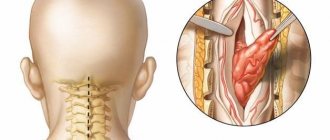
Most often, specialists use craniovertebral decompression. The operation involves removing a bone fragment with widening of the foramen magnum. Specialists also eliminate signs of compression of neurological structures, remove the cerebellar tonsils and parts of the first two cervical vertebrae. To restore the normal flow of cerebrospinal fluid, artificial materials and transplants are used, which are sutured into the area of the dura mater as patches.
Carrying out bypass operations
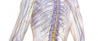
In neurosurgery, shunt systems are actively used to eliminate hydrocephalus. The purpose of this surgical intervention is to restore the outflow of cerebrospinal fluid. Shunts have one-way valves. In the future, it is possible to replace these systems or revise them during repeated neurosurgical interventions.
The outflow of cerebrospinal fluid is achieved by draining the ventricles of the brain. The shunt system can be connected to the right atrium or the abdominal cavity. It is possible to use modern endoscopic methods of surgical treatment. They are characterized by minimally invasiveness and a short rehabilitation period.
With a highly qualified neurosurgeon, operations take place without any complications. The risk of infection increases if the basic rules of infection safety are not followed, the shunt is disconnected, or a sharp decrease in intracranial pressure. By using special shunt systems, such adverse consequences can be avoided.
Causes of Arnold-Chiari malformation type I
The exact causes of Arnold-Chiari malformation type I are not known. The problem may arise during fetal development due to some defect, presumably associated with exposure of the fetus to harmful substances. But it is possible that the appearance of the disease is caused by heredity and genetic mutations.
Arnold-Chiari malformation type I occurs after birth. Its causes are the flow of cerebrospinal fluid into the lumbar or thoracic spine. This usually occurs due to injury, infection, or exposure to harmful substances.
Causes of the disease
According to a number of authors, Chiari disease is an underdevelopment of the cerebellum, combined with various abnormalities in parts of the brain. Arnold Chiari malformation grade 1 is the most common form. This disorder is a unilateral or bilateral descent of the cerebellar tonsils into the spinal canal. This can occur as a result of downward movement of the medulla oblongata; often the pathology is accompanied by various disorders of the craniovertebral border.
Clinical manifestations can occur only in the 3rd–4th decade of life. It should be noted that the asymptomatic course of ectopia of the cerebellar tonsils does not require treatment and often appears accidentally on MRI. To date, the etiology of the disease, as well as the pathogenesis, are poorly understood. A certain role is assigned to the genetic factor.
No ads 1
There are three links in the development mechanism:
- genetically determined congenital osteoneuropathy;
- trauma to the stingray during childbirth;
- high pressure of cerebrospinal fluid on the walls of the spinal canal.
The disease may be caused by hereditary disorders
Symptoms and manifestations of Arnold-Chiari type malformation
The main manifestations of malformation are:
- Syrinx. This formation is a fluid-filled cavity in the spinal cord (syringomyelia) or cysts. As it grows, this formation puts more and more pressure on the spinal cord. In some cases, this can lead to impaired neuromuscular functions and weakness of the limbs. Difficulty moving or breathing. To clarify this diagnosis, patients are prescribed magnetic resonance imaging.
- Scoliosis. In children under 16 years of age, in whom the formation of the spine has not yet completed, as a result of the appearance of cavities, lateral curvature of the spine (scoliosis) may occur.
- Headache . Children up to and including adolescence with undiagnosed Arnold-Chiari malformation type 1 may experience headaches localized to the back of the head and neck, aggravated by exercise.
- Sleep apnea. With this symptom, a short-term cessation of breathing is observed during the patient’s sleep. The presence of this symptom can be determined during a sleep study of the patient.
Other symptoms include hoarseness, difficulty breathing, rapid side-to-side eye movements, muscle weakness, difficulty maintaining balance, abnormal reflexes, and neurological problems including paralysis.
Tests and diagnostics
At the first signs, it is necessary to undergo a neurological examination and assess the severity of clinical and functional disorders. To confirm the diagnosis, the following are most often used:
- neuroimaging techniques, most preferably MRI;
- electroencephalography;
- radiography of the skull;
- myelography , which allows you to identify defects in the upper cervical spinal cord, brain stem, as well as the localization of the cerebellum below the Chamberlain line in the foramen magnum.
MRI results for Arnold Chiari malformation with syringomyelic cyst and normal
Diagnosis of Arnold-Chiari malformation type I
If the disease is asymptomatic, it can be detected during examinations (computer or magnetic resonance imaging) prescribed by a doctor for other diseases. If there are symptoms and suspicion of Arnold-Chiari malformation type 1, the doctor will refer the patient for a CT or MRI. A CT scan creates a three-dimensional image of an area of the body using X-rays, while an MRI uses a powerful magnetic field for this purpose. The resulting images are processed using a computer, creating three-dimensional visualization.
Get an MRI of the brain in St. Petersburg
Treatment of Arnold-Chiari malformation type I
Typically, the treatment method for the disease depends on the symptoms and their severity, the general health of the patient and his age:
- If there are no symptoms, doctors carefully monitor the patient's health with frequent examinations or regular MRIs.
- If symptoms occur, your doctor may prescribe pain medication or suggest surgery to relieve pressure on the brain and restore normal flow of cerebrospinal fluid.
- If you have few or no symptoms but have fluid-filled cavities, your doctor may order an MRI with contrast. This type of MRI is excellent for examining the flow of cerebrospinal fluid and identifying areas where these flows are blocked. If symptoms worsen, surgery may be suggested.
- If there are signs of sleep apnea, a sleep study is performed, based on which the doctor develops a further treatment plan.
Treatment of Chiari malformation
Treatment of Chiari malformation can be conservative or surgical. In some cases, when the pathology is not accompanied by severe symptoms, treatment may not be required at all. If patients who have been diagnosed with pathology complain of pain in the neck and occipital region, as well as other unpleasant symptoms, then they are prescribed drug treatment. Therapy for Chiari malformation involves taking muscle relaxants, analgesics and anti-inflammatory drugs.
However, conservative treatment is not always effective. Usually, if a patient has neurological disorders such as impaired muscle tone and sensitivity, disorders of the cranial nerves, or paresis, doctors are forced to resort to surgical treatment of the disease. One of the most common operations used to treat pathology is craniovertebral decompression.
Second opinion for Arnold-Chiari malformation type I
Despite the fact that the malformation is clearly visible on MRI and CT, errors often occur during these examinations. Some of them are related to the use of outdated equipment or its poor performance. These are objective reasons. Subjective reasons include the fact that doctors often cannot make the correct diagnosis based on MRI and CT. For example, the diagnosis of Arnold-Chiari malformation is often made in children with a simple low location of the cerebellar tonsils (a normal variant), or can be confused with other anomalies of the craniospinal junction. In addition, the description of MRI images may be performed inaccurately and incorrectly by the radiologist.
If we talk about experience, then in a city of half a million there may be an average of 25 people with Arnold-Chiari malformation, and doctors rarely observe this condition. Therefore, when looking at the results of CT and MRI, they may assume diseases that are more familiar to them, which will lead to incorrect treatment, wasted money, wasted time and health.
In order to avoid misdiagnosis, it is vital for the patient to obtain a second opinion on the scan results and it is advisable that this opinion be from a doctor of higher qualifications than the doctor who gave the first opinion.
The National Teleradiological Network (NTRS) provides every patient with free access to obtain an alternative opinion from leading specialists in the field of radiology (radiodiagnostics) and magnetic resonance imaging. No matter where you are, all you need is access to the Internet and scan results in electronic form. Upload them to our server and within a day you will receive the most qualified conclusion that can be obtained in our country.
NTRS is your opportunity to avoid misdiagnosis and treatment.
Sonographic diagnosis of Arnold-Chiari malformation in the fetus
Ultrasound machine HM70A
Expert class at an affordable price.
Monocrystal sensors, full-screen display mode, elastography, 3D/4D in a laptop case. Flexible transformation into a stationary scanner with a cart.
The material for this study was 55 ultrasound examinations in 55 pregnant women at 15-40 weeks of gestation with verified Arnold-Chiari syndrome in the fetus. A number of features have been identified in the echographic image of the brain in the presence of this anomaly at different stages of gestation. An isolated analysis of echographic criteria for the defect was carried out depending on the scanning plane and gestational age. The significance of the known echographic signs of meningomyelocele in the fetus (“banana”, “lemon”) has been shown to decrease with increasing gestational age. New, in the author's opinion, more informative criteria for Arnold-Chiari malformation, identified in horizontal planes, have been proposed (difficult visualization of the cerebellum, expansion of the suprapeal recess of the 3rd ventricle, a special shape of the posterior sections of the body of the lateral ventricle, as well as ventriculomegaly without expansion of the anterior sections of the 3rd ventricle ventricle). The special information content of the sagittal scanning plane is indicated, which allows in 95% of cases to identify the main morphological sign of Arnold-Chiari anomaly - herniation of the cerebellum into the foramen magnum and elongation of the brain stem. The exceptional importance of examining the fetal brain for identifying Spina Bifida has been shown.
Introduction
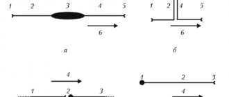
An anomaly of brain development - Arnold-Chiari malformation (syndrome) was first described in 1986 [1]. Modern pathomorphology identifies three main types of this anomaly: I - penetration of the cerebellar tonsils into the cervical spinal canal; II - herniation of the dysplastic cerebellum into the foramen magnum in combination with elongation of the brain stem; III - isolated total displacement of the hindbrain structures into the enlarged foramen magnum, accompanied by the formation of a hernia (Fig. 1). Type I is usually not accompanied by damage to the spinal cord and is detected more often in adults using CT and NMR [2]. Types II and III of the defect are characterized by high mortality in the perinatal period or early childhood [2, 3]. According to autopsy data, in children with meningomyelocele, type II Arnold-Chiari malformation is detected in 95-100% of cases [2, 3] (Fig. 2, 3).
Rice. 1.
Diagram of different types of Arnold-Chiari malformation in the sagittal plane.
Rice. 2.
Pregnancy 23 weeks. Arnold-Chiari malformation type II. View of the posterior cranial fossa in the fetus after elimination (marked by arrows).
Rice. 3.
Fetus with Arnold-Chiari malformation at 19 weeks. Meningocystocele in the sacral region of the fetus after elimination.
The exact frequency of Arnold-Chiari syndrome is unknown, but meningomyelocele occurs in 1-4 cases per 1000 births [4, 5], occupying one of the first places in the structure of central nervous system anomalies. Negative results in attempts to treat children with meningomyelocele may be explained by the presence of brain dysontogenesis [2]. In this regard, the role of prenatal determination of not only spinal cord herniation, but also assessment of brain development in the fetus in order to identify or exclude Arnold-Chiari defect is increasing.
Most publications by both domestic and foreign authors devoted to the ultrasound diagnosis of CNS defects in the fetus note significant difficulties in identifying meningomyelocele, especially in the second trimester of pregnancy [6-8]. When mentioning the Arnold-Chiari malformation, significant difficulties in typing and diagnosing the defect are also usually indicated [8, 9]. At the present stage, possible echographic signs of Arnold-Chiari anomaly include internal hydrocephalus [8, 9], as well as the typical “lemon” shape of the brain skull and the “banana” image of the cerebellum [4, 9]. However, according to some researchers, these signs are not highly specific in relation to Arnold-Chiari malformation [7]. The purpose of this study was to determine reliable ultrasound criteria for diagnosing Arnold-Chiari malformation in the fetus, as well as to develop rational methodological scanning techniques when this defect is suspected.
Materials and methods
The material for this study was 55 ultrasound examinations in 55 pregnant women at 15-40 weeks of gestation with verified Arnold-Chiari syndrome in the fetus. Mothers' ages ranged from 18 to 38 years (mean age 26.4 years). Fathers' ages ranged from 21 to 46 years (mean age 29.6 years). In 23 cases (41%) the patients were primigravida. Multigravida primiparas - 5 cases. Multiparous women accounted for 49.1% (27 people), of which 9 children had a history of developmental defects of the central nervous system (anencephaly, Spina Bifida, Smith-Lemli-Opitz syndrome, hydrocephalus). 18 women had a history of healthy children.
After an echographic examination, in 50 cases the pregnancy was terminated for medical reasons at 20-34 weeks of pregnancy. In 5 women, pregnancy ended in the birth of a live child with meningomyelocele. In all cases of live births, children died between 12 days and 5 months of age. In all cases, a pathological examination was performed. Spina Bifida was noted in 54 cases, of which lumbosacral localization was 52 (94.5%), cervical - 3 cases (5.5%). Internal hydrocephalus was found in 44. 2 fetuses had agenesis of the corpus callosum, 1 had clubfoot, diaphragmatic hernia and intestinal atresia, 2 had microcephaly, 2 had external hydrocephalus, and 3 had porencephalic cysts of the brain. Abnormal development of the posterior cranial fossa and cerebellum, characteristic of Arnold-Chiari malformation, was noted in all cases.
Ultrasound examination of fetuses was carried out using various ultrasound devices using both transabdominal and transvaginal approaches with sensors with a frequency of 3.5-7.5 MHz. After a routine obstetric ultrasound examination, all fetuses had their brain scanned using three mutually perpendicular planes (horizontal, frontal, sagittal), and the features of the echographic picture were noted in each plane.
results
To clarify the possible diversity of echographic images of the brain in fetuses with Arnold-Chiari malformation at different stages of pregnancy using three scanning planes, all examinations were divided into three groups: 1 - fetuses during pregnancy 15-22 weeks of gestation (n=18), 2 - 23 -28 weeks (n=17), 3 - 28-40 weeks (n=20). The identified features in the echographic image of the brain in fetuses with Arnold-Chiari syndrome using the horizontal, frontal and sagittal planes in separate groups are presented in Table. 1-3.
During an echographic examination of the spinal cord, Spina Bifida was detected in 53 fetuses (3 - cervical localization, 50 - lumbosacral localization). The size of the defect ranged from 1 to 5 cm in length. However, no relationship was found between the size of the spinal cord defect and the echographic image of the brain. In 1 case, a spinal defect in the fetus at 17 weeks of pregnancy was not detected due to the patient’s severe obesity and the position of the fetus in the posterior view. Taking into account the anatomy of the posterior cranial fossa, as well as the degree of brainstem elongation, the study showed the possibility of diagnosing the type of Arnold-Chiari malformation.
Type II Arnold-Chiari malformation was found in 52.1 types - in type 2 and type III - in 1 fetus (Fig. 4-6). In this case, the sagittal plane was the most informative. The accuracy of echographic detection of the type of defect in all observations coincided with the pathological data. When diagnosing type III Arnold-Chiari malformation, the main criterion, along with pronounced elongation of the brain stem, was the identification of a hernia in the occipital region, which was formed due to the expansion of the foramen magnum. When examining fetuses with the presence of type I syndrome, the severity of elongation of the brainstem was insignificant; only the absence of the cistern magna and subarachnoid space in the posterior part of the upper parts of the spinal canal attracted attention. Diagnosis of type II defect was the most accessible due to the pronounced elongation of the brainstem, as well as the absence of images of the cistern magna and the subarachnoid space of the upper part of the cervical spinal canal.
Rice. 4.
Pregnancy 37 weeks. Arnold-Chiari malformation type I. Sagittal scanning. Absence of a large tank (marked by an arrow).
Rice. 5.
Pregnancy 23 weeks. Arnold-Chiari malformation type II. Enlarged posterior parts of the 3rd ventricle (marked with an arrow).
Rice. 6.
Pregnancy 17 weeks. Arnold-Chiari malformation type II. Frontal scanning. Deformation of the interhemispheric fissure (1). The presence of an additional lateral wall of the lateral ventricle (2).
As a result of echographic examinations of the brain of fetuses with Arnold-Chiari malformation in horizontal planes (see Table 1), ventriculomegaly, namely an increase in the width of the posterior sections of the body of the lateral ventricle, was found in 41 cases. It should be noted that the expansion of the bodies of the lateral ventricles in our studies was not always accompanied by the expansion of the 3rd ventricle at 36-40 weeks of pregnancy. In all observations, the expansion of the anterior sections of the 3rd ventricle was combined with an expansion of the width of the posterior sections of the body of the lateral ventricle by more than 2 cm.
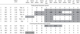
Table 1.
Echographic detection of some features of the brain in a fetus with Arnold-Chiari malformation in horizontal planes.
| Sign of a defect (feature) | Number of cases and percentage to the number of observations in groups | ||
| 1 (n=18) | 2 (n=17) | 3 (n=20) | |
| "Banana" | 3 (16,6%) | 2 (11,7%) | 0 (0%) |
| "Lemon" | 5 (27,7%) | 2 (11,7%) | 0 (0%) |
| "Banana" + "Lemon" | 3 (16,6%) | 0 (0%) | 0 (0%) |
| Ventriculomegaly (width of the posterior parts of the body of the lateral ventricle > 1.0 cm) | 11 (61,0%) | 13 (76,4%) | 17 (85,0%) |
| Dilatation of the 3rd ventricle: anterior parts of the posterior parts | 0 (%) 9 (50,0%) | 1 (5,8%) 7 (41,1%) | 5 (25,0%) 10 (50,0%) |
| Lanceolate shape of the posterior parts of the body of the ventricle | 8 (44,0%) | 7 (41,1%) | 11 (55,0%) |
| Difficulty visualizing the cerebellum | 12 (66,0%) | 15 (88,2%) | 19 (95,0%) |
Enlargement of the posterior portions of the 3rd ventricle was found in 26 fetuses in the present study. The detection of a rhomboid structure in the area of the quadrigeminal cistern (see Fig. 5), corresponding to the expansion of the suprapeal recess of the 3rd ventricle, was combined with either the complete absence of hydrocephalus or with ventriculomegaly, when the width of the posterior sections of the body of the lateral ventricle was less than 2 cm. It should be noted that that expansion of the posterior sections of the 3rd ventricle in fetuses with Arnold-Chiari malformation was noted in half of the cases in each group (see Table 1).
Without a clear correlation with the gestational age in fetuses with Arnold-Chiari malformation, horizontal scans revealed a characteristic, pointed posterior (“lanceolate”) shape of the posterior sections of the body of the lateral ventricle, which in the absolute number of observations correlated with characteristic changes in the shape of the lateral ventricles during frontal scans.
When examining the fetal brain in the frontal planes, the body of the lateral ventricle along the length from the foramen of Monro to the triangle acquired an additional lateral wall. It should be noted that there is a tendency for the appearance of this sign to become more frequent in fetuses with increasing gestational age (see Table 2). A similar trend was also observed in the echographic detection of asymmetry in the location of the choroid plexuses of the lateral ventricles (Fig. 6). Curvature of the corpus callosum and asymmetry of the grooves of the medial surfaces of the hemispheres were more often observed in fetuses in groups 2 and 3. The absence of septum pellucidum did not correlate with gestational age.
Table 2.
Echographic detection of some features of the brain in a fetus with Arnold-Chiari malformation in the frontal planes.
| Sign of a defect (feature) | Number of cases and percentage to the number of observations in groups | ||
| 1 (n=18) | 2 (n=17) | 3 (n=20) | |
| Atypicality of the subarachnoid spaces of the frontal lobes | 6 (33,3%) | 13 (76,4%) | 16 (80,0%) |
| Change in the shape of the lateral ventricles | 8 (44,4%) | 12 (70,5%) | 19 (95,0%) |
| Asymmetry of the location of the choroid plexuses of the lateral ventricles | 11 (61,1%) | 13 (76,4%) | 15 (75,0%) |
| Curvature of the corpus collosum and asymmetry of the grooves of the medial surfaces of the hemispheres | 3 (16,6%) | 5 (29,4%) | 9 (45,0%) |
| Lack of transparent partition | 2 (11,1%) | 3 (17,6%) | 3 (15,0%) |
Frontal scans through the frontal lobes of fetuses with Arnold-Chiari syndrome in groups 2 and 3 (see Table 2) revealed frequent atypical images of the lateral subarachnoid spaces of the frontal lobes, which manifested themselves in a sharp narrowing or even their complete absence at gestational ages of less than 34 weeks.
A sharp elongation of the cerebral peduncles and pons, noted during sagittal scans in fetuses with Arnold-Chiari malformation, in the vast majority of cases (see Table 3) was characterized by an increase in the distance (2 cm or more) between the posterior surface of the splenium of the corpus callosum and the superior surface of the cerebellar vermis (see Fig. 5). It should be noted that in the presence of type I Arnold-Chiari malformation, the lengthening of the cerebral peduncles and pons was insignificant (see Fig. 4). In all cases of diagnosis of type I Arnold-Chiari malformation, it was revealed, first of all, the absence of the cistern magna, as well as the other signs of this anomaly noted above.
Table 3.
Echographic detection of some features of the brain in a fetus with Arnold-Chiari malformation in the sagittal plane.
| Sign of a defect (feature) | Number of cases and percentage to the number of observations in groups | ||
| 1 (n=18) | 2 (n=17) | 3 (n=20) | |
| Absence of the cisterna magna due to displacement of the cerebellum | 12 (66,6%) | 16 (94,1%) | 19 (95,0%) |
| Sharp lengthening of the cerebral peduncles and pons | 17 (94,4%) | 15 (88,2%) | 19 (95,0%) |
Discussion
Ultrasound diagnosis of CNS malformations in the fetus has significantly increased its potential over the past decade [13]. According to Nelson NL et al. [10], open forms of spina bifida can be diagnosed in the fetus in 88% of cases, and closed forms - in 76%. However, these same authors emphasize that the absolute possibility of ultrasound detection of Spina Bifida during prenatal examinations ranges from 22 to 0%. Apparently, this situation in the diagnosis of one of the most common defects of the central nervous system in the fetus is due to the currently established general system of approach to identifying this anomaly, which includes the echographic detection of signs of “lemon” and “banana”, as well as ventriculomegaly in the horizontal plane in combined with longitudinal and transverse visual examination of the spine.
Analyzing the results of this study (see Table 1), when using horizontal scanning planes, the cumulative detection of such signs of Arnold-Chiari malformation as “lemon” and “banana” was no more than 45% for gestational age up to 22 weeks and 24% for pregnancy up to 28 weeks. Ventriculomegaly (an increase in the width of the posterior sections of the body of the lateral ventricle or atrium by more than 1.0 cm) occurred in 61, 76 and 85% (in groups 1, 2 and 3, respectively). However, according to the American Institute of Ultrasound in Medicine, ventriculomegaly when the width of the posterior sections of the body of the lateral ventricle is more than 1.5 cm is absolutely accurately determined only in 53% of cases [10]. Difficult identification or complete absence of visualization of the cerebellum in combination with the identification of the “lanceolate” shape of the posterior parts of the body of the lateral ventricles and the expansion of the suprapeal recess of the 3rd ventricle, in our opinion, in their information content significantly exceeds other criteria for Arnold-Chiari malformation and meningomyelocele in horizontal scans ( see table 1). Determining ventriculomegaly with no signs of dilatation of the anterior sections of the 3rd ventricle can also provide sufficient assistance in diagnosing Arnold-Chiari malformation. This is indicated both by the data of the present study and by the results of ultrasound examinations in newborns with meningomyelocele [12].
The lack of data in the literature on the use of frontal and sagittal scanning planes in fetuses with Spina Bifida or Arnold-Chiari syndrome makes it somewhat difficult to interpret the results obtained in this study (see Tables 2, 3). However, they do not contradict both pathomorphological ideas about the macroscopic organization of the defect and data from postnatal brain studies using various visual methods [2, 3,11,12].
The results of echographic examinations are given in table. 2 indicate the presence of a number of brain features that can be detected using frontal scans in fetuses with meningomyelocele. Despite the high detectability (see Table 2) of such signs as atypicality of the subarachnoid spaces of the frontal lobes, changes in the shape of the lateral ventricles and asymmetry of the choroid plexuses and grooves, these criteria apparently reflect more secondary general dysontogenetic changes in the brain associated with various variants of abnormal development of the central nervous system [14]. At the same time, identifying such features can contribute to a more accurate diagnosis of an abnormal brain and complement the prognosis picture in each specific case.
The frequency of echographic detection of such signs as the absence of the cisterna magna and elongation of the cerebral peduncles (see Table 3) convincingly showed the advantages of the sagittal plane of brain scanning for diagnosing the main macroscopic sign of Arnold-Chiari malformation - herniation of parts of the cerebellum into the foramen magnum. Obtaining an image of the fetal brain in the sagittal plane in the second trimester of pregnancy, in our opinion, is not a difficult technical task. In this regard, the use of the sagittal plane of brain scanning can be proposed as one of the most informative planes for diagnosing or excluding Arnold-Chiari malformation in the fetus in both the second and third trimesters of pregnancy.
Detection of signs of Arnold-Chiari malformation in the fetus may be an indication to exclude developmental defects of the spine (including invasive ones), while even the complete absence of ultrasound information about the condition of the spine indicates the presence of Spina Bifida in more than 95% of cases.
Thus, the study showed that the use of not only horizontal, but also other scanning planes (frontal, sagittal) makes it possible to identify a number of highly informative criteria for Arnold-Chiari malformation in the fetus. The echographic diagnosis of this defect, using a combination of planes and taking into account the signs listed above, should not present difficulties in the vast majority of cases after the 16th week of gestation.
Literature
- Chiari H. Veber Veranderungen des Kleinhirns, des Pons und der Medulla oblongata in Folge von genitaler Hydrocephalic des Grosshirns. Denkschr Acad Wiss Wein. - N 63. - 1896. - S. 71-116.
- Carmel PW The Chiari malformations and Syringomyelia/Disorders of Developing Nervous System: Diagnosis and Treatment/Ed. Hoffman HV - Boston. - 1986. - pp. 133-135.
- Mc Lone DG, Naidich TP Myelomeningocele / Disorders of the Developing Nervous System: Diagnosis and Treatment/Ed. Hoffman HJ - Boston. — 1986.- pp. 87-107.
- Nicolaides KH, Campbell S, Gabbe SG, Guidetti R. Ultrasound Screening for Spina Bifida: Cranial and Cerebellar Signs. The Lancet. - Vol. 2. - 1986. - pp. 72-74.
- Pilu G., Bovicelli L. Sonography of the Fetal Cranium/Diagnosis and Therapy of Fetal Anomalies/ Ed. Hobbins JC, Benaceraff BR - NY - 1989. - pp. 221-258.
- V. N. Demidov, A. M. Stygar, B. I. Zykin, M. V. Medvedev, A. G. Sichinava, M. A. Fuks, G. L. Doronin. Ultrasound diagnostics in obstetrics // Ultrasound diagnostics. Regulatory materials and methodological recommendations. -M.: 1990.- p. 401-418.
- Pilu G., Romero R., Rizzo N., Gabrielli S., Perot o A., Bovicelli L. Prenatal Diagnosis of Cerebro-Spinal Anomalies / The Principles and Practice of Ultrasonography in Obstetrics and Gynecology / Ed. Fleischer AC - 1991.- pp. 211-228.
- Steven M., Edwards V., Filly R. Diagnosis and Management of Fetal Disorders of the Central Nervous System / Disorders of the Developing Nervous System: Diagnosis and Treatment / Ed. Hoffman HJ - Boston. — 1986.-pp. 67-70.
- Charvenak F. A., Isaacson G., Streltzoff J. Normal Anatomy and Malformations of the Fetal Neural Axis / An Atlas of Ultrasonography in Obstetrics and Gynecology / Ed. Kurjak A. 1992. - pp. 33-48.
- Nelson NL, Filly RA, Goldstein RV, Callen RW The AIUM/ACCR Antepartum Obstetrical Sonographic Guidelines: Expectations for Detection of Anomalies. J. Ultrasound in Med., Vol. 12. - N4. — 1993.-pp. 189-196.
- Voevodin S. M., Ozerova O. E. Echographic anatomy of the brain in newborns of different gestational ages // Obstetrics. and gynecol. - 1991, - N 6. - p. 33-42.
- Voevodin S. M. Sonographic diagnosis of brain malformations in newborns and infants // Pediatrics. - 1990. - N 9. - p. 45-51.
- Pilu G. Prenatal Diagnosis of Central Nervous System Anomalies - 20 Years after. Ultrasound Obstet. Gynecol. - 1993. - Vol. 3. - N 3. - pp. 231-233.
- Voevodin SM Echographic Signs of Cerebral Cortex Anomalies in the Fetus. Ultrasound Obstet. Gynecol. — 1994, Vol. 4. - Suppl. 1 - p. 209.
Ultrasound machine HM70A
Expert class at an affordable price.
Monocrystal sensors, full-screen display mode, elastography, 3D/4D in a laptop case. Flexible transformation into a stationary scanner with a cart.