The vast majority of more than 120 types of primary brain tumors are benign tumors. Their main common differences from malignant ones are slow growth, absence of metastases and relapses. Moreover, they are formed in different places from different types of cells and manifest themselves differently. Accordingly, treatment approaches can also vary significantly.
Benign brain tumors include pituitary adenomas, well-differentiated ependomas, chondromas, schwannomas, meningiomas, choroid plexus papillomas, cysts, lipomas.
Pituitary adenomas
They make up about 10% of primary brain tumors and are most often found in the anterior third of the gland. They can appear at any age, but usually occur in older people. Women of childbearing age develop more often than men of the same age group. There are hormonally active (secreting) and hormonally inactive pituitary adenomas.
Pituitary adenoma
Most formations are hormonally active. In turn, they are classified according to the type of hormone produced, an excess of which in the body leads to the appearance of characteristic symptoms.
Classification of brain tumors by occurrence
There are different types of brain tumors in adults, which are classified according to certain criteria.
First of all, all brain tumors are divided into primary and secondary.
A primary tumor (for example, primary CNS lymphoma) is formed by the brain tissue itself and adjacent to it: tissue of the cranial nerves, meninges, pineal gland, lymphoid tissue or pituitary tissue. The development of these neoplasms is associated with mutations that lead to the appearance of abnormalities in the DNA of brain cells.
The occurrence of secondary brain tumors is most often associated with a metastatic process from other organs affected by cancer.
Meningiomas
They grow not from brain tissue, but from the soft membranes covering it (meninx), which is why they got their name. They make up about a third of all primary neoplasms. Most often observed in women, starting from middle age (2 times more often than in men). The incidence rate increases after 60 years. They are rare in children. The typical location is in the upper regions, but can also form at the base of the skull. They usually grow inward, causing compression of adjacent parts of the brain. Sometimes they grow outward, which is accompanied by thickening of the cranial bones in the problem area. May contain calcifications, fluid-filled cavities (cysts), and vascular nodes.
Schwannomas
Develop from Schwann cells of the auditory nerve, also called the 8th cranial, acoustic or vestibulocochlear nerve.
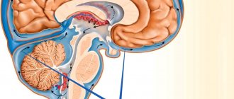
Places of formation of schwannomas
Usually located between the cerebellum and the pons, in the region of the posterior fossa. They grow very slowly, are quite rare (no more than 8%), and develop in middle age. The risk of the disease is twice as high in women.
Choroid plexus papillomas
They arise from mutated cells of the choroid plexuses, which are part of the structure of the membranes lining the ventricles of the brain and producing cerebrospinal fluid (CSF). Typically diagnosed in children and young people.
Traditional sites of formation of choroid plexus papillomas
In children with brain tumors under the age of one year, choroid plexus papillomas range from 10 to 20%, from 1 year to 1 year – 2-4%. At the same time, depending on age, the localization of the lesion also differs: the older the patient, the lower the tumor is located.
Reasons for development
As with cancer cells, the reasons for the development of benign tumors are not fully understood. Predisposing factors are:
- genetic failures in tissues and cells;
- external damaging factors - temperature, radiation;
- exposure to chemicals;
- hypoxia.
However, despite similar risk factors, the mechanism of pathogenesis and growth of benign and malignant tumors is very different, which is the reason for such a large difference in the risk of diseases.
In cancer, tumor growth is invasive, or invading other tissues (spreading). In the case of a benign neoplasm, the growth will be expansive (swollen). If we draw an analogy, in the first case the process is similar to the infection of surrounding tissues by atypical cells, and in the second, it is similar to a local process within the primary tissue.
At the cellular level, malignant tumors have a more atypical structure; the cells are not differentiated and lose their antigens on the membrane. In a benign process, the cells may have a normal appearance, characteristic of the tissue, and grow slowly.
The classification of benign tumors is entirely based on their location - depending on where the tumor is located. Let's look at the most common organs in which tumors can develop.
Cysts
Although they are considered benign, they can cause serious problems when located in structures that control vital body functions.
Cysts can appear in a variety of areas of the brain
They are varied, the most common are arachnoid (filled with fluid), colloid (filled with a thick iron-like substance), dermoid and epidermoid (filled with soft tissue) cysts.
Symptoms of a benign brain tumor
It is necessary to immediately make a reservation: it is impossible to determine by any external signs that a benign and not a malignant neoplasm has appeared in the brain. Moreover, certain disturbances in well-being can only cause the doctor to suspect a tumor process. Only diagnostics can confirm or refute these suspicions. At the same time, based on the patient’s complaints, the neurologist makes preliminary conclusions and draws up an examination plan.
“Even a benign tumor that grows in the head is potentially dangerous,” says Robert Fenstermaker, MD, chairman of the department of neurosurgery at Roswell Park Cancer Center (USA). “The space inside the skull is not unlimited, and even if a brain tumor is benign and grows slowly, it will eventually begin to compress the brain, symptoms will develop and this can be life-threatening.”
Benign brain tumors manifest themselves in a variety of symptoms, the nature of which is primarily determined depending on the location of the lesion and its size.
- An extremely slowly growing chondroma may not show itself in any way for a long time. When the tumor grows in size, it begins to compress nearby structures, which most often leads to headaches, hearing impairment, and visual hallucinations.
- With choroid plexus papillomas, general cerebral symptoms are observed, including characteristic headaches and other signs of increased intracranial pressure.
- It is impossible to identify any general symptoms for cysts due to the wide variety of neoplasms of this type and the possibility of their appearance in a variety of places.
- Ependiomas are characterized by irritability, vomiting, insomnia, and headache. In children under one year of age, one of the first symptoms is often an abnormal increase in head size.
- The symptoms of benign adenoma and malignant adenocarcinoma of the pituitary gland are identical, therefore, when making a diagnosis, other features are taken into account, including the absence of metastases and growth into neighboring sections, scan data, etc. Among the most common manifestations are headache, behavioral disturbances and visual disturbances. Signs of this type of hormone-producing tumors depend on the type of hormone produced.
- Meningiomas grow slowly and can become large before they begin to interfere with normal brain function. Symptoms depend on the location of the tumor. Most often, patients complain of headache, weakness in the arm or leg. Seizures, personality changes, and vision problems may also occur.
- In the vast majority of cases, lipomas do not cause any health problems.
- Schwannomas are characterized by unilateral hearing loss and noise or ringing in the ear. Less often, patients complain of dizziness. If the process affects the 7th cranial nerve, its paralysis may develop. Sometimes there is difficulty swallowing, changes in taste, unsteady gait, and disturbances in eye movements.
Brain tumors: symptoms, risk factors, diagnosis and treatment
Neurosurgery March 23, 2017
A brain tumor is a growth or abnormal growth of cells in or near the brain. There are several types of brain tumors. They can be noncancerous (benign) or cancerous (malignant). Tumors can also develop from brain tissue (primary) or form in other parts of the body and then spread to the brain (secondary or metastatic).
The rate of tumor growth in the brain can vary greatly. The growth rate, as well as the location of the tumor in relation to other brain structures, determines its effect on the functioning of the nervous system.
Treatments for brain tumors depend on the type of tumor, as well as its size and location.
MAIN SYMPTOMS
Signs and symptoms of a brain tumor can vary greatly depending on its size, location, and growth rate. Main signs and symptoms caused by a brain tumor may include:
- The appearance or change of pain in the head
- Headaches that become more frequent and severe
- Nausea or mouth for no apparent reason
- Vision problems such as blurred vision, double vision, or loss of peripheral vision
- Gradual loss of feeling or ability to move arms or legs
- Balance problems
- Speech problems
- Disorientation in time and space
- Personality and behavior changes
- Seizures, especially if they have not happened before
- Hearing problems
When should you see a doctor?
Make an appointment with your doctor if you have persistent signs and symptoms that bother you.
CAUSES OF BRAIN TUMORS
Tumors that arise directly in the brain
Primary tumors arise directly in the brain or in tissues located close to it—the membranes covering the brain (the meninges), the cranial nerves, the pituitary gland, and the pineal gland. Primary tumors form when errors (mutations) appear in the DNA of normal cells. Mutations allow cells to grow, divide quickly, and survive when healthy cells die. The result is a collection of abnormal cells that form a tumor.
Primary brain tumors are less common than secondary tumors, in which cancer appears in other parts of the body and spreads to the brain.
There are several types of primary tumors and each has its own name, depending on the cells that form it. For example:
- Gliomas. These are tumors that form in the brain or spinal cord and include astrocytomas, ependymomas, glioblastomas, oligodendrogliomas, and oligoastrocytomas.
- Meningiomas. A meningioma is a tumor that develops in the membranes surrounding the brain and spinal cord (the meninges). Most meningiomas are cancerous.
- Acoustic neuromas (schwannomas). These are benign tumors that grow on the nerves that control balance and hearing and carry nerve impulses from the inner ear to the brain.
- Pituitary adenomas. These are mostly benign tumors that form in the pituitary gland at the base of the brain. These tumors can affect pituitary hormones, which affect the entire body.
- Medulloblastoma. It is the most common malignant brain tumor in children. Medulloblastoma forms in the lower part of the brain and spreads through the cerebrospinal fluid. These tumors are less common in adults but do occur occasionally.
- Primitive neuroectodermal tumors are rare malignant tumors that develop in embryonic (fetal) cells in the brain. They can appear in any area of the brain.
- Germ cell tumors. Germ cell tumors can occur in childhood, in the area where the reproductive organs form. But sometimes germ cell tumors move to other parts of the body, such as the brain.
- Craniopharyngiomas. These are rare benign tumors that arise near the pituitary gland, which produces hormones and controls many body functions. While a craniopharyngioma slowly grows, it can affect the pituitary gland and other structures located near the brain.
Cancer that starts in other areas of the body and spreads to the brain
Secondary brain tumors (metastases) are tumors that form against the background of a cancerous tumor that has arisen in another organ and spreads (metastasizes) to the brain.
Secondary brain tumors are most common in people with cancer. But, in rare cases, a metastatic brain tumor may be the first sign of cancer in any organ.
Secondary brain tumors are much more common than primary ones. Although any malignant tumor can metastasize to the brain, the most common metastases are:
- Breast cancer
- Colon cancer
- Kidney cancer
- Lungs' cancer
- Melanoma
RISK FACTORS
In most patients with primary brain tumors, their cause is unclear. But doctors have identified a group of factors that increase the risk of developing a brain tumor. Risk factors include:
Age. As you age, your risk of developing brain tumors increases. They are most common in older people. However, the tumor can appear at any age. There are even some types of brain tumors that occur almost exclusively in children. Exposure to radiation. Individuals exposed to ionizing radiation have an increased risk of developing brain tumors. Examples of ionizing radiation are radiation therapy used to treat cancer and radiation exposure from the explosion of atomic bombs. Other types of radiation, such as electromagnetic fields from high-voltage power lines and radiation from cell phones and microwave ovens, have not been scientifically proven to be associated with brain tumors. Family history. A small proportion of brain tumors occur in people with a family history of tumors or genetic syndromes that increase the risk of developing brain tumors.
DIAGNOSTICS
If a brain tumor is suspected, your doctor may recommend a series of tests and procedures, including:
Neurological examination. A neurological examination may include tests of vision, hearing, balance, coordination, stamina and reflexes, among other things. Abnormalities in one or more functions may indicate parts of the brain are being affected by the tumor.
Medical imaging. Magnetic resonance imaging (MRI) is often used to diagnose brain tumors. In some cases, a contrast agent may be injected through a vein in the arm during the examination.
A number of specialized MRI components —including functional MRI, perfusion MRI, and magnetic resonance spectroscopy—can help the doctor evaluate tumors and determine treatment plans.
Other medical imaging studies may include computed tomography (CT) and positron emission tomography (PET).
Tests done to detect cancer in other organs . If it is suspected that a brain tumor may be caused by cancer in other organs, your doctor may order a series of tests and procedures to determine the origin of the cancer. An example would be a CT scan of the chest to look for signs of lung cancer.
Collection and examination of abnormal tissues (biopsy). A biopsy may be part of surgery to remove a brain tumor or may be performed as a needle puncture.
A stereotactic biopsy can be done to examine tumors in hard-to-reach or sensitive areas of the brain that are easily damaged by surgery. The neurosurgeon makes a small hole in the skull and inserts a thin needle through it. The tissue is then removed using a needle, often guided by CT or MRI (neuronavigation).
The collected material is examined under a microscope to determine whether it contains cancerous or benign cells. This is the most important information for making an accurate diagnosis and prognosis, and, most importantly, for determining further treatment.
TREATMENT
Treatment for a brain tumor depends on its type, size and location, as well as the patient's health and preferences. Surgical intervention.
If the location of the brain tumor allows surgery, the neurosurgeon will remove as much of it as possible.
In some cases, tumors may be small and easily separated from surrounding tissue, allowing for complete surgery. In other cases, the tumors cannot be separated from surrounding tissue or are located near sensitive parts of the brain, making surgery very risky. Then the neurosurgeon removes the maximum part of the tumor located in the safe zone.
Even removing part of the tumor helps relieve signs and symptoms of the disease
Surgery to remove a brain tumor carries certain risks, such as infection and bleeding. Other risks depend on the location of the tumor. For example, removing a tumor near the optic nerve risks vision loss.
Radiation therapy
Radiotherapy uses ionizing radiation—X-rays or protons—that destroy the tumor. The beams may come from a machine outside the body (external beam radiation) or, in very rare cases, the radiation source is placed inside the body near a brain tumor (brachytherapy).
The external beam of radiation can be focused only on the tumor localization area or directed to the entire brain. Whole-brain radiation is most often used to treat cancer that has spread from other organs. The side effects of radiation therapy depend on the type and dose of radiation received. Common side effects during or after radiation include fatigue, headache, nausea, and vomiting.
Chemotherapy
Chemotherapy uses drugs that destroy cancer cells. Medicines may be given orally as tablets or may be given into a vein (intravenously). The most commonly used drug to treat brain tumors is temozolomide (Timodal), available in tablet form. Other types of chemotherapy drugs are also available, prescribed depending on the type of tumor.
The side effects of chemotherapy depend on the type and dose of drugs the patient receives. Chemotherapy can cause nausea, vomiting, and hair loss.
RECOVERY AFTER TREATMENT
Because brain tumors can occur in the parts of the brain that control motor skills, speech, vision, and thinking, rehabilitation may be a necessary part of recovery.
A neurosurgeon can use the following rehabilitation methods:
- Physical therapy to restore lost motor skills or muscle strength
- Occupational therapy to help a patient return to normal daily activities and work after treatment for a brain tumor or other illness
- Speech therapy performed by a speech therapist in case of speech impairment (speech pathology).
Author: Danu Adrian, neurosurgeon, highest category
Treatment of benign brain tumor: basic principles and choice of method
In some cases, the doctor may choose active surveillance. This applies to patients with arachnoid cysts and meningiomas, as well as lipomas, which do not cause significant health problems and, as a rule, are detected by chance or during preventive cancer screening.
If a patient is diagnosed with a benign brain tumor that is “unfortunately” located, interferes with the quality of life for other reasons, or has a high risk of malignancy, treatment is aimed at removing the tumor (surgery) or destroying it using modern radiosurgical methods.
For example, some meningiomas can be removed by traditional surgery, while others are inoperable. In the latter case, the tumor can be destroyed using CyberKnife, Gamma Knife, TrueBeam installations. Depending on the characteristics of the tumor, tomotherapy or proton therapy may also be the optimal treatment method.
Operable colloid, epidermoid and dermoid cysts are most often removed in the traditional way or using endoscopic or microsurgical technologies. If indicated, additional antibacterial drug therapy may be prescribed. Sometimes cyst removal is complicated, impossible, or only partially possible.
Radical removal of chondroma, choroidal papilloma, ependyoma, pituitary adenoma can be carried out surgically or radiosurgically.
When treating patients with schwannomas, microsurgical methods or stereotactic radiosurgery are usually used. According to indications, medication, hormonal and radiation therapy are additionally prescribed.
Leading neurosurgeons have high hopes for intraoperative fluorescent control for the purpose of total resection of tumors, in particular pituitary adenomas, since incomplete removal of neoplasm leads to relapses in 20% of patients. But although pituitary adenomas are rarely malignant, they can cause serious problems by compressing nearby areas of the brain, which can lead to Cushing's disease, gigantism, blindness and death.
At the medical school. Perelman University of Pennsylvania developed the first organ-specific marker OTL38, which consists of two parts: folate and an infrared-luminescent dye. Since folic acid is actively used for the development and growth of neoplasms, they are characterized by a high content of folate receptors on the cell surface (which is sometimes more than 20 times higher than the normal level). By binding to receptors, OTL38 causes luminescence of the affected tissues, making it possible to accurately identify the boundaries of the tumor and completely remove it.
As noted by Yu.K. Lee, MD, PhD, professor in the Department of Neurosurgery at the University of Pennsylvania, said, “This study heralds a new era in personalized tumor surgery. Surgeons can now see the molecular characteristics of a patient’s tumors, not just light absorption or reflectivity.”
If you need a second opinion to clarify your diagnosis or treatment plan, send us an application and documents for consultation, or schedule an in-person consultation by phone.
+7 499 490-24-13
Expert opinion
Types of benign skin tumors
New growths on the skin do not necessarily originate from epidermal or dermal cells. Most often this is the growth of adipose, vascular tissue or sebaceous gland. Let's look at these types of tumors in more detail.
Lipoma
Lipoma is a benign tumor of adipose tissue. It is visually localized on the skin, but is located in the subcutaneous fatty tissue. Usually it reaches several centimeters in diameter and growth stops, however, there are known cases of the development of huge lipomas, which significantly reduce the patient’s quality of life. It very rarely becomes malignant, but it is better to remove it even for aesthetic reasons.
Atheroma
A benign tumor that develops due to blockage of the sebaceous gland. Localized on the skin, usually in places rich in sebaceous and sweat glands - armpits, head, back. It does not become malignant, but can fester. The size of the atheroma is on average from a centimeter to 5.
Papilloma
A benign tumor that develops from skin epithelial cells. It is purely viral in nature and is caused by HPV (human papillomavirus). Papilloma usually ranges in size from a few millimeters to 0.5 cm, and is white, flesh-colored, or discolored. The surface may be uneven.
Pigmented nevus
The neoplasm originates from pigment cells - melanocytes. People call them moles or warts. Nevus is dangerous because it can become malignant and turn into melanoma - an extremely dangerous and aggressive oncological disease.
Keratoma
Just like papilloma, it comes from the epithelium, but has a different nature; here the epithelial cells become keratinized and grow. The causes of development are exposure to external aggressive factors on the skin - temperature, chemical burns, and so on. May develop into skin cancer.
Fibroma
A benign tumor arising from the growth of connective tissue under the skin. It mainly occurs due to skin trauma or insect bites. It looks like a dense nodule under the skin, ranging in size from 1 millimeter to half a centimeter.