Causes
Pathology most often develops in newborns, as well as people of retirement age, although people who have not reached it are also at risk of developing this problem. The maximum chance of signs of hydrocephalus appearing is in those who become ill during fetal development. For every 1000 births there are approximately 1-2 such cases.
Several pathological conditions contribute to an increased volume of cerebrospinal fluid (cerebrospinal fluid):
- disruption of outflow due to the formation of traffic jams in the outflow tract;
- deterioration of fluid absorption by arachnoid granulations;
- too much cerebrospinal fluid production.
So far, all the reasons for the development of pathology have not yet been sufficiently studied. Congenital hydrocephalus is often a consequence of a brain defect, due to which there is a deterioration in the circulation of cerebrospinal fluid for the following reasons:
- hemorrhage into the cerebral ventricles of the fetus;
- incomplete healing of the spinal column;
- intracranial hemorrhage;
- infectious diseases;
- traumatic brain injury;
- congenital genetic pathologies;
- central nervous system tumors;
- arachnoid cysts.
Acquired hydrocephalus can develop in any person due to certain diseases or mechanical injuries:
- intracerebral hemorrhage;
- formation of venous blood clots in the brain;
- stroke;
- meningitis;
- traumatic brain injury;
- development of a tumor in the brain.
Sometimes the vessels through which cerebrospinal fluid flows are narrowed at birth. This pathology can be compensated thanks to the internal reserves of the body. Then hydrocephalus does not appear in the newborn, causing signs of concern only as the child grows older.
In older people, hydrocephalus is called a normotensive disease. This is caused by the situation that the volume of cerebrospinal fluid increases without causing an increase in its pressure. In this condition, intracranial edema occurs, which impairs the functioning of the brain. Conditions in which the natural absorption of cerebrospinal fluid is disrupted increase the risk of developing pathology, although more often the mechanism of development of the disease is unclear. There is a higher risk of infectious meningitis in people who have suffered a traumatic brain injury.
Symptoms and diagnosis
Acutely developed occlusive hydrocephalus is manifested by symptoms of increased intracranial pressure:
- headache;
- nausea and vomiting;
- drowsiness;
- congestion of the optic discs;
- symptoms of axial displacement of the brain.
Headache is most pronounced upon awakening in the morning due to an additional increase in intracranial pressure during sleep. This is facilitated by the expansion of cerebral vessels due to the accumulation of carbon dioxide, which is accompanied by blood flow, stretching of the dura mater of the brain in the area of the base of the skull and the walls of blood vessels. Nausea and vomiting worsen and sometimes lead to a decrease in headaches. The most dangerous sign of increased intracranial pressure is drowsiness. It appears on the eve of a sharp and rapid deterioration of neurological symptoms.
With increased pressure in the subarachnoid space, congestion of the optic discs develops. Manifestations of dislocation syndrome are rapid depression of the patient’s consciousness to a deep coma, oculomotor disorders, and forced head position. When the medulla oblongata is compressed, breathing and cardiac activity are inhibited.
The main signs of chronic dysresorptive hydrocephalus are a triad of symptoms: dementia, paresis of both lower extremities and impaired walking, urinary incontinence. The first symptoms appear 3 weeks after a traumatic brain injury, hemorrhage, or meningitis. Initially, the sleep cycle is disrupted - patients become drowsy during the day with disturbances in night sleep. Over time, their overall activity level drops sharply. Patients become spontaneous, lacking initiative, and inert. Short-term memory is impaired, patients lose the ability to remember numbers. In the later stages of the disease, intelligence is impaired, patients cannot take care of themselves, they answer questions asked inadequately, in monosyllables with long pauses.
Walking impairment is manifested by apraxia. The patient can freely pretend to walk or ride a bicycle in a lying position, but in an upright position this ability is immediately lost. A person walks uncertainly, with his legs spread wide apart, and his gait becomes shuffling. In the later stages of hydrocephalus, paresis of the lower extremities develops. The most late and variable symptom is urinary incontinence.
Neurologists at the Yusupov Hospital diagnose occlusive hydrocephalus using computed tomography and magnetic resonance imaging. In chronic dysresorptive hydrocephalus, tomograms reveal a symmetrical expansion of the ventricular system with a balloon-like enlargement of the anterior horns, the subarachnoid fissures are not visualized, and there is a diffuse bilateral change in the white matter of the cerebral hemispheres in the form of a decrease in its density, most often around the lateral ventricles. Computed tomography also makes it possible to clarify the presence and extent of concomitant ischemic brain damage in patients with subarachnoid hemorrhages.
Patients undergo a lumbar puncture and at least 40 ml of cerebrospinal fluid are removed. She is sent to the laboratory for research. Improvement in patients' condition after the procedure is a good predictor of the patient's recovery after surgery.
Symptoms of hydrocephalus
Typically, the manifestations of the problem depend on the age of the patient and the type of hydrocephalus. Newborns have noticeable characteristic appearance features:
- increased head diameter;
- the skin is thin, the veins are visible;
- the fontanel protrudes;
- eyes are directed downwards.
Clinically, this pathology manifests itself:
- frequent vomiting;
- lack of appetite;
- excessive irritability;
- drowsiness;
- decrease or increase in muscle tone of the limbs.
The acquired pathology is manifested by frequent headaches, especially severe after a night's sleep. This is due to the fact that when positioned horizontally, cerebrospinal fluid flows more slowly, it accumulates, bursting the brain tissue. The condition improves after standing up, although this improvement gradually disappears as the disease progresses.
Other manifestations include:
- neck pain;
- drowsiness;
- mental disorders;
- memory impairment;
- the appearance of a “veil” before the eyes;
- lack of coordination;
- nausea;
- walking disorder;
- epilepsy;
- urinary incontinence, sometimes involuntary bowel movements.
Normal pressure hydrocephalus is a disease of older people. It is characterized by the following symptoms:
- walking disorders;
- deterioration of intellectual abilities;
- urinary incontinence.
The first thing to break is gait. It becomes difficult to start moving. As the disease progresses, instability becomes more pronounced and a person may suddenly fall when changing direction. Walking disorder is accompanied by urinary incontinence: first there is a frequent urge that requires immediate emptying, then control of this function is completely lost.
The disease also affects thought processes:
- reaction worsens;
- the patient is not able to quickly respond to changing situations;
- a failure occurs when trying to process the received information.
Forecast and consequences
A favorable outcome for hydrocephalic syndrome is observed in newborns and infants. This is due to the fact that the level of intracranial pressure gradually stabilizes, and the secretion of cerebrospinal fluid slows down. If you are treated promptly and competently, the likelihood of recovery is high.
A common complication of HGS in newborns is bulging fontanel. With this pathology, the sutures on the skull do not close completely, which is why the brain becomes vulnerable and susceptible to external negative influences.
In adolescents and adults, complications of HGS depend on the causes of its development. In some cases, the pathology provokes serious consequences.
These include:
- developmental delays;
- blurred vision;
- deafness;
- paralysis of individual muscle groups;
- involuntary bowel movements and urination;
- development of epilepsy.
In severe cases, hydrocephalus syndrome can cause extensive paralysis, coma, and death.
Pathogenesis
The disease provokes expansion of the cerebral ventricles for various reasons. The main provocateur is a violation of the outflow of cerebrospinal fluid.
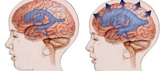
The process of cerebrospinal fluid circulation
The choroid plexuses produce approximately 600–700 ml of cerebrospinal fluid per day. It enters the central canal through special openings, washing the brain and then the spinal cord. Reabsorption occurs through the cerebral hemispheres through the venous sinuses.
Brain damage process
Excessive pressure of the cerebrospinal fluid provokes its leakage into the surrounding substance, which causes the development of edema. The dilated ventricles begin to compress the brain tissue.
The white matter takes the brunt of the attack, causing destruction of the myelin sheaths that transmit nerve impulses to the body.
Up to a certain point, such changes are reversible—after timely surgery, it is possible to almost completely restore neurological functions. Otherwise, atrophy of the brain substance develops, causing spastic paralysis, and the person ceases to control the functioning of the pelvic organs.
In infants, under the influence of such excess pressure, there is a rupture of adhesions inside the subarachnoid space, which opens the way for the outflow of cerebrospinal fluid and self-healing. In advanced situations, brain tissue ruptures. Timely treatment can prevent the development of neurological disorders.
Classification
Based on the openness of the cerebrospinal fluid outflow pathways, hydrocephalus occurs:
- closed;
- communicating;
- substitutive.
According to the characteristics of the deformation of the outflow tract:
- internal;
- external;
- mixed.
By type of cerebrospinal fluid pressure:
- hypotensive;
- hypertensive;
- normotensive.
By etiology:
- congenital;
- after the inflammatory process;
- tumor;
- vascular;
- idiopathic.
Other types of classification:
- with the flow;
- by compensation phase;
- by activity;
- according to the speed of development.
Complications
Excess cerebrospinal fluid dilates the ventricles, compresses the medulla - swelling occurs. A closed cranial cavity provokes the development of irreversible changes in blood vessels - in the absence of the necessary treatment, this condition is life-threatening.
Children who develop hydrocephalus are mentally behind their peers. With complications, mental disorders, mental retardation, personality disorders, and inhibited reactions are likely. Irritability, attacks of euphoria or anger appear. Due to brain damage, speech problems, vision or hearing problems occur.
Adult patients complain of a bursting headache and nausea. Only vomiting sometimes alleviates the condition. The organs of vision experience internal pressure, and there is a feeling as if sand has gotten into the eyes. As pressure increases, drowsiness occurs.
Chronic hydrocephalus provokes an increase in intracranial pressure. The pathology develops slowly, appearing several months after the onset of the disease. Its main symptoms:
- cognitive disorders;
- walking disorders.
Treatment of hypertensive-hydrocephalic syndrome
Drug treatment is prescribed aimed at reducing intracranial pressure, reducing cerebrospinal fluid production, symptomatic therapy (anticonvulsants, sedatives), microcurrent reflexology, massage. Treatment of hypertensive-hydrocephalic syndrome should begin as early as possible. Our Center’s specialists will conduct a comprehensive examination of your child and prescribe comprehensive treatment.
Make an appointment
The Neurology Center accepts patients from Russia and the CIS countries, including Kazakhstan. You can reach us by ground transport. Travel time from Kostanay, Uralsk, Aktobe will be 4-10 hours. We are also waiting for patients from Astana, Almaty, and other cities of Kazakhstan.
You can learn about the work of the Center and ask questions about the treatment of hypertensive-hydrocephalic syndrome by requesting a call back. To do this, fill out the form below and we will call you back within one business day. You can also write to us at the email indicated in contacts.
We remind you that we provide assistance to out-of-town patients in finding rental housing near the Center.
Diagnosis of hydrocephalus
Signs of the disease are often detected while the fetus is still in the womb during ultrasound. Then the disease is diagnosed by a doctor after the baby is born. The main indicator for determining pathology is the large diameter of the skull. In older children, as in adults, the doctor evaluates the patient’s thinking, his gait, the presence of signs of urinary incontinence, and the condition of the ventricles using MRI images.
To verify the presence of congenital hydrocephalus, CT or MRI are used, which allow:
- assess the state of the brain;
- identify excess cerebrospinal fluid volume;
- detect increased cerebrospinal fluid pressure;
- see structural changes in brain tissue.
Normal pressure hydrocephalus is more difficult to identify because its symptoms are largely similar to neurodegenerative processes. This variant of pathology is diagnosed according to the following signs:
- walking disorder;
- mental disorders;
- urinary incontinence;
- excess volume of cerebrospinal fluid.
With any variant of the disease, it is important to make an accurate diagnosis in a timely manner so as not to be late with surgical intervention that will eliminate the severity of symptoms or reduce their intensity.
To find out how effective the operation will be, some additional measures are necessary:
- Lumbar puncture. A small amount of cerebrospinal fluid is removed through a puncture made in the lower back. The cerebrospinal fluid pressure is then measured. The procedure allows you to slightly reduce it in the cerebral ventricles in order to neutralize severe symptoms.
- Lumbar drainage. It is necessary when the previous procedure was ineffective. A catheter is installed between the lumbar vertebrae, through which the cerebrospinal fluid comes out for several days. All this time the patient is observed in the hospital. Antibiotics are administered to prevent infection.
- Infusion test. This is a stress diagnostic option that helps assess how much the brain is able to absorb cerebrospinal fluid.
- ICP measurement. To perform the procedure, a hole is drilled into the skull bone. A sensor in the form of a fiber-optic cable is installed through it. The event is carried out while the patient is in the hospital for at least 24 hours. The sensor detects surges in intracranial pressure, sending values to a recording device.
When an event involves the need to measure ICP, various sensor options are used:
- Intraventricular catheter. This is the most accurate way to measure. The device is brought to the lateral cerebral ventricle through a hole drilled in the skull bone. The instrument helps to simultaneously ensure the drainage of excess fluid.
- Subdural sensor. The device is attached under the dura mater of the brain. It is used if there is an urgent need to check ICP. The method allows you to quickly identify changes in ICP.
- Epidural sensor. The instrument is attached between the dura mater of the brain and the cranial bone, having previously drilled a hole in it. The procedure is less traumatic than others, but has a significant drawback - excess cerebrospinal fluid cannot be drained during the procedure.
When installing sensors, local anesthesia is required. It is also sometimes necessary to additionally inject the patient with sedative medications in order to reduce anxiety and relax the patient as much as possible.
Typically, sensors used to measure ICP are administered to people in intensive care, often in the most critical condition. The necessary indications for such an event are severe trauma to the skull, pathologies of the brain that cause swelling and depression of consciousness up to a coma. In patients who have undergone brain surgery, such a monitoring sensor helps detect increasing swelling.
Drainage of cerebrospinal fluid also helps to reduce high ICP. It is performed by installing a ventricular catheter, intravenous injection of certain medications, and changing the method of ventilation (in a situation where the patient breathes through a tube inserted into the trachea). Normal ICP is 1–20.
Installing sensors is a risky undertaking, as it can cause the following complications if done incorrectly:
- bleeding;
- irreversible damage in a situation with prohibitive ICP values;
- “herniation” of the brain;
- damage to brain tissue during catheter insertion;
- infectious complications;
- inability to accurately determine the location of the ventricle in order to place a catheter to it;
- other risks present during general anesthesia of the patient.
Diagnostics
If symptoms of HGS occur, you should contact a pediatric neurologist. You may also need to consult a neurosurgeon and ophthalmologist. To make a preliminary diagnosis, anamnesis is collected and the baby is examined. Subsequently, diagnostic procedures are prescribed to confirm the diagnosis and find out the cause of the disease.
Diagnostic methods:
- ultrasound examination (ultrasound);
- radiography;
- magnetic resonance imaging (MRI);
- lumbar puncture (taking a sample of cerebrospinal fluid for laboratory analysis);
- echoencephalography.
Based on the results of the diagnostic examination of the child, further treatment is prescribed.
Treatment of hydrocephalus
The option of surgical intervention for hydrocephalus is quite effective. Two types of operations are used: neuroendoscopy or liquor shunting. They are performed by a neurosurgeon, a specialist who can correct problems in the brain or spinal cord, as well as the peripheral nerve ending system.
Bypass surgery
In this type of surgery, a thin tube is inserted into the brain ventricle. Excess cerebrospinal fluid flows through it to another anatomical zone of the body. If the second end of the drainage system is placed in the peritoneum, the shunt is called ventriculoperitoneal. Then the excess cerebrospinal fluid is absorbed by the abdominal cavity and then enters the blood.
When the draining end is directed into the cardiac chamber, the shunt is called ventriculoatrial. This option is practiced during surgery for children, because their increase in height does not affect the functioning of the organ as much, so the drainage does not have to be changed as often as the ventriculoperitoneal system.
Occasionally, lumboperitoneal shunts are also used. With the help of such drainage, cerebrospinal fluid is drained from the lumbar region into the peritoneal cavity.
Any drainage system contains a valve that is set to a certain capacity. It is selected for a specific patient, taking into account the patient’s cerebrospinal fluid pressure. The doctor performing the surgery determines these values in advance by installing a suitable valve, which then looks like a “bump” protruding under the skin on the head.
Currently, neurosurgeons have two types of valves for drainage systems:
- With predefined characteristics that define a specific throughput.
- Adjustable magnetic fixtures. When choosing them, the doctor remotely, using special equipment, without performing additional incisions, changes the pressure in the device to achieve an optimal result. Thanks to this, it is possible to neutralize adverse side effects in the form of insufficient or excessive cerebrospinal fluid pressure.
Bypass surgery is performed under general anesthesia. The procedure lasts 1–2 hours and after it the patient is kept in the hospital for several days. If the outflow through the shunt is disrupted or the patient becomes infected, a repeat procedure is sometimes performed.
Neuroendoscopy
This operation is an excellent alternative to bypass surgery. Without taking steps to install drainage, the neurosurgeon forms a hole directly in the wall of the cerebral ventricle, after which he creates a bypass path from the vessel through which cerebrospinal fluid flows freely to the surface. There it is absorbed by the brain tissue without obstacles.
This procedure is not universal - it cannot be done on all patients, although this method is often used if the natural cerebrospinal fluid pathways are blocked. In this case, the cerebrospinal fluid flows through the created hole, successfully bypassing all blocked vessels.
The endoscopic procedure is done under general anesthesia. First, the neurosurgeon makes a burr hole with a diameter of 1 cm on the skull. Then, using an endoscope, he examines the inside of the cerebral ventricles. Through the internal channel of the instrument, surgical instruments are inserted, which are used to perform an operation inside the brain. When the endoscope reaches the third ventricle, the doctor creates a hole in its lower wall through which the cerebrospinal fluid penetrates into the subarachnoid cavity. After removing the device, first the aponeurosis is sutured, and then the skin on the skull. The duration of the operation is approximately an hour.
The risk of infection with such an intervention is significantly lower than with cerebrospinal fluid bypass. Neuroendoscopy does not have significant advantages over drainage surgery over long periods of patient observation. After such an intervention, hydrocephalus can also develop again after a few years.
Actions for normal pressure hydrocephalus
When normal pressure hydrocephalus (a common occurrence in older people) is diagnosed, the patient's condition can be improved by performing a cerebrospinal fluid shunt. Although surgery is not effective for every patient. Due to the considerable risks that accompany all operations, certain tests are required to assess how much the potential benefit outweighs the risk of negative consequences.
Almost 80% of patients diagnosed with normal pressure hydrocephalus experienced significant improvement after preliminary testing, so ventriculoperitoneal shunting was recommended for them. Significant clinical postoperative improvement occurs after a few weeks, sometimes even months. Only a timely correct diagnosis guarantees successful recovery even with many years of development of hydrocephalus.