Headache occurs in the form of episodic attacks or worries the patient constantly. Attacks of headaches may be accompanied by pain in the eyes, ripples before the eyes, and the sensation of flies flying. These are symptoms of serious diseases that doctors at the Yusupov Hospital identify using modern instrumental and laboratory research methods.
Neurologists, therapists, and oncologists treat patients. Professors and doctors of the highest category at a meeting of the expert council discuss all cases of severe headaches and eye pain that are not amenable to drug therapy. Leading cephalgologists (specialists in the field of headache treatment) collectively develop further tactics for patient management.
Types and causes of pain in the head and eyes
Most often, headaches in the forehead and eyes occur after emotional breakdowns. Many patients experience it in the evening, when the work day ends. In most cases, the cause of pain is simple overwork.
Often the headache radiates to the eyes. Sometimes, first there are ripples in the eyes, then a headache. The eyeballs may first become tense, and then the pain moves to the forehead. Floaters, flickering before the eyes and headache are symptoms of various diseases:
- glaucoma (increased intraocular pressure);
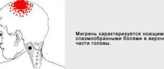
- migraine;
- myopia (myopia);
- arterial hypertension (high blood pressure);
- damage to soft tissues and bones of the head;
- traumatic brain injury.
A headache in the forehead area can occur due to eating unhealthy foods. These are chocolate, tea, coffee, which contain a large amount of caffeine, nuts, cheese, and processed meats. Headache pressing on the eyes after drinking alcohol and smoking. Head pain is caused by the following diseases:
- migraine;
- hypertonic disease;
- diseases of the organs of vision;
- brain tumors;
- Horton's syndrome;
- vegetative-vascular dysfunction;
- encephalitis;
- stroke.
Causes of eye pain
Eye burn
A burn, regardless of type (chemical, thermal), leads to severe pain, the intensity of which increases over time. The patient cannot open his eyes on his own. Associated symptoms include photophobia, increased lacrimation, eyelid edema, and conjunctival chemosis. In severe burns, hyperemia is replaced by blanching of the eye tissue.
Eye injuries
Eye injury results in severe pain. The patient is unable to open his eyelids without the help of a doctor. The surrounding tissues are swollen and hyperemic. Symptoms are extremely pronounced when the integrity of the cornea is violated. The most common types of traumatic eye injuries are:
- Foreign bodies.
They can be located on the ocular surface or have intraocular localization. In the practice of an ophthalmologist, foreign bodies of the conjunctiva, eyelids and cornea are more often encountered. The location of foreign particles under the upper eyelid injures the cornea when blinking, causing pain. - Corneal erosion.
The appearance of erosive defects is often caused by microtrauma when applying makeup or being hit by a branch. Symptoms are especially noticeable upon awakening and include soreness, flushing, lacrimation, blurred vision and photophobia.
Conjunctivitis
Redness and soreness of the eye are common signs of inflammation of the conjunctiva, regardless of etiology. Therefore, it is very important to pay attention to the specific symptoms of individual forms. Differential diagnosis should primarily be carried out among the following types of conjunctivitis:
- Bacterial.
Pain syndrome is of moderate intensity, most pronounced in the morning. Purulent discharge may appear, which is yellow in color and has a viscous consistency. Patients are unable to open their eyes after sleep. - Allergic.
Inflammation of the conjunctiva is accompanied by itching, pain, and lacrimation. Discomfort increases upon contact with an allergen. In the conjunctival fornix, large follicles are often formed in a “cobblestone” type. - Adenoviral.
This conjunctivitis is characterized by increasing chemosis, hyperemia and pain. The course of the disease is protracted (3 or more weeks). Often the pathological process spreads to the cornea.
Keratitis
The leading symptom of keratitis is severe pain, which is combined with conjunctival hyperemia, swelling of surrounding tissues and photophobia. Common causes of corneal inflammation: contact with infectious patients, failure to comply with personal hygiene rules. The main forms of keratitis:
- Herpetic.
Accompanied by acute pain, blepharospasm, photophobia. The conjunctiva and soft tissues of the eyelids are swollen and hyperemic. Pathognomonic signs are tree-like defects on the surface of the cornea and a decrease in its sensitivity. - Bacterial.
The disease is characterized by an acute onset. The clinical picture includes pain, pericorneal or mixed injection of conjunctival vessels, and photophobia. Mucopurulent discharge is characteristic of keratoconjunctivitis. - Adenoviral.
Patients note decreased visual acuity, pain and swelling of surrounding tissues. Corneal damage is represented by “coin-shaped” subepithelial opacities that persist for a long time (from 2-3 months to 1-1.5 years).
Uveitis
Uveitis is an inflammation of the choroid (uveal tract). Depending on the location of the pathological process, anterior (iridocyclitis) and posterior uveitis are distinguished. Symptoms include pain, decreased visual acuity, and redness of the eye. Anisocoria, lacrimation, and photophobia may develop. With iridocyclitis, patients often report blurred vision.
Ophthalmohypertension
Increased intraocular pressure above 30 mm. rt. Art. may lead to pain in the eye that radiates to the head. Due to diurnal fluctuations in IOP, pain is most pronounced in the morning. An associated manifestation is congestive conjunctival injection. When looking at a light source, rainbow circles appear. A similar clinical picture is observed with angle-closure glaucoma.
Neurological disorders
Pain syndrome is a common sign of pathology of the central nervous system. In most cases, eye pain is associated with intracranial hypertension, in which patients' vision deteriorates, diplopia, photophobia and blurred vision may occur. At the same time, a decrease in intracranial pressure leads to positional pain, photophobia and double vision. Also, pain in the eye occurs with the following pathologies:
- Trigeminal neuralgia.
Short-term attacks of intense pain or prolonged burning pain on the affected side are typical. The pain is clearly limited to the zone of innervation. The clinic increases with irritation of trigger points. - Supraorbital neuralgia .
The affected area is limited to the supraorbital region, the brow ridge and the lower part of the forehead. The pain is paroxysmal or constant. Lacrimation is determined only on the side of neuralgia. - Optic neuritis.
Patients complain of pain, which increases with eye movements. The clinical picture is represented by deterioration or blurred vision. The anterior part of the eyeball is unchanged. - Ophthalmoplegic migraine.
This is one of the forms of associative migraine, characterized by transient paresis of one or more oculomotor nerves. Pain in the eye spreads to the corresponding half of the head and is combined with diplopia, ptosis, and anisocoria.
How to help yourself with a headache
If a headache radiates to the eyes after hard work, you should rest well, take a walk in the fresh air, and give yourself an acupressure massage. You can take a warm bath with chamomile infusion added to the water. Take a break from working at the computer or watching TV
Give yourself a head massage. Relax, drink warm milk with honey, tea with lemon balm. Do simple exercises to relieve headaches:
- sit on a chair, keep your back straight and your head free;
- without effort, just under the influence of gravity, tilt it towards your chest;
- stay in this position for twenty seconds; take a break for 30 seconds;
- Bend again for 20 seconds.
Repeat the exercise 15-16 times.
The second exercise is done in the following sequence:
- sitting or standing, raise your hands to your head;
- the thumbs of each hand are pressed to the zygomatic arches, with the remaining fingers clasping the back of the head;
- look up;
- while inhaling, try to throw your head back for 10 seconds, while holding it with your hands;
- while exhaling, look down for 6-8 seconds;
- tilting your head to your chest as much as possible, stretch but do not strain the neck muscles.
Repeat the inhale-exhale cycle 5-6 times.
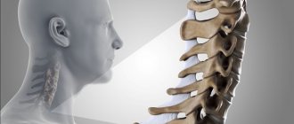
To relieve headaches coming from the cervical spine, rehabilitation experts recommend performing the following exercise:
- sitting on a chair, with one hand clasp your head from above on the side in which the pain is felt more strongly;
- Place the index finger at the level of the beginning of the ear;
- With a little hand effort, turn your head to the “healthy” side;
- Press your free palm from below to your chin and cheek;
- while inhaling for 10 seconds, looking down, press your chin to your lower palm against its resistance;
- As you exhale, relax for 6-8 seconds and look up.
- repeat the exercise 5-6 times, slightly changing the turn of the head.
Diagnostics
Severe pain in the eye leads to blepharospasm, which makes ophthalmological examination difficult. To alleviate the patient's condition, instillation of analgesics is recommended, after which diagnosis begins. It is important to assess the condition of the eyelids, the shape of the palpebral fissure and the position of the eyes. The following are specific studies:
- Visometry.
Visual acuity is determined at the beginning of the examination of the patient. In the absence of object vision, it is necessary to study light projection. Visometry is performed with and without distance correction. - Non-contact tonometry.
Penetrating eye injuries are often accompanied by hypotension. With iridocyclitis, intraocular pressure increases. It is important to compare the IOP in both eyes and also measure the central corneal thickness. - Biomicroscopy of the eye.
First, a detailed examination of the anterior segment of the eyeball is carried out with mandatory eversion of the upper eyelid. Next, fluorescein staining and examination with a cobalt filter are performed, which allows visualization of small erosive defects. - Ophthalmoscopy.
Fundus examination is carried out after cycloplegia, if intraocular pressure is compensated. The ophthalmologist evaluates the transparency of the optical media and the condition of the retina down to the dentate line. - Ultrasound of the eye.
Ultrasound examination is used when there are difficulties in visualizing the structures of the eyeball due to miosis, corneal edema, hyphema or hemophthalmos. The advantage of this method is the ability to detect x-ray negative foreign bodies. - Radiography.
It is performed in case of severe injuries in order to prevent damage to the bone walls of the orbit. The special Komberg-Baltin prosthesis makes it possible to determine the location of intraocular radiopaque foreign bodies.
Eye examination by an ophthalmologist
Flickering in the eyes and headache
The main cause of flicker in the eyes and headaches is retinal detachment and rupture. Sharp flashes of light are the result of tension in the eye membrane. After a rupture of the capillaries and retina, a person sees many black dots.
Vitreous detachment also causes tension on the retina. It comes in back and front. The vitreous body is divided into a gel and a liquid part, which passes under the membrane and “disconnects” it from the retina. The main sign of the pathology is “flying flies”. They can be seen when looking at the sun, snow in sunny weather, or blue sky.
Chorioretinitis is inflammation of the vessels of the retina of the eye. The development of the disease is caused by:
- infectious diseases;
- increased levels of radiation;
- allergy;
- intoxication;
- injuries and damage to the organs of vision;
- autoimmune conditions;
- decreased immunity after long-term treatment or in HIV-infected patients.
Chorioretinitis can be congenital. Symptoms of the disease include darkening of the eyes and decreased visual acuity. The patient cannot determine the size of objects; it is difficult for him to navigate in low light. Ophthalmologists at the Yusupov Hospital use the latest diagnostic equipment from leading global manufacturers to examine patients and apply innovative treatment methods.
Eye spots and headaches
The main reason why a person experiences ripples in the eyes, and then a headache, is the compression of the blood vessels. This happens when the weather changes, atmospheric pressure changes, or oxygen starvation. Floaters “fly” before the eyes and a headache appears after stress. The cause of headaches and dizziness is nervous exhaustion, deficiency of essential substances and microelements, and the presence of bad habits.
In this case, the pain goes away after rest. But ripples and flickering of spots before the eyes, a headache may be the first signs of a tumor, diseases of the cardiovascular system, or vegetative-vascular dysfunction. If, after rest, the ripples and flickering of spots before your eyes, the headache does not go away, make an appointment by calling the Yusupov Hospital. In each case of headache, neurologists first determine the type of pain, identify its cause and eliminate it, stop the pain attack and carry out therapy, the purpose of which is to prevent subsequent headache attacks.
Causes of headaches with eye pressure
The symptom of a headache that radiates into the eyes is very common and can indicate a number of problems. The most common reasons:
- Fatigue, eye strain. This is typical for people whose occupation involves constantly sitting at a computer, after watching TV for a long time, or reading books from electronic “readers”.
- Lack of sleep. Glasses or contact lenses that were not fitted correctly. The symptom gets worse throughout the day. Diopters that are not suitable for specific eyes have a negative impact on the optic nerve.
Subscribe to our INSTAGRAM account!
- If discomfort occurs when sneezing and coughing, this indicates high blood pressure.
- Nervous overstrain. “Floaters” may appear before the eyes. Combined with pressing pain in the eyes, this occurs due to spasms of the blood vessels supplying the face and neck.
- After bruises and blows to the head, a migraine with pressure in the eyes in this case may indicate a concussion.
- Glaucoma, migraine and other diseases.
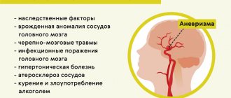
- Constant jerking pain occurs in dangerous conditions: meningitis, sarcoma, encephalitis, aneurysm.
- Allergic reaction.
The list of causes of the symptom is very large, so it is important to consult a doctor and not self-medicate, especially if the phenomenon does not go away for a long time.