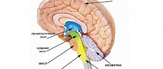
Laws of axon work
When conducting a nerve impulse along axons, four main laws apply:
- Law of anatomical and physiological integrity. Conduction is possible only through intact neuron processes. This rule also applies to damage resulting from changes in membrane permeability (under the influence of drugs or poisons).
- Excitation isolation law. One axon – conduction of one excitation. Axons do not share nerve impulses with each other.
- Law of unilateral conduct. The axon conducts impulses either centrifugally or centripetally.
- The law of no loss. This property is non-decremental - when an impulse is carried out, it does not subside or change.
How does a neuron work?
Neurons exchange information with each other at axonal and dendritic terminals. These terminals form a special structure known as a synapse (Greek "syn" = together + "haptein" = "to interlock"). Communication between two neurons begins with a stream of electrical impulse, known as an action potential, in one of the neurons. This action potential travels down the axon and reaches the synaptic terminal. Here it triggers the release of neurotransmitters at the synaptic cleft, the tiny space between the terminals of two interacting neurons. The released neurotransmitter then binds to receptors on the synaptic terminal of another neuron and also induces an action potential in that neuron. Now the electrical impulse will travel through the neuron.
The location of these nerve cells
Pyramidal neurons can be found at various points in the nervous system, but they are much more common in some specific areas. Among them the following stand out.
Cortex
Pyramidal neurons are found primarily in the cerebral cortex, forming part of most of it, and are found in five of the six layers that make up this area of the brain. In particular, they can be observed in the granular and pyramidal layers, both external and internal.
They are especially prominent in the third and fifth layers (which are actually called the outer pyramidal and inner pyramidal), and they are larger the deeper into the cortex. There are also areas within the cortex where its existence has been discovered more frequently.
Motor cortex
In the motor cortex we can find a large number of pyramidal neurons, especially those associated with motor control. In this area of the cortex there are many giant pyramidal neurons known as Betz cells that transmit motor information from the brain to areas of the spinal cord, where they synapse with motor neurons that activate movement.
Prefrontal cortex
Pyramidal neurons can also be found in the prefrontal cortex, influencing higher mental processes. It is believed that these cells are the main primary excitations of prefrontal neurons, participating in numerous functions and considering themselves primordial for the existence of behavioral control.
Corticospinal tract
Pyramidal neurons are especially visible along the corticospinal tract, which sends motor information from different brain nuclei responsible for motor control to motor neurons that will cause muscle contraction as they pass through the spinal cord.
Hippocampus
Not only in the cortex we can find pyramidal neurons, but we can also find in subcortical structures. One of them is the hippocampus, associated with aspects such as memory and orientation.
Article on the topic: “Hippocampus: functions and structure of the memory organ”
Amygdala
Another structure in which these neurons are found is in the amygdala, an area of the limbic system associated with emotional memory.
Motor neuron function
The motor neuron is the final structural unit of the reflex arc. It has a large body enclosed in the anterior horns of the spinal cord. Those nerve cells that innervate skeletal muscles are called these motor elements. Other efferent neurocytes enter the secreting cells of the glands and cause the release of corresponding substances: secretions, hormones. In involuntary, that is, unconditionally reflex acts (swallowing, salivation, defecation), efferent neurons extend from the spinal cord or from the brain stem. To perform complex actions and movements, the body uses two types of centrifugal neurocytes: central motor and peripheral motor. The body of the central motor neuron is located in the cerebral cortex, near the Rolandic fissure.
The bodies of peripheral motor neurocytes, innervating the muscles of the limbs, trunk, and neck, are located in the anterior horns of the spinal cord, and their long processes - axons - emerge from the anterior roots. They form the motor fibers of 31 pairs of spinal nerves. Peripheral motor neurocytes innervating the muscles of the face, pharynx, larynx, and tongue are located in the nuclei of the vagus, hypoglossal and glossopharyngeal cranial nerves. Consequently, the main function of the motor neuron is the unhindered conduction of excitation to the muscles, secreting cells and other working organs.
Classification
Structural classification
Based on the number and arrangement of dendrites and axons, neurons are divided into axonless neurons, unipolar neurons, pseudounipolar neurons, bipolar neurons, and multipolar (many dendritic arbors, usually efferent) neurons.
Axonless neurons
- small cells, grouped near the spinal cord in the intervertebral ganglia, which do not have anatomical signs of division of processes into dendrites and axons. All processes of the cell are very similar. The functional purpose of axonless neurons is poorly understood.
Unipolar neurons
- neurons with a single process, present, for example, in the sensory nucleus of the trigeminal nerve in the midbrain. Many morphologists believe that unipolar neurons do not occur in the body of humans and higher vertebrates.
Bipolar neurons
- neurons with one axon and one dendrite, located in specialized sensory organs - the retina, olfactory epithelium and bulb, auditory and vestibular ganglia.
Multipolar neurons
- neurons with one axon and several dendrites. This type of nerve cells predominates in the central nervous system.
Pseudounipolar neurons
- are unique in their kind. One process extends from the body, which immediately divides in a T-shape. This entire single tract is covered with a myelin sheath and is structurally an axon, although along one of the branches the excitation goes not from, but to the body of the neuron. Structurally, dendrites are branches at the end of this (peripheral) process. The trigger zone is the beginning of this branching (that is, it is located outside the cell body). Such neurons are found in the spinal ganglia.
Functional classification
Based on their position in the reflex arc, afferent neurons (sensitive neurons), efferent neurons (some of them are called motor neurons, sometimes this not very accurate name applies to the entire group of efferents) and interneurons (interneurons) are distinguished.
Afferent neurons
(sensitive, sensory, receptor or centripetal). Neurons of this type include primary cells of the sensory organs and pseudounipolar cells, whose dendrites have free endings.
Efferent neurons
(effector, motor, motor or centrifugal). Neurons of this type include the final neurons - ultimatum and penultimate - non-ultimatum.
Association neurons
(interneurons or interneurons) - a group of neurons communicates between efferent and afferent ones.
Secretory neurons
- neurons that secrete highly active substances (neurohormones). They have a well-developed Golgi complex, the axon ends at axovasal synapses.
Morphological classification
The morphological structure of neurons is diverse. Several principles are used to classify neurons:
- take into account the size and shape of the neuron body;
- number and nature of branching of processes;
- axon length and the presence of specialized sheaths.
According to the shape of the cell, neurons can be spherical, granular, stellate, pyramidal, pear-shaped, fusiform, irregular, etc. The size of the neuron body varies from 5 μm in small granular cells to 120-150 μm in giant pyramidal neurons.
Based on the number of processes, the following morphological types of neurons are distinguished:
- unipolar (with one process) neurocytes, present, for example, in the sensory nucleus of the trigeminal nerve in the midbrain;
- pseudounipolar cells grouped near the spinal cord in the intervertebral ganglia;
- bipolar neurons (have one axon and one dendrite), located in specialized sensory organs - the retina, olfactory epithelium and bulb, auditory and vestibular ganglia;
- multipolar neurons (have one axon and several dendrites), predominant in the central nervous system.
From the editor: Classification and methods of treating nervous tics
Types of neurons
Neurons are usually classified in one of the following ways:
- depending on the number of processes from the cell body,
- depending on the direction of transmission of information regarding the brain and spinal cord,
- depending on the type of neurotransmitter (chemical messengers that neurons use to communicate with each other) the neuron uses.
Bipolar and multipolar
Depending on the number of processes extending from the cell body, a neuron can be classified as bipolar, pseudounipolar, or multipolar.
- A bipolar neuron has an axon and dendrites.
- A pseudounipolar neuron has two axons.
- A multipolar neuron has one axon but several dendritic processes extending from its cell body.
Sensory and motor
Depending on the direction of information flow, neurons are divided into three types - sensory neurons, motor neurons and interneurons.
- Sensory neurons carry signals from sensory receptors such as the skin, ears, eyes, tongue, nose, etc. to the brain or spinal cord. It sends information to the brain about various sensations such as temperature, pain, sound, smell and vision.
- Motor neurons work in the opposite direction: they transmit signals from the brain or spinal cord to the glands or muscles of the body. In this way, the brain can control the various muscles of the body and the activities of various glands.
- Interneurons provide information transfer between sensory neurons and motor neurons in the brain and spinal cord.
Neurotransmitters
Although neurons transmit information through electrical impulses, they also communicate with each other through chemical messengers known as neurotransmitters. Each neuron predominantly uses one type of neurotransmitter, and this makes it possible to classify these neurons.
Although neurons transmit information through electrical impulses, they also communicate with each other through chemical messengers known as neurotransmitters.
Some of the commonly used neurotransmitters are:
- dopamine
- GABA
- serotonin
- acetylcholine
Neuroglia
Neurons are not able to divide, which is why the statement appeared that nerve cells do not regenerate. That is why they should be protected with special care. Neuroglia cope with the main function of the “nanny”. It is located between the nerve fibers.
These small cells separate neurons from each other and hold them in place. They have a long list of features. Thanks to neuroglia, a constant system of established connections is maintained, the location, nutrition and restoration of neurons is ensured, individual mediators are released, and genetically foreign substances are phagocytosed.
Thus, neuroglia perform a number of functions:
- supporting;
- delimiting;
- regenerative;
- trophic;
- secretory;
- protective, etc.
In the central nervous system, neurons make up the gray matter, and outside the brain they accumulate in special connections, nodes - ganglia. Dendrites and axons create white matter. At the periphery, it is thanks to these processes that the fibers that make up the nerves are built.
Integumentary tissues
Integumentary tissues are also called epithelial tissues
Integumentary tissues line not only the surfaces of the body, but also the cavities of internal organs. So the stomach, intestines, oral cavity, bladder, etc. are lined with integumentary tissues from the inside.
There is almost no intercellular substance in epithelial tissues. Their cells adhere tightly to each other and form from one to several layers.
The main functions of the epithelium are protection, secretion production, gas exchange, absorption, and excretion.
is expressed in protecting the deeper tissues of the animal from damage, temperature changes, and the entry of harmful microorganisms. This function is performed by the skin.
epithelium is characteristic of the intestine. Here nutrients are absorbed into the blood using the intestinal villi.
of the animal's integumentary tissue is observed in the stomach, where its cells secrete mucus. There are also various glands in the skin.
carried out by the epithelium of the lungs; in some animals the skin also takes part in gas exchange.
performs the epithelium of the excretory organs.
Structure of neurons
File:Neuron-rus.svg Neuron diagram
Cell body
The body of a nerve cell consists of protoplasm (cytoplasm and nucleus), and is externally bounded by a membrane of a double layer of lipids (bilipid layer). Lipids consist of hydrophilic heads and hydrophobic tails, arranged with hydrophobic tails facing each other, forming a hydrophobic layer that allows only fat-soluble substances (eg oxygen and carbon dioxide) to pass through. There are proteins on the membrane: on the surface (in the form of globules), on which growths of polysaccharides (glycocalyx) can be observed, thanks to which the cell perceives external irritation, and integral proteins that penetrate the membrane through, in which ion channels are located.
A neuron consists of a body with a diameter of 3 to 130 µm, containing a nucleus (with a large number of nuclear pores) and organelles (including a highly developed rough ER with active ribosomes, the Golgi apparatus), as well as processes. There are two types of processes: dendrites and axons. The neuron has a developed and complex cytoskeleton that penetrates its processes. The cytoskeleton maintains the shape of the cell; its threads serve as “rails” for the transport of organelles and substances packaged in membrane vesicles (for example, neurotransmitters). The cytoskeleton of a neuron consists of fibrils of different diameters: Microtubules (D = 20-30 nm) - consist of the protein tubulin and stretch from the neuron along the axon, right up to the nerve endings. Neurofilaments (D = 10 nm) - together with microtubules, provide intracellular transport of substances. Microfilaments (D = 5 nm) - consist of actin and myosin proteins, especially expressed in growing nerve processes and in neuroglia. A developed synthetic apparatus is revealed in the body of the neuron; the granular ER of the neuron is stained basophilically and is known as the “tigroid”. The tigroid penetrates the initial sections of the dendrites, but is located at a noticeable distance from the beginning of the axon, which serves as a histological sign of the axon. Neurons vary in shape, number of processes, and functions. Depending on the function, sensitive, effector (motor, secretory) and intercalary are distinguished. Sensory neurons perceive stimuli, convert them into nerve impulses and transmit them to the brain. Effector (from the Latin effectus - action) - generate and send commands to the working organs. Intercalary neurons - communicate between sensory and motor neurons, participate in information processing and the generation of commands.
There is a distinction between anterograde (away from the body) and retrograde (toward the body) axon transport.
Dendrites and axon
Main articles: Dendrite
,
Axon
File:Complete neuron cell diagram ru.svg Diagram of the structure of a neuron
An axon is usually a long extension of a neuron that is adapted to conduct excitation and information from the body of the neuron or from the neuron to the effector. Dendrites are, as a rule, short and highly branched processes of a neuron, serving as the main site of formation of excitatory and inhibitory synapses influencing the neuron (different neurons have different ratios of axon and dendrites lengths), and which transmit excitation to the body of the neuron. A neuron may have several dendrites and usually only one axon. One neuron can have connections with many (up to 20 thousand) other neurons.
Dendrites divide dichotomously, while axons give off collaterals. Mitochondria are usually concentrated at branching nodes.
Dendrites do not have a myelin sheath, but axons may have one. The place of generation of excitation in most neurons is the axon hillock - a formation at the point where the axon departs from the body. In all neurons, this zone is called the trigger zone.
Synapse
Main article: Synapse
Synapse
(Greek σύναψις, from συνάπτειν - hug, clasp, shake hands) - the place of contact between two neurons or between a neuron and the effector cell receiving the signal. It serves to transmit a nerve impulse between two cells, and during synaptic transmission the amplitude and frequency of the signal can be adjusted. Some synapses cause depolarization of the neuron, others cause hyperpolarization; the former are excitatory, the latter are inhibitory. Typically, stimulation from several excitatory synapses is necessary to excite a neuron.
Editorial: What foods best clean blood vessels?
The term was introduced in 1897 by the English physiologist Charles Sherrington.
Evolution of fabric
The main property of a living organism is irritability or sensitivity. The type of nervous tissue is determined by the phylogenetic position of the animal and is characterized by wide variability, becoming more complex in the process of evolution. All organisms require certain parameters of internal coordination and regulation, proper interaction between stimulus for homeostasis and physiological state. The nervous tissue of animals, especially multicellular ones, the structure and functions of which have undergone aromorphoses, contributes to survival in the struggle for existence. In primitive hydroids, it is represented by stellate, nerve cells scattered throughout the body and connected by thin processes intertwined with each other. This type of nerve tissue is called diffuse.
The nervous system of flat and roundworms is stem, scalene type (orthogonal) consists of paired cerebral ganglia - clusters of nerve cells and longitudinal trunks extending from them (connectives), interconnected by transverse cords-commissures. In the rings, from the peripharyngeal ganglion, connected by cords, the abdominal nerve chain departs, in each segment of which there are two close nerve ganglia connected by nerve fibers. In some soft-bodied animals, nerve ganglia are concentrated to form the brain. Instincts and spatial orientation in arthropods are determined by the cephalization of the ganglia of the paired brain, the peripharyngeal nerve ring and the ventral nerve cord.
In chordates, the nervous tissue, the types of tissues of which are strongly expressed, is complex, but such a structure is evolutionarily justified. Different layers arise and are located on the dorsal side of the body in the form of a neural tube, the cavity is the neurocoel. In vertebrates, it differentiates into the brain and spinal cord. As the brain forms, swellings form at the anterior end of the tube. If in lower multicellular organisms the nervous system plays a purely connecting role, then in highly organized animals it stores information, retrieves it when necessary, and also ensures processing and integration.
In mammals, these cerebral swellings give rise to the main parts of the brain. And the rest of the tube forms the spinal cord. Nervous tissue, the structure and functions of which are unique in higher mammals, has undergone significant changes. This is the progressive development of the cerebral cortex and all parts of the nervous system, which determine complex adaptation to environmental conditions and the regulation of homeostasis.
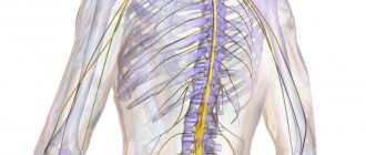
Nervous system
Most cells in the body have a similar structure. They have a compact shape enclosed in a shell. Inside there is a nucleus and a set of organelles that perform the synthesis and metabolism of necessary substances. However, the structure and functions of the neuron are different. It is a structural unit of nervous tissue. These cells provide communication between all body systems.
The basis of the central nervous system is the brain and spinal cord. These two centers secrete gray and white matter. The differences are related to the functions performed. One part receives a signal from the stimulus and processes it, while the other is responsible for carrying out the necessary response command. Outside the main centers, the nervous tissue forms bundles of clusters (nodes or ganglia). They branch, spreading a signal-conducting network throughout the body (peripheral nervous system).
Structure and functions of a neuron[edit | edit code]
A. Structure and functions of a nerve cell
Excitable cells respond to stimuli by changing the state of their membranes. There are two types of excitable cells: nerve cells, which conduct and transduce impulses in the nervous system, and muscle cells, which contract either in response to nerve impulses or autonomously.
Nervous system
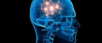
The human body consists of more than 1010 nerve cells, or neurons.
A neuron
is a structural and functional unit of the nervous system. A typical neuron (motoneuron, A1) consists of a soma, or cell body, and two types of processes - axon and dendrites. In addition to the usual cellular organelles, such as the nucleus and mitochondria (A2), the neuron contains neurofibrils and neurotubules. A neuron receives afferent signals (excitatory and inhibitory) from several and sometimes several thousand neighboring neurons through dendrites (usually tree-like), and the signals are summed along the neuron body on the cell membrane (summation).The axon starts from the axon hillock of the neuron body: it carries out transmission of efferent nerve signals to nearby or distant effectors (muscle and secretory cells) and nearby neurons. Axons often have branches (collaterals), which branch further and end in swellings - synaptic vesicles or synaptic endings. If the total potential at the axon hillock exceeds a certain threshold, an action potential is generated and transmitted down the axon, where it reaches the next synapse through the synaptic terminal (A1, 3), described below.
Vesicles containing various substances (proteins, lipids, sugars and neurotransmitter molecules) are transported from the Golgi complex in the soma to the synaptic terminal and to the tips of dendrites by rapid axonal transport (40 cm/day). This type of anterograde (forward-directed) transport along neurotubules is carried out by kinesin (myosin-like protein), and the energy required for this is supplied by ATP. Endogenous and exogenous substances, such as nerve growth factor (NGF), herpes virus, polio virus, and tetanus toxin, are transported retrogradely from peripheral sites to the soma at a rate of ~25 cm/day. Slow axonal transport (~1 mm/day) plays an important role in the treatment of severe neuritis.
The plasma membrane of the soma continues along the axon and is called the axolemma (A1, 2).
In the central nervous system (CNS), the axolemma is surrounded by oligodendrocytes, and in the peripheral nervous system, by Schwann cells (A1, 2). A nerve fiber consists of an axon and its sheath. In some neurons, Schwann cells form a multilayered myelin sheath of double phospholipid layers (A1, 2) around the axon, which insulates the axon from ionic currents. The myelin sheath is interrupted approximately every 1.5 mm at the nodes of Ranvier (A1). The conductivity of myelinated nerve fibers is much higher than that of unmyelinated nerve fibers and increases with the diameter of the nerve fiber.
A synapse (A3) is the site where a neuron's axon interacts with effectors or other neurons. Synaptic transmission in almost all mammals is carried out by chemical compounds rather than by electrical signals. In response to an electrical signal in the axon, neurotransmitters are released from vesicles on the presynaptic membrane by exocytosis. The transmitter diffuses through the synaptic cleft (10-40 nm) to the postsynaptic membrane, where it connects with receptors that create new electrical signals (AZ). Depending on the type of neurotransmitter and receptor involved in the process, the neurotransmitter has either an excitatory (for example, acetylcholine in skeletal muscle) or an inhibitory effect (for example, glycine in the central nervous system) on the postsynaptic membrane. Because the postsynaptic membrane does not normally release neurotransmitters (there are only a few exceptions), nerve impulses can only pass through the synapse in one direction. Thus, the synapse acts as a valve that ensures orderly signal transmission. Synapses are also sites at which the transmission of a nerve impulse can be converted by other (excitatory or inhibitory) neurons.
From the editor: Books for the comprehensive development of the brain
Neuron and its structure
You can often hear that a person’s mental abilities are guaranteed by the presence of gray matter. What is this substance and why is it gray? This is the color of the cerebral cortex, which consists of microscopic cells. These are neurons or nerve cells that ensure the functioning of our brain and control of the entire human body.
How does a nerve cell work?
A neuron, like any living cell, consists of a nucleus and a cell body called the soma. The size of the cell itself is microscopic - from 3 to 100 microns. However, this does not prevent the neuron from being a real repository of various information. Each nerve cell contains a complete set of genes - instructions for producing proteins. Some of the proteins are involved in the transmission of information, others create a protective shell around the cell itself, others are involved in memory processes, others provide mood changes, etc.
Even a small malfunction in one of the programs for the production of a certain protein can lead to serious consequences, illness, mental impairment, dementia, etc.
Each neuron is surrounded by a protective sheath of glial cells; they literally fill the entire intercellular space and make up 40% of the brain substance. Glia or a collection of glial cells performs very important functions: it protects neurons from unfavorable external influences, supplies nerve cells with nutrients and removes their waste products.
Glial cells guard the health and integrity of neurons, and therefore prevent many foreign chemicals from entering nerve cells. Including medications. Therefore, the effectiveness of various drugs designed to enhance brain activity is completely unpredictable, and they affect each person differently.
Dendrites and axons
Despite the complexity of the neuron, it itself does not play a significant role in the functioning of the brain. Our nervous activity, including mental activity, is the result of the interaction of many neurons exchanging signals. The reception and transmission of these signals, more precisely, weak electrical impulses, occurs with the help of nerve fibers.
The neuron has several short (about 1 mm) branched nerve fibers - dendrites, so named because of their resemblance to a tree. Dendrites are responsible for receiving signals from other nerve cells. And the axon acts as a signal transmitter. A neuron has only one fiber, but it can reach a length of up to 1.5 meters. Connecting with the help of axons and dendrites, nerve cells form entire neural networks. And the more complex the system of relationships, the more complex our mental activity.
Neuron operation
The most complex activity of our nervous system is based on the exchange of weak electrical impulses between neurons. But the problem is that initially the axon of one nerve cell and the dendrites of another are not connected; between them there is a space filled with intercellular substance. This is the so-called synaptic cleft, and the signal cannot cross it. Imagine that two people reach out to each other and just barely reach each other.
This problem is easily solved by a neuron. Under the influence of a weak electric current, an electrochemical reaction occurs and a protein molecule, a neurotransmitter, is formed. This molecule blocks the synaptic cleft, becoming a kind of bridge for the passage of the signal. Neurotransmitters also perform another function - they connect neurons, and the more often a signal passes along this nerve chain, the stronger this connection. Imagine a ford across a river. Walking along it, a person throws a stone into the water, and then each subsequent traveler does the same. The result is a strong, reliable transition.
This connection between neurons is called a synapse, and it plays an important role in brain activity. It is believed that even our memory is the result of the work of synapses. These connections provide a high speed of passage of nerve impulses - the signal along the chain of neurons moves at a speed of 360 km/h or 100 m/sec. You can calculate how long it takes for a signal from a finger that you accidentally pricked with a needle to reach your brain. There is an old riddle: “What is the fastest thing in the world?” Answer: "Thought." And this was very accurately noted.
Types of neurons
Neurons are not only found in the brain, where they interact to form the central nervous system. Neurons are located in all organs of our body, in muscles and ligaments on the surface of the skin. There are especially many of them in receptors, that is, sensory organs. The extensive network of nerve cells that permeates the entire human body is the peripheral nervous system, which performs no less important functions than the central one. The entire variety of neurons is divided into three main groups:
- Affector neurons receive information from the sensory organs and deliver it to the brain in the form of impulses along nerve fibers. These nerve cells have the longest axons, since their body is located in the corresponding part of the brain. There is strict specialization, and sound signals arrive exclusively in the auditory part of the brain, smells - in the olfactory part, light signals - in the visual part, etc.
- Intermediate or intercalary neurons process information received from affectors. After the information is evaluated, the interneurons send commands to the sensory organs and muscles located on the periphery of our body.
- Efferent or effector neurons transmit this command from intermediate neurons in the form of a nerve impulse to organs, muscles, etc.
The most complex and least understood is the work of interneurons. They are responsible not only for reflexive reactions, such as withdrawing your hand from a hot frying pan or blinking when a light flashes. These nerve cells provide such complex mental processes as thinking, imagination, and creativity. And how does the instant exchange of nerve impulses between neurons turn into vivid images, fantastic stories, brilliant discoveries, and just thoughts about a hard Monday? This is the main mystery of the brain, which scientists have not yet come close to solving.
The only thing that was found out is that different types of mental activity are associated with the activity of different groups of neurons. Dreams about the future, memorizing a poem, perceiving a loved one, thinking about purchases - all this is reflected in our brain as bursts of nerve cell activity at various points in the cerebral cortex.
Let's sum it up
All our automatic and reflex actions occur under the supervision of the spinal cord. The only exceptions are those that are controlled by the brain itself. For example, when we perceive what we see using the optic nerve, which goes directly to the brain, we change the angle of vision using the muscles of the eyeball, which are already controlled by the spinal cord. By the way, we also cry on the orders of the spinal cord - it is he who “commands” the lacrimal glands. Our conscious actions begin in the brain, but as soon as they become automatic, their control passes to the spinal cord. You could say that our inquisitive brains love to learn. And when he has already learned, he becomes bored and gives the “reins of power” to his brother, who is more ancient in evolutionary terms.