Definition of pathology
A brain cyst is a fluid localized lesion inside the brain or its lining. Small lesions are usually asymptomatic and are discovered incidentally, for example, during neuroimaging of the skull for another reason.
Large cysts with a lack of space inside the skull begin to compress the surrounding tissues, which leads to the formation of hypertension. The presence of cysts can be detected at any age - from infancy to the elderly. The congenital cystic form most often manifests itself between the ages of 30 and 50 years. Regardless of the causes of occurrence, neurological practice provides for a position of passive observation if the brain cyst is non-progressive or slightly growing.
Classification of cystic formations
Depending on the location of the brain cyst, it can be:
- arachnoid - located in the meningeal tissues, appears as a result of the accumulation of cerebrospinal fluid;
- cerebral - occupies the internal structure of the brain matter in place of cells that died for various reasons.
In addition, cystic formations are classified separately:
- on the pineal gland - they affect the epiphysis, with a diameter > 1 cm they can manifest clinically;
- in the area of the choroid plexus, the intervascular space is filled with fluid. The clinic is rarely given, sometimes epileptic or hypertensive signs appear;
- colloid - formed in the ventricles of the brain, have a viscous structure, symptoms are based on manifestations of fetal hydrocephalus;
- dermoid - abnormal development of the embryonic medulla. The liquid contents also contain ectodermal elements. After birth, the baby rapidly progresses in growth and requires removal.
The last two are only congenital in origin. Acquired brain cysts include formations that arise against the background of infections, injuries, echinococcal lesions, and cerebral stroke.
What does a cyst look like on an MRI of the brain?
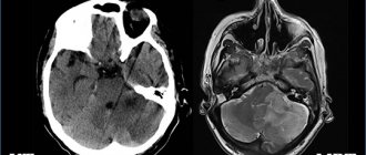
Cystic structure of brain metastases on MRI
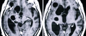
The brain is studied using high-field equipment with a power of 1.5 Tesla, which allows creating detailed and clear images. With magnetic resonance imaging, monochrome sections of the area under study are obtained in increments of 1-2 mm. On MRI images, brain cysts appear as round formations of varying diameters with clear contours. The deviation can be determined by the change in the signal. The intensity of the latter will be different compared to healthy areas. The difference is clearly visible in the photographs. Special scanning modes allow you to obtain more detailed information about the cyst and determine its type:
- Epidermoid. It is usually located in the area of the cerebellopontine angle, prepontine cistern, quadrigeminal tract, ventricles, and less commonly in the hemispheres. Growth is slow, with the risk of compression of the brain stem, entrapment of nerves and blood vessels. There are signs of inflammation around the epidermoid cyst. The intensity of the signal from the formation is usually heterogeneous, but corresponds to the cerebrospinal fluid (if there is no fat inside). On DWI and FLAIR images, the cyst appears lighter than the brain fluid. Epidermoids with fat inside T1-weighted images give a hyperintense signal, on T2 - hypointense (compared to the cerebrospinal fluid).
- Dermoid. Usually the formation is located in the midline of the skull. Due to the high lipid content, T1 imaging gives a hyperintense signal. Unlike the epidermoid, it always has a heterogeneous structure.
- Lipoma. Fatty cysts are most often located in the area of the corpus callosum, interhemispheric fissure, pituitary infundibulum and hypothalamus. MRI scans reveal clear contours of the tumor. Mass effect and swelling of surrounding tissues are not observed. On T1 WI the cyst is sharply hyperintense. On T2 VI it is the opposite (compared to cerebrospinal fluid). Calcifications and vessels look like hypointense areas in the structure of the formation.
- Ependymal. A rare type of cyst that forms under the membrane lining the brain cavity. On MR scans it looks like a formation with clear contours, the signal intensity corresponds to the cerebrospinal fluid.
- Arachnoid. It is formed as a result of the accumulation of cerebrospinal fluid between the sheets of the arachnoid membrane of the brain, most often in the middle cranial fossa, interhemispheric fissure, cerebellopontine angle, at the level of the quadrigeminal. On T1, the VI looks somewhat lighter than the cerebrospinal fluid. In FLAIR mode it produces a hypointense signal.
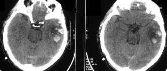
Arachnoid cyst (indicated by arrow) on an MRI scan
- Neuroglial. Found in the cerebral parenchyma, in the area of the choroid plexus of the ventricles. In the photographs it looks like a round formation with liquor contents, often combined with anomalies of brain development.
- Colloidal. Usually located in the anterior superior part of the third ventricle, between the foramina of Monroe. It has a round shape, a fibrous capsule, and clear contours. In the presence of protein in the contents, it gives a hyperintense signal on T1 VI and a hypointense signal on T2 VI. Contrast does not accumulate.
- Rathke's pouch cyst. Rarely seen. Located in the area of the sella turcica. The signal intensity depends on the nature of the content. When serous, it gives a typical fluid response. Mucoid contents are characterized by a hyperintense signal on T1-weighted images. When contrasting, accumulation of the substance is observed in the capsule area.
Arachnoid cyst (circled in red) on MRI
An experienced radiologist knows exactly what a cyst looks like in an MRI photo. An ordinary person will not be able to independently identify and differentiate education. In doubtful cases, it makes sense to show the research results to several specialists.
Causes of cysts
Congenital formations can be a consequence of an unfavorable pregnancy, complicated by fetal hypoxia, taking teratogenic drugs, Rh conflict and other factors that have a negative impact on the fetus. This also includes the intoxicating effects of drugs, alcohol, and nicotine.
An acquired brain cyst is primarily the result of degenerative processes against the background of:
- TBI;
- abscesses;
- acute brain disorders;
- parasitic infection, etc.
Cyst-forming complications are possible after intracranial surgery. Sometimes an existing cyst is provoked to growth by cranial injuries, infections, inflammations, vascular pathologies, and hydrocephalus.
"Scary" names
What is a cyst? A cyst (its other name is “cerebrospinal fluid cyst”) is a cavity filled with liquid, having a capsule that isolates it from other liquor-containing spaces.
with a retrocerebellar cyst consult a neurosurgeon . And although this phrase frightens and even makes parents panic, it must be said that with these words radiologists describe a variant of the norm. Most often, this is not a cyst at all (that is, not an isolated cavity filled with fluid), and it does not require surgical treatment.
Children under two years of age diagnosed with a retrocerebellar cyst should be shown to a neurosurgeon to avoid the development of hydrocephalus.
Another normal variant is a pineal gland cyst . Perhaps the most common incidental finding on MRI. Does not require surgical treatment.
An epidermoid or dermoid cyst is not a true cyst. It is filled not with liquid, but with skin appendages - follicles, sebaceous glands, hair, cartilage tissue, etc. In its structure, it is more similar to a tumor and requires the same treatment as for a tumor - removal.
A porencephalic cyst usually occurs in the place where part of the brain has died as a result of hypoxia or hemorrhage; this place is filled with fluid. In itself, such a cyst does not interfere with development and does not require mandatory surgical treatment.
Pseudocyst - looks like a cyst, but is not a cyst, a cavity that is not limited by anything, it has communication with other parts of the skull. Does not require surgical treatment.
a cyst of the transparent septum is a normal variant, but if it is large in size, it can lead to impaired circulation of the cerebrospinal fluid, which requires surgical treatment.
For children in whom the cerebrospinal fluid cyst rapidly enlarges, causes increased intracranial pressure, neurological symptoms or seizures, surgical treatment is indicated.
If your child has a photo taken and a cyst is found, don’t panic! Having received a description of the x-ray image, first of all, pay attention to the conclusion. If the report says: “There are no pathological abnormalities,” most likely you will not need any surgical intervention. Even if you see the word “cyst” somewhere, most likely the radiologist needed it to describe a normal condition. Just in case, check with him whether you need a consultation with a neurosurgeon.
Symptoms of cystic tumors
Symptoms of intracranial hypertension, most characteristic of the clinical picture of a brain cyst:
- cephalalgia (headache);
- feeling of nausea of non-food origin;
- low performance;
- sensation of “squeezing out” the eyeballs;
- sleep disorders;
- visual dysfunctions;
- ataxia;
- tremor of the limbs;
- fainting states.
With high intracranial pressure, vomiting is repeated.
In some cases, the first symptoms of a brain cyst are expressed by epileptic manifestations, which subsequently recur regularly. The formation can be complicated by ruptures, compression of brain tissue, hemorrhages from ruptured vessels, and persistent epilepsy. In childhood, cysts, accompanied by hypertension or epileptic paroxysm, provoke mental retardation up to mental retardation.
Healing procedures
Conservative methods of treating brain cysts are not effective. A positive result is achieved quickly. However, active therapy is not required if the formation is small and does not increase over time. The patient is under observation with regular instrumental diagnostics to monitor the condition.
Neurosurgical intervention is required if the cyst:
- manifested by signs of hydrocephalus;
- actively growing;
- compresses the tissues inside the skull;
- complicated by hemorrhages or ruptures.
The method of performing the operation is determined by the neurosurgeon based on the patient’s condition: endoscopy, trepanation, external ventricular drainage. Drug treatment is included at the rehabilitation stage. The recovery complex includes exercise therapy, physiotherapy, massage, etc.
Cyst in the brain in an adult
A cyst is a cavity containing fluid located in the brain itself. The cyst can be located anywhere in the skull, but most often it is localized in the cobweb-like mesh covering the cortex of the cerebral hemispheres. These hemispheres are most susceptible to damage and various types of inflammation.
This disease may not manifest itself in any way, but may cause pain. It all depends on the size of the cyst and its location. If the cyst is small and does not grow over time, a person may have it all his life and not be aware of its presence.
There is no need to treat such a disease, but you should be examined regularly, since the cyst can begin to grow at any time. If the diagnosis has been established, the patient must adhere to all the doctor’s instructions, and, if necessary, agree to surgical intervention.
Forecast and preventive measures
For inactive brain cysts of small volumes, the prognosis is usually favorable if they do not grow and do not cause concern to the patient. The same can be said for significant formations that have undergone adequate surgical intervention in a timely manner.
Formed adhesions after surgical operations can provoke the resumption of epileptic seizures. Moderate manifestations of hypertension may also be residual.
For acquired cysts, correctly formulated therapy (using neuroprotectors and absorbable drugs) is required, taking into account the treatment of the disease that provoked the brain cyst. Prevention of congenital cysts means proper management of pregnancy, obstetric care, and preservation of the fetus from negative factors.
Click the “SIGN UP” button and come to the Consultative and Diagnostic Center (formerly the National Diagnostic Center) for diagnosis and treatment.